Transcription
BIBLIOGRAPHIC INFORMATION SYSTEMJournal Full Title: Journal of Biomedical Research &Environmental SciencesJournal NLM Abbreviation: J Biomed Res Environ SciJournal Website Link: https://www.jelsciences.comJournal ISSN: 2766-2276Category: MultidisciplinarySubject Areas: Medicine Group, Biology Group, General,Environmental SciencesTopics Summation: 128Issue Regularity: MonthlyReview Process type: Double BlindTime to Publication: 7-14 DaysIndexing catalog: Visit herePublication fee catalog: Visit hereDOI: 10.37871 (CrossRef)Plagiarism detection software: iThenticateManaging entity: USALanguage: EnglishResearch work collecting capability: WorldwideOrganized by: SciRes Literature LLCLicense: Open Access by Journal of Biomedical Research& Environmental Sciences is licensed under a CreativeCommons Attribution 4.0 International License. Based on awork at SciRes Literature LLC.Manuscript should be submitted in Word Document (.doc orOnline Submissionform or can be.docx) throughmailed to support@jelsciences.comVisionJournal of Biomedical Research & Environmental Sciences main aim is to enhance the importance of science and technology to the scientific community and also toprovide an equal opportunity to seek and share ideas to all our researchers and scientists without any barriers to develop their career and helping in their developmentof discovering the world.
CASE REPORTFluid Dynamic Transcrestal SinusFloor Elevation Using a New SurgicalInstrument, Flusilift and HyaluronicAcid as Only Biomaterial: A CaseReportAlessandro Scarano1*, Roberto Luongo2, Mario Rampino3, EugenioPedulla4 and Calogero Bugea5Private Practice Francavilla Fontana (BR), ItalyPrivate Practice Bari, Italy3Private Practice Brindisi, Italy4Department of General Surgery and Medical-Surgical Specialties, University of Catania, Italy5Private Practice Gallipoli, Italy12*Corresponding authorABSTRACTDespite validated surgical techniques and the development of biomaterials, the proceduresaimed at increasing the maxillary bone volume by sinus floor elevation have complications withvarious degrees of relevance.The perforation of the Schneiderian membrane is one of the most frequent events whileperforming the detachment of the membrane and it can increase the risk of iatrogenic sinusitis,impairment of functional homeostasis, dispersion of the graft material in the antral cavity as well asits bacterial colonization with a subsequential failure of the procedure.This report presents a case where transcrestal sinus lift was performed using Flusilift (Sweden& Martina, Due Carrare PD), a new instrument that allows fluid dynamic elevation of the sinus floorusing saline solution to detach the Schneider’s membrane in an atraumatic way without using a sinuselevator and obtain an adequate alveolar ridge regeneration using hyaluronic acid in gel formulationto support an implant placement.Hyaluronic acid seems to play a key role in wound healing and contributes to a faster boneneoformation in bone regeneration procedures.INTRODUCTIONAlessandro Scarano, Private Practice, ViaMons Alberico Semeraro, 19 72021 FrancavillaFontana (BR), ItalyE-mail: alessandroscarano82@gmail.comORCID ID: 0000-0002-4390-7994DOI: 10.37871/jbres1381Submitted: 17 Decmeber 2021Accepted: 27 December 2021Published: 29 December 2021Copyright: 2021 Scarano A, et al. Distributedunder Creative Commons CC-BY 4.0OPEN ACCESSKeywords Transcrestal sinus floor elevation Bone regeneration Flusilift Hyaluronic acidThe Sinus Floor Elevation technique, first described by Boyne and James in1980 [1], aims to obtain an adequate bone regeneration in order to perform implantsurgery and place implants within maxillary sinus in atrophic maxillary alveolarridge.The procedure is performed by osteotomy of the lateral wall of the maxillarysinus, followed by Schneiderian membrane elevation and placement of autogenousparticulate bone graft between the membrane and the alveolar ridge. Boneaugmentation is generally provided by grafting the sinus cavity with autogenousbone, bone substitutes, or a combination of these biomaterials [2].MEDICINE GROUPSURGERYDENTISTRYORTHOPEDICSVOLUME: 2 ISSUE: 12 - DECEMBERHow to cite this article: Scarano A, Luongo R, Rampino M, Pedulla E, Bugea C. Fluid Dynamic Transcrestal Sinus Floor Elevation Usinga New Surgical Instrument, Flusilift and Hyaluronic Acid as Only Biomaterial: A Case Report. J Biomed Res Environ Sci. 2021 Dec 29;2(12): 1267-1273. doi: 10.37871/jbres1381, Article ID: JBRES1381, Available at: https://www.jelsciences.com/articles/jbres1381.pdf
Transcrestal Sinus Floor Elevation (tSFE) was firstsubstitutes are used in sinus augmentation procedures.described by Tatum [3] and subsequently modified byMany biomaterials have been developed and suggested as aSummers [4,5].valid option to the autogenous bone, such as xenogenic andIn 1994, Summers introduced the osteotome technique,a minimally invasive modification to sinus lift that canbe used to place implants in the same sinus surgery or toprepare a future implant placement site.Osteotome-root analog instruments are used to preparethe crestal osteotomy site. The osteotome is inserted throughthe edentulous alveolar crest at the lower border of themaxillary sinus floor and produces a fracture at the corticalbone of the sinus floor, leaving the Schneiderian membraneintact. Then, the condensing of the bone graft materialapplies lateral and apical pressure, resulting in separationof the Schneiderian membrane from the floor of the sinus,creating new space for the graft between the membrane andthe sinus floor and allowing the placement of an implant ofadequate length.The crestal approach is highly effective, with an eccellentimplant and prosthetic survival rate, low intraoperatorycomplications and low morbidity compared to the lateralapproach. However, tSFE proved to be safe only when anelevation threshold of 5 mm without bone graft and implantplacement was estimated [6-8].A further development of the technique proposed thepreparation of the implant site through drills with depthstops which avoid sinus floor perforation during thepenetration phase and allows a subsequently biomaterialgraft placement in the site.In 2002, Fugazzotto [9] suggested that the pristine boneat sites of implant placement could be drilled up to the sinusfloor with a trephine bur and used to fracture the sinus floorby hydraulic pressure through osteotomes. Since then, manysurgical techniques with specially designed instrumentsfor the transcrestal approach have been reported in theliterature.In literature there are surgical techniques whichsuggest the elevation of the Schneiderian’s membrane byusing hydraulic pressure, following the Pascal principle [10].The introduction of a pressured fluid tends to detach themembrane in a controlled and homogeneous way, avoidingperforations even in alveolar ridge with reduced height. Theprocedure is followed by the insertion of a bone substitutebiomaterial graft within the sinus and by the placement ofan implant in the same appointment.Autogenous bone grafts are considered the goldstandard in bone regeneration techniques. Boyne, Jamesand Tatum first reported the use of autogenous grafts insinus floor elevation. To reduce the volume of autogenousbone to harvest and the morbidity of the donor area, boneallogenic grafting materials, other natural and syntheticbiomaterials. Autogenic, allogenic, xenogenic, and syntheticbiomaterials are currently on-the-board options for adental bone grafting process. Absence of immunologicalresponses and a high-volume augmented bone should belisted as the main advantages of autogenic grafts, on theother side they showed a higher infection rate. Other naturalbiomaterials such as xenogenic grafts can also be employeddue to their feeble induction of inflammatory reactions andhigh durability. Synthetic biomaterials such as bioactiveglasses are also another promising way to perform boneaugmentation considering their notable neosynthesizedbone and low amount of residual graft.Over the years, allografts, alloplasts, and xenograftsof various types have been used alone, or in combinationwith autografts. These grafting materials were reported asosteogenetic, osteoconductive or osteoinductive [11,12].However, these biomaterials are not exempt fromcomplications linked to their placement or their nature.For example, during bone like biomaterial graft packing,in cases where a perforation with rents or tears 10 mm hasbeen produced, there is a risk of partial or complete loss ofthe graft material into sinus cavity, that can lead to ostiumobliteration, postoperative sinusitis or sinus teinfection, which can spread to the graft. Scarano, et al. [13]examined bacterial proliferation into the grafted biomaterialin sinus cavities. The sample was sent for a histopathologicexamination that detect the spread of infection from animplant surface to the entirety of the graft in the maxillaryantrum. Complete removal of all infected bone graft materialis the treatment to choose in such cases.Unlike particulate biomaterials, hyaluronic acid ingel formulation can avoid these complications due to theseverely reduced risks of perforation during the membraneelevation procedure and, moreover, due to its fluidity andsolubility that makes ostium obliteration very unlikely incase of gel graft displacement [14-17].MATERIALS AND METHODSKit miseThe M.I.S.E. EVO Kit (Sweden & Martina, Due CarrarePD) is a system that allows the maxillary sinus to beatraumatically and gradually augmented to a height of 5-10mm above its initial level. The bending of the cortical boneand the overcoming of the phase of elastic deformation untilthe bone breaks to allow the insertion of a reconstructionbiomaterial and an implant are guaranteed by gradual andScarano A, et al. (2021) J Biomed Res Environ Sci, DOI: https://dx.doi.org/10.37871/jbres13811268
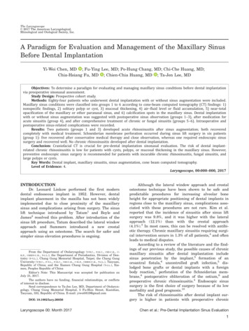
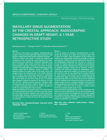
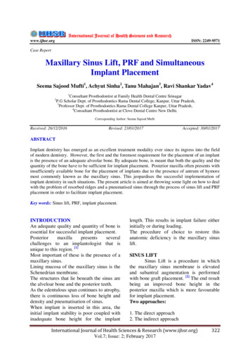
atraumatic series of steps of 1 mm each, using depth stops.The significant advantage of this system with respect toconventional osteotomy techniques is the use of drills that,when used with depth stops, make it possible to graduallyand predictably raise the Schneiderian membrane in steps of1 mm at a time, conserving its integrity. This technique alsoavoids the need to open a lateral window [18-24].FlusiliftFlusilift is a device that allows a fluid dynamic transcrestalsinus lift elevation, using a constant pressure created byFlusilift handpiece by the extrusion of a low viscosity fluid(reticulated hyaluronic acid) that gently detach the way fromthe membrane and, at the same time, fill the newly formedspace between the alveolar ridge and the membrane.The kit is composed by a handpiece, a ferrule and threetips of different diameters.Following the Pascal Law, the extruded fluid applies ahomogeneous pressure on the Schneiderian’s membranewhich permits a gentle elevation in every clinical situationwithout perforation risks linked to sinus elevators.The instrument has the shape of an osteotome with arounded tip, hollow inside on which a syringe is insertedthrough a plastic junction. Once injected, the biomaterialgel or the saline solution flows within the Flusilift andextrudes from two holes placed 1 mm below the rounded tip,on the lateral side and opposite to each other. In this way,the injected biomaterial uplifts the membrane and remainswithin the sinus, promoting bone neoformation.Hyaluronic acidTo our knowledge, there are not any articles that evaluatethe effectiveness of hyaluronic acid as a surrogate for bonederived biomaterials on humans, but there are many papersthat assess the association between hyaluronic acid andbiomaterials. These studies highlight how hyaluronic acidcould boost bone regeneration through a faster mesenchymalcells’ differentiation along with a facilitated mineralizationof the cellular matrix.This paper shows how a minimally invasive approach,in association with an osteoinductive biomaterial, couldminimize the patient morbidity, risks, intra or postoperativecomplications and costs while speeding up the surgicaltiming.CASE PRESENTATIONChief complaintA 37-year-old female, non-smoker, who was medicallyfit and had no bruxism, consulted our practice with acomplaint of a missing upper molar (Figure 1). The tooth wasextracted several years earlier due to a periodontal problem.Figure 1 Preoperative view.History of present illnessMissing upper molar with adequate dimensions andkeratinized gingiva in #17 area were found for furtherprosthetic restoration.History of past illnessShe had no significant medical history.Imaging examinationsCone-Beam Computed Tomography (CBCT) showed asevere vertical bone loss with a mean Residual Bone Height(RBH) of 3 mm and a thickness of the Schneider’s membraneof about 6 mm (Figures 2,3).FINAL DIAGNOSISThe final diagnosis was severe vertical bone loss with aRBH lower than 4 mm.TREATMENTBefore the surgery, the patient signed an informedconsent form. The developed treatment plan, based on thepatient’s condition, was discussed with the patient. Theprocedure began by patient rinsing her mouth with 0.12%chlorhexidine mouthwash for 1 minute. Articaine 4% withadrenaline 1:100000 (PIERREL) was administered as localanesthesia. Antibiotic therapy with Amoxicillin 1 g every 12hours for 6 days, rinses with chlorhexidine 0.12% for 10 daysstarting the day prior the surgery and dexibuprofen (Seractil400 mg) after the surgery and every 12 hours if needed.A palatal incision with intrasulcular extension on adjacentteeth was chosen as the best design to enhance visibility andto avoid overlap between the suture and the implant site andpossible exposures of the surgical site (Figure 4). After thefull thickness flap elevation, the implant site was preparedwith MISE kit (Sweden & Martina, Due Carrare PD) followingthe producer indications.Scarano A, et al. (2021) J Biomed Res Environ Sci, DOI: https://dx.doi.org/10.37871/jbres13811269
the cylindrical drills, was measured using the depth gauge ofthe MISE kit.Then we used the Chamfered drill with ø 3.0 mm (C3.0),fitting it with the depth stop (2 mm) corresponding to thedepth measured with the depth gauge, which would bringthe working length to 1 mm from the cortical floor. We usedthe handpiece at a speed of 800 rpm, ensuring adequateexternal irrigation. Then we moved on to the second depthstop (3 mm), which would increase working height by 1 mmwith respect to the first stop.Figure 2 Cross sectional CBCT view.Proceeding with this sequence, we were able to feelthe cortical floor with the third depth stop (4 mm). Then,we fitted the fourth stop (5 mm). During these final steps,a substantial bending of the cortical bone was generatedwithout breakage, lifting the maxillary sinus floor by about2 mm.Given the particular shape of the tip, the Chamfereddrill is able to not only to deform the cortical bone of thesinus floor, but also to break it if it is particularly thin. Atthe moment of the breakage of the cortical bone, the stopguarantees extremely limited penetration beyond the sinusfloor, of about 0.5 mm on average. This avoids significantdamage to the Schneiderian membrane. In this case thebreakage occurred with the fifth stop (6 mm) onto thechamfered drill.Figure 3 CBCT views.After breaking through the cortical bone, we proceededwith the boring of the fractured cortical bone at low speed(100 rpm), using the Rounded drill with ø 3.0 mm (R3.0) andfitting the same stop used in the phase of breakage (6 mm)first, and then the 7 mm stop (Figure 6).Then, inserting Flusilift for 8 mm (2 mm over the lengthat which we broke of the sinus floor), we injected 0.5 cc ofsaline solution beneath the sinus to evaluate, through theaspect of the suctioned saline, the integrity of the sinusmembrane. Then 2.4 cc of cross linked high molecularweight Hyaluronic Acid (xHya) (Hyadent, Regedent, Zurich)Figure 4 Handpiece with stop.The initial drill was used with ø 2 mm stop at a speed of800 rpm to ream the guide hole for the following drills. Anadequate depth stop (2 mm) was chosen to ensure that thepreliminary preparation reached 1 mm from the maxillarysinus floor (Figure 5). Then the intermediate drill with ø2.50 mm at 800 rpm with the same depth stops was usedto enlarge the site preparation. The stop guaranteed thatthe residual bone thickness of 1 mm beneath the sinus floorremained unaltered. Then the implant socket, prepared withFigure 5 Occlusal view of the preparation.Scarano A, et al. (2021) J Biomed Res Environ Sci, DOI: https://dx.doi.org/10.37871/jbres13811270
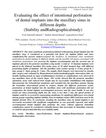
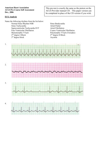
Intraoral periapical x-rays were performed to evaluatethe sinus elevation and filling at 7, 8 and 12 months as aradiographic follow-up (Figure 11).After 7 months a CBCT was performed, the implantwas uncovered, and the rehabilitation was finalized with amonolithic zirconium screwed crown (Figures 12,13).Figure 6 Occlulsal view, note the membrane.Figure 9 Occlusal view of the implant.Figure 7 Flusilift in action.Figure 10 Suture.Figure 8 Impant insertion.was injected (Figure 7). Contextually, a 3.8 x 13 mm Sweden &Martina CSR-DAT implant with a Zir-Ti surface was insertedwith an implant handpiece (Figures 8,9) and the final torquewas recorded with a dynamometric key (30 Ncm), an ISQof 72 was detected, a cover screw was positioned and thenthe wound was sutured with a 4/0 monofilament polyamidesuture (Figure 10).Figure 11 Periapicall x-rays postoperative (left), healing abutment (middle),follow up (right).Scarano A, et al. (2021) J Biomed Res Environ Sci, DOI: https://dx.doi.org/10.37871/jbres13811271
Figure 12 CBCT after healing.Figure 13 CBCT cross sections.RESULTSThe follow up showed radiopacity at 0 and -1 mm fromthe fixture apex, so we obtained an Endo Sinus Bone Gainof 9/10 mm in comparison to the pre-operative situation.Moreover, it was possible to detect bone neoformation allCONCLUSIONThe future goal of oral surgery is to obtain the samequality and amount of bone regeneration with less invasivetechniques and, therefore, fewer complications.or bleeding.The fluid dynamic technique described in this case report,in association with the use of hyaluronic acid as biomaterial,aims to shorten the surgical times and to obtain great bonevolume regeneration with a better bone quality comparedto the bone quality obtained with granules biomaterials.Furthermore, this technique can be less invasive, with fewerintra and postoperative risks. Further studies are requiredto fully evaluate the features of the procedure in a variety ofclinical situations.DISCUSSIONACKNOWLEDGEMENTSaround the implant and even above it. Thus, considering thefact that no other graft biomaterials apart from hyaluronicacid were used during the surgery and knowing thathyaluronic acid has a high resorbable rate and is radiolucent,we can hypothesize that the radiopaque area surroundingthe implant is newly formed bone all around. The patientdid not complain of pain or show signs of edema, bruisingThe crestal and fluid dynamic approach, together withthe use of Hyaluronic acid as biomaterial, could be a validalternative to the lateral approach to obtain great newbone volumes in the maxillary sinus, in patients with lowresidual bone height ( 4 mm). The great endo-sinus bonegain is probably correlated to the high implant lengthprotrusion (10 mm), as already demonstrated in literature[20]. The high thickness of the Schneider’s membrane didnot influence the bone regeneration that occurred up to theThe authors would like to thank Dr. Cinzia Spagnolo, Dr.Elisabetta Guerrazzi and Marilisa Prezioso for their help andcontribution.References1.Boyne PJ, James RA. Grafting of the maxillary sinus floor with autogenous marrowand bone. J Oral Surg. 1980 Aug;38(8):613-6. PMID: 6993637.2.Mc Gowan DA, Baxter PW, James J. The maxillary sinus and its dental implications.Oxford, UK: Wright, Butterworth-Heinemann Ltd; 1993. https://bit.ly/3EwUWKA3.Tatum H Jr. Maxillary and sinus implant reconstructions. Dent Clin North Am. 1986Apr;30(2):207-29. PMID: 3516738.4.Summers RB. A new concept in maxillary implant surgery: the osteotome technique.Compendium. 1994 Feb;15(2):152, 154-6, 158 passim; quiz 162. PMID: 8055503.5.Summers RB. The osteotome technique: Part 4--Future site development. Compendapex of the implant. The absence of pain, edema, bruising orbleeding could be correlated to the mini-invasive nature ofthe surgery.Scarano A, et al. (2021) J Biomed Res Environ Sci, DOI: https://dx.doi.org/10.37871/jbres13811272
Contin Educ Dent. 1995 Nov;16(11):1090, 1092 passim; 1094-1096, 1098, quiz 1099.PMID: 8598008.14. Giovanni Abatangelo, Paola Brun. Integrated Biomaterials Science. Tissue Engineering6.Farina R, Franceschetti G, Travaglini D, Consolo U, Minenna L, Schincaglia GP, RiccardiO, Bandieri A, Maietti E, Trombelli L. Morbidity following transcrestal and lateral sinusfloor elevation: A randomized trial. J Clin Periodontol. 2018 Sep;45(9):1128-1139. doi:10.1111/jcpe.12985. Epub 2018 Aug 3. PMID: 29992594; PMCID: PMC6175473.15. Cardaropoli G, Araújo M, Lindhe J. Dynamics of bone tissue formation intooth extraction sites. An experimental study in dogs. J Clin Periodontol. 2003Sep;30(9):809-18. doi: 10.1034/j.1600-051x.2003.00366.x. PMID: 12956657.7.Gargallo-Albiol J, Tattan M, Sinjab KH, Chan HL, Wang HL. Schneiderian membraneperforation via transcrestal sinus floor elevation: A randomized ex vivo study withendoscopic validation. Clin Oral Implants Res. 2019 Jan;30(1):11-19. doi: 10.1111/clr.13388. Epub 2018 Dec 23. PMID: 30450593.8.Al-Moraissi EA, Altairi NH, Abotaleb B, Al-Iryani G, Halboub E, Alakhali MS. What Isthe Most Effective Rehabilitation Method for Posterior Maxillas With 4 to 8 mm ofResidual Alveolar Bone Height Below the Maxillary Sinus With Implant-SupportedProstheses? A Frequentist Network Meta-Analysis. J Oral Maxillofac Surg. 2019Jan;77(1):70.e1-70.e33. doi: 10.1016/j.joms.2018.08.009. Epub 2018 Aug 22. PMID:30243705.9.Fugazzotto PA. Immediate implant placement following a modified trephine/osteotome approach: success rates of 116 implants to 4 years in function. Int J OralMaxillofac Implants. 2002 Jan-Feb;17(1):113-20. PMID: 11858567.10. Sotirakis EG, Gonshor A. Elevation of the maxillary sinus floor with hydraulic pressure.J Oral Implantol. 2005;31(4):197-204. doi: 10.1563/1548-1336(2005)31[197:EOTMSF]2.0.CO;2. PMID: 16145848.11. Scarano A, Degidi M, Iezzi G, Pecora G, Piattelli M, Orsini G, Caputi S, Perrotti V,Mangano C, Piattelli A. Maxillary sinus augmentation with different biomaterials: acomparative histologic and histomorphometric study in man. Implant Dent. 2006Jun;15(2):197-207. doi: 10.1097/01.id.0000220120.54308.f3. PMID: 16766904.12. Stacchi C, Spinato S, Lombardi T, Bernardello F, Bertoldi C, Zaffe D, Nevins M.Minimally Invasive Management of Implant-Supported Rehabilitation in the PosteriorMaxilla, Part I. Sinus Floor Elevation: Biologic Principles and Materials. Int JPeriodontics Restorative Dent. 2020 May/Jun;40(3):e85-e93. doi: 10.11607/prd.4497.PMID: 32233183.13. Scarano A, Cholakis AK, Piattelli A. Histologic Evaluation of Sinus Grafting MaterialsAfter Peri-implantitis-Induced Failure: A Case Series. Int J Oral Maxillofac Implants.2017 Mar/Apr;32(2):e69-e75. doi: 10.11607/jomi.5303. PMID: 28291856.New York: Kluwer Academic/Plenum; 2002.16. Sasaky T, Watanabe C. Stimulation of osteoinduction in bound wound healing by highmolecular hyaluronic acid. Bone 1995;16:9-15.doi:10.1016/8756-3282(95)80005-B17. Brunel G, Piantoni P, Piotrowski B, Baysse E. Action of Hyaluronic acid on the woundhealing following extraction. Dent Inform 2004;7:385-91.18. Lopez MA, Casale M, Candotto V, Papalia R, Bressi F, Carinci F. The use of hyaluronicacid as a support of two different micronized biomaterials in crestal sinus liftprocedures. A report on two case studies with volume comparison. J Biol RegulHomeost Agents. 2017 Dec 27;31(4 Suppl 2):129-138. PMID: 29202573.19. Sisti A, Canullo L, Mottola MP, Iannello G. A case series on crestal sinus elevation withrotary instruments. Eur J Oral Implantol. 2011 Summer;4(2):145-52. PMID: 21808764.20. Esposito M, Grusovin MG, Coulthard P, Worthington HV. The efficacy of variousbone augmentation procedures for dental implants: a Cochrane systematic reviewof randomized controlled clinical trials. Int J Oral Maxillofac Implants. 2006 SepOct;21(5):696-710. PMID: 17066630.21. Carusi G. The Minimal Invasive Sinus Elevation Technique. Scientific Poster, 15 European Association for Osseointegra- Tion, 5-7 Oct 2006; Scientific Poster, 12 International FRIADENT Symposium, 24-25 March 2006.22. Cosci F, Luccioli M. A new sinus lift technique in conjunction with placement of265 implants: a 6-year retrospective study. Implant Dent. 2000;9(4):363-8. doi:10.1097/00008505-200009040-00014. PMID: 11307560.23. Bernardello F, Righi D, Cosci F, Bozzoli P, Soardi CM, Spinato S. Crestal sinus lift withsequential drills and simultaneous implant placement in sites with 5 mm of nativebone: a multicenter retrospective study. Implant Dent. 2011 Dec;20(6):439-44. doi:10.1097/ID.0b013e3182342052. Erratum in: Implant Dent. 2013 Apr;22(2):202. Carlo,Maria Soardi [corrected to Soardi, Carlo Maria]. PMID: 21989240.24. Suk-Arj P, Wongchuensoontorn C, Taebunpakul P. Evaluation of bone formationfollowing the osteotome sinus floor elevation technique without grafting using conebeam computed tomography: a preliminary study. Int J Implant Dent. 2019 Aug1;5(1):27. doi: 10.1186/s40729-019-0181-7. PMID: 31367919; PMCID: PMC6669224.How to cite this article: Scarano A, Luongo R, Rampino M, Pedulla E, Bugea C. Fluid Dynamic Transcrestal Sinus Floor Elevation Using a New Surgical Instrument, Flusilift and Hyaluronic Acidas Only Biomaterial: A Case Report. J Biomed Res Environ Sci. 2021 Dec 29; 2(12): 1267-1273. doi: 10.37871/jbres1381, Article ID: JBRES1381, Available at: Scarano A, et al. (2021) J Biomed Res Environ Sci, DOI: https://dx.doi.org/10.37871/jbres13811273
described by Tatum [3] and subsequently modi ed by fi Summers [4,5]. In 1994, Summers introduced the osteotome technique, a minimally invasive modi cation to sinus lift that can fi be used to place implants in the same sinus surgery or to prepare a future implant placement site. Osteotome-root analog instruments are used to prepare