Transcription
SL Dentistry, Oral Disorders And TherapySpecial Issue Article: Dental ImplantsResearch ArticleAssessment the Effectiveness Balloon-Assisted Maxillary Sinus Floor Elevation andTraditional Sinus Floor Elevation in the Posterior Maxillary AreaGagik Hakobyan1*, Asdghig Boyadjian2, Ashot Jilavyan2, Vilen Seyranyan2, Ani Khachatryan2 and SonaGevorgyan21Headof Department of Oral and Maxillofacial Surgery, Yerevan State Medical Universi ty, Armenia2Assistantprofessor of Department of Oral and Maxillofacial Surgery, Yerevan State Medical University, ArmeniaARTICLE INFOReceived Date: February 15, 2021Accepted Date: March 09, 2021Published Date: March 10, 2021KEYWORDSABSTRACTBackground: The posterior maxillary area sometimes has insufficient bone mass fordental implants. The augmentation of the sinus floor allows the implant to be placed inthe posterior of the upper jaw.Purpose: The aim of this study was to compare the effectiveness antral balloonassisted maxillary sinus elevation and traditional sinus floor elevation followed byTraditional maxillary sinus liftBalloon sinus liftBone grafbone graft and delayed implant placement the posterior maxillary area.Material and methods: A total of 68 patients, (aged 27 to 56 years,32 women and 36 men),Copyright: 2021 Gagik Hakobyanet al., SL Dentistry, Oral Disorders AndTherapy. This is an open access articledistributedundertheCreativeCommons Attribution License, whichpermits unrestricted use, distribution,and reproduction in any medium,provided the original work is properlycited.without any systemic diseases,withunilateral/bilateralmissing teeth and atrophy of the posterior maxillary area, who required anenlargement of the sinus prior to implant placement, whom the location of the sinusfloor from the crest was 3-5mm, width 5 mm were included in the study between2018 and 2021. Patients underwent a thorough clinical examination according to thegenerally accepted scheme. All patients were selected after meticulous evaluation oftheir medical histories and dental examinations, including OPG and dental ComputedTomography (CT) scans.Results: The sinus lift using balloon technique was performed successfully in patients 1Citation for this article: GagikHakobyan, Asdghig Boyadjian, AshotJilavyan, Vilen Seyranyan, AniKhachatryan and Sona Gevorgyan.Assessment the Effectiveness BalloonAssisted Maxillary Sinus Floor Elevationand Traditional Sinus Floor Elevation inthe Posterior Maxillary Area. SLDentistry, Oral Disorders And Therapy.2021; 4(1):117group, with no complications. In 9 patients 2 group, perforation of the sinus membraneoccurred during the operation, sinusitis in 4 patients, graft failure in 3 patients.Regardless of the approach used, both approaches showed significant increases inbone mass gain. Though not statistically significant difference, balloon -assistedprocedure showed more mean bone gain (8.4 mm) compared to osteotome -assistedprocedure (8.1 mm). The mean amount of Marginal Bone Loss (MBL) in patients 1group 3 years follow-up was 0.86 mm in patients 2 group showed significantly lessmarginal bone loss 1,16mm. The implant survival rate 3 years follow-up was inpatients 1 group was 97.62%, in patients 2 groups was 95.2%.Conclusions: Research has shown that the balloon sinus lifting offers predictable, safeCorresponding author:Gagik Hakobyan,Head of Department of Oral andMaxillofacial Surgery, Yerevan StateMedical University, Armenia, Tel:( 37410)271146;Email: prom hg@yahoo.comand effective results, and eliminates the complications associated with traditional sidewindow techniques. However, further controlled clinical trials are needed to evaluatethe efficacy and safety of these technique for their appropriate implementation in thefield of oral implanotology.01Assessment the Ef f ectiveness Balloon-Assisted Maxillary Sinus Floor Elev ation and Traditional Sinus Floor Elev ation in thePosterior Maxillary Area. SL Dentistry , Oral Disorders And Therapy . 2021; 4(1):117.
SL Dentistry, Oral Disorders And TherapyINTRODUCTIONperforation [9-11]. H. Tatum in 1986 Transcrestal Sinus FloorConventional removable dentures have multiple drawbacks likeElevation (TSFE) was performed by lifting the sinus floor vialack of stability, minimal retention, discomfort while chewingsequential crestal bone preparations [12]. Later, in 1994,and compromised aesthetic outcome. Dental implants willsummers introduced the osteotomy sinus floor elevation, which isresolve the problems associated with conventional dentures.a minimally invasive technique to localize the elevation of theSufficient alveolar bone to for placement 10 mm long and amaxillary sinus through the alveolar ridge [13]. This approachdiameter of 3.5-4 mm implants has traditionally beenis supposed to offer more patient comfort, more primaryconsidered the minimum requirement to allow bone placementstability, and less morbidity. However, this method has beenof the implant. Due to the extraction of teeth into the segmentshown to be effective only when the crest height exceeds 6 mm.molar and pneumatization of the maxillary sinus, the verticalPerforation of the sinus membrane will result in deposition orheight of the bone in the posterior edentulous upper jawinterruption of the sinus lift procedure. Various modificationsdecreases thus limiting the installation of dental implants [1].have been proposed to prevent complications associated withBone density greatly affects primary implant stability andthe summers sinus floor elevation method [14]. Over the pastsuccess, since implants in areas of lower bone quality aredecade, many authors have developed minimally invasive sinusassociated with a high failure rate [2]. It is important to placelift techniques to overcome the postoperative complicationsimplants in locations with good primary stability, which cannotassociated with traditional sinus lift procedures. Muronoi et al.be acquired in regions with low bone density. Posterior maxillafor the first time it was proposed to enlarge the maxillary sinusoften presents type III or type IV bone quality according tofloor using a balloon [15]. The technique of balloon lift of theLekholm and Zarb‘s classification [3]. Increased pneumatizationantral membrane was introduced through the lateral approachof the maxillary sinus and the quality of the III or IV types of[16]. Thereafter, a technique was described for minimallybone in the posterior part of the upper jaw-all this emphasizesinvasive balloon elevation of the antral membrane using athe need for additional procedures that increase the qualitytranscrestal approach, which included the use of a balloonand quantity of bone [4,5]. One solution in these clinical casesdevice through a 3 mm osteotomy [17-19]. Approach to theis to use shorter implants, which sometimes leads to anantrum through the lateral window and elevation of theunfavorable crown-to-root ratio. Maxillary sinus augmentationShneidar membrane with an antral balloon is the method thathas become the most common surgical procedure that involveshas shown the lowest of membrane perforation. It elevates thedetaching the Schneider membrane from the floor of themembrane easily and makes the antral floor accessible formaxillary sinus, creating a space filled with a bone graft toaugmentation with grafting materials. The development offacilitate vertical bone augmentation in the maxillary sinusminimally invasive sinus lift surgery includes progress incavity, allowing future dental implants to be restored [6].endoscopy, development of intraoperative navigation forBoyne and James in 1980 proposed a conventional sinusmaxillofacial surgery. Decision making includes diagnostic andaugmentation procedure that involves direct visualization andtherapeutic indications, patient preferences and values, andmanipulation of the Schneider membrane through a lateralcost considerations. After the sinus membrane lifting a varietywindow osteotomy (modified Caldwell-Luc approach) [7].of bone grafting materials can be used [20-22]. Since differentAlthough these procedures often ensured high implant survivaltechniques sinus lifting were evaluated in different trials, forand stability of bone tissue levels over time [8]. However theyimplant failures and complications. Based on relevance questionwere not always well accepted by patients due to their highin focus in this study is the antral membrane balloon elevationcost, increased postoperative morbidity, high risk of infectiontechnique effective in the terms of sinus augmentation success(fistula with pus or abscess, often caused by infection of therate, survival rate of dental implants, bone gain, andgraft material) and a long healing time. In addition to being ancomplication rate compared with the traditional sinus floorinvasive surgical procedure, it also presents with post-operativeelevation technique. The aim of this study was to compare theconditionseffectiveness antral balloon-assisted maxillary sinus ent the Ef f ectiveness Balloon-Assisted Maxillary Sinus Floor Elev ation and Traditional Sinus Floor Elev ation in thePosterior Maxillary Area. SL Dentistry , Oral Disorders And Therapy . 2021; 4(1):117.
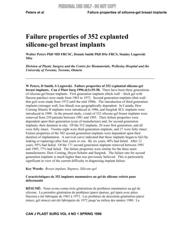
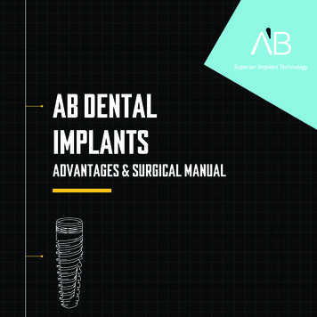
SL Dentistry, Oral Disorders And Therapyand traditional sinus floor elevation followed by bone graftmaxillary area. In 32 patients of 2 group, implantation wasand delayed implant placement the posterior maxillary area.performed after traditional sinus augmentation procedureMATERIAL AND METHODS(Boyne and James method) a that involves direct visualizationyears,and manipulation of the Schneider membrane through a lateralwithwindow osteotomy followed by bone graft and delayedunilateral/bilateral missing teeth and atrophy of the posteriorimplant placement the posterior maxillary segment. Patientsmaxillary area, who required an enlargement of the sinus priorwere started on prophylactic antibiotic treatment 24 hoursto implant placement, were included in the study betweenbefore surgery.Atotalof68patients,32 women and 36 men), without(aged27to56any systemic diseases,2018 and 2021. All patients presented functional and estheticcomplaints. Written informed consent was obtained from allpatients explaining the possible side effects of the procedure.Indications of the technique in the studyThe study included patients in whom the location of the sinusfloor from the crest was 3–5 mm, width 5 mm (from the floorof the sinus to the crest of the bone, as determined radiographically), a minimum follow-up period of 1-year loading.Contraindications of the technique in the studyFigure: 1 Preoperative radiograph.Contraindications included any systemic condition that couldinterfere with physiological wound healing, orofacial cancer,radiation / chemotherapy to the head and neck area,Advanced medical conditions, patients who consumed oralbisphosphonates for more than three years, excessive smoking,alcohol or substance consumption, psychological problems.Local contraindications of sinus lift surgery included untreatedactive periodontal disease, maxillary sinus infections andpathological lesions, chronic sinusitis, alveolar scar possibility,odontogenic infections, allergic rhinitis. Patients underwent athorough clinical examination according to the generallyFigure 2: Bone scraper is used to collect autogenous bone fromsite of the operaration.accepted scheme. All patients were selected after meticulousIn patients 1 group аfter local anesthesia was injected into theevaluation of their medical histories and dental examinations,edentulous ridge, after reflecting a mucoperiosteal flap boneincluding OPG and dental Computed Tomography (CT) scansscraper is used to collect autogenous bone from site of the(Figure 1). The initial height of the bone from the alveolaroperation (Figure 2). Opening a window on lateral wall of theridge to the sinus floor, the width of the ridge and themaxillary sinus by round diamond bur and separating themesiodistal diameter of the edentulous area were measuredShneider membrane from the bony walls of maxillary sinususing CT. To assess the volume of new bone and to monitor(Figure 3). Balloon-assisted maxillary sinus floor elevation wasmaxillary sinus re-pneumatization, CT scans were taken, Thesecarried out using a Zimmer balloon (Zimmer, USA) (Figure 4).tests were conducted after the functional loading of implantsInsertion of Zimmer balloon (Zimmer, USA) in between the sinusand repeated after 1,2,3 years. Patients were divided into 2membrane and the bony walls to detach the remaining part ofgroups, group distribution was performed randomly. In 36the sinus membrane from its bony walls (Figure 5). The balloonpatients of 1 group, implantation was performed after lateralwas then slowly inflated with saline (1 cm3 of salineapproach antral balloon technique for sinus elevation followedcorresponds to 6 mm membrane elevation) until the desiredby bone graft and delayed implant placement the posteriorelevation (usually 11 mm) was achieved. The balloon was03Assessment the Ef f ectiveness Balloon-Assisted Maxillary Sinus Floor Elev ation and Traditional Sinus Floor Elev ation in thePosterior Maxillary Area. SL Dentistry , Oral Disorders And Therapy . 2021; 4(1):117.
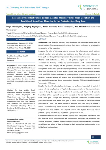
SL Dentistry, Oral Disorders And Therapythen slowly deflated and removed. Mixing the autogeneosmaintaining periosteal integrity. The superoiorinferior andbone which is collected from the site of the operation by aanteroposterior borders of the lateral window are determinedscraper withCerasorb (Curasan, German) cristals along withby the sinus volume, which is preoperatively examined by CT.the patient’s blood Platelet-Rich fibrin (Figure 6). Bone graftmaterial and Platelet-Rich fibrin were inserted into the sinusunder the antral membrane, placement of Resodont (ResorbaWundversorgung GmbH&Co.KG,Germany),resorpablemembrane to cover the lateral wall of the sinus and separate itfrom the mucuperiosteal flap after which the flap wasrepositioned and sutured using 3-0 silk sutures (Figure 7,8,9).Radiographs were taken to assess the degree of sinus floo relevation in the surgical site after the procedure (Figure 10).The augmentation evaluated by CT scans. Patients wereadvised to strictly follow the postoperative instructions. Post-Figure 4: Zimmer balloon used in balloon sinus lift surgery (Zimmer,USA).operative patient reactions including swelling, discoloration,discomfort, hematomas. Implant placement was initiated 6months post-operatively and reviewed at frequent intervals.Loading of the implants was carried out after 6 months. Afterremoving the cover screw, healing plugs were installed andafter 10 days the prosthetic stage of treatment was started(Figure11,12,13).Allpatients wereevaluated radio-graphically after prosthetics (Figure 14) and 6th month, 1, 2, 3years after prosthetics. The crestal bone height was maintainedand verified by subsequent radiographs. In 32 patients ofgroup 2 sinus lift procedure was performed using thetraditional lateral approach method using bone graft materialResodont and Platelet-Rich fibrin.Figure 3: Opening a window on lateral wall of the maxillary sinusby round diamond bur.Figure 5: Insertion of Zimmer balloon in between the sinusmembrane and the bony walls and inflating it with saline solution todetach the remaining part of the sinus membrane from its bonywalls.Figure 6: the autogeneos with Cerasorb (Curasan,German) cristals along with the patient’s blood PlateletRich fibrin.The shape of the osteotomy window rectangular or oval andMidcrestal incision is made in the mesiodistal direction alongoutlined with a size of 10 20 mm. The size of the window cantheincrease or decrease, according to the size of the isionsarecrestand anterior‐ andmade.Afull‐thicknessmucoperiosteal flap with a trapezoid base is elevated whileaugmentation for implant placement. The inferior border of thebony window should be 2-5 mm superior to the sinus floor.04Assessment the Ef f ectiveness Balloon-Assisted Maxillary Sinus Floor Elev ation and Traditional Sinus Floor Elev ation in thePosterior Maxillary Area. SL Dentistry , Oral Disorders And Therapy . 2021; 4(1):117.
SL Dentistry, Oral Disorders And TherapyThe elevation sinus membrane performed using broad‐basedfreers or curettes. The prepared graft material Resodont andPlatelet-Rich fibrin is placed by pieces into the drilled hole,followed by a 6‐month wait. After the bone is placed in thesinus, Resorpable Resodont membrane to cover the lateralwall of the sinus and the mucoperiosteal flap is positioned andprimary closure is achieved. Patients instructed about sinusprecautions, which are avoiding anything that can causesudden pressure changes in the sinus, such as nose blowing withFigure 7: Augmentation of the sinus with bone graft and PlateletRich fibrin.nostrils pinched closed and sneezing with a closed mouth. Atotal of 118 implants were installed in patients 1 group and 96implants were installed in patients 2 group. An implant wasconsidered to have failed (clinical or absolute failure) if it hadany of the following conditions: pain on function, mobility,radiographic bone loss 1/2 the length of the implant,uncontrolled exudate, or was no longer in the mouth.Postsurgical change in Marginal Bone Loss (MBL) was assess arison), 1, 2, 3 years after prosthesis loading. Statisticswere used to calculate and analyze the mean marginal boneFigure 8: Resorpable Resodont membrane to cover the lateralwall of the sinus and separate it from the mucuperiosteal flap.Figure 9: Suturing of the flap.loss of implants.Figure 11: Radiograph after implant placement.Statistical analysesStatistical analyses were performed using SPSS software ver.22.0 (IBM, Armonk, NY, USA), and MedCalc program forWindows. To test the significance of variations in the BOP,PPD, MBL, the t-test was used. The minimum level of statisticalsignificance was set at a value of less than 0.05.Figure 10: Postoperative radiograph.05Assessment the Ef f ectiveness Balloon-Assisted Maxillary Sinus Floor Elev ation and Traditional Sinus Floor Elev ation in thePosterior Maxillary Area. SL Dentistry , Oral Disorders And Therapy . 2021; 4(1):117.
SL Dentistry, Oral Disorders And Therapyfailure in 3 patients. Regardless of the approach used, bothapproaches showed significant increases in bone mass gain.Though not statistically significant difference, balloon-assistedprocedure showed more mean bone gain (8.4 mm) comparedto osteotome-assisted procedure (8.1 mm). The mean amount ofMarginal Bone Loss (MBL) in patients 1 group 3 years follow-upwas 0.86 mm in patients 2 group showed significantly lessmarginal bone loss 1,16mm. In our study, delayed implantplacement was performed due to insufficient initial bone height,as well as to ensure sufficient graft maturation. The implantsurvival rate 3 years follow-up was in patients 1 group wasFigure 13: Intraoral view with abutments before fixation of theprosthetic construction.97.62%, in patients 2 groups was 95.2%.Table 1: The sinus lift complications in1 and 2 groups.Complications1 group2 groupperf oration of the sinus membranesinusitisgraf t f ailure000943Table 2: Bone mass gain after sinus lift procedures in1 and 2groups.Bone mass gain.mm1 group8,4mm2 group8,1mmTable 3: Mean of marginal bone loss (MBL) in patients 1and 2groups 3 years follow-up.Figure 13: Final restoration with metal ceramic bridge.Mean marginal bone loss (MBL)mm1 group2 group0,86mm1,16mmDISCUSSIONSInsufficient bone volume is a common problem encountered inthe rehabilitation of the edentulous posterior maxillae withimplant supported prostheses. Alveolar bone quantity andquality are the most important parameters primarily affectingthe success of implant treatment. The choice of the method ofrehabilitation with an implant for upper jaw atrophy is ofFigure 14: Final postoperative radiograph with restorations.decisive importance. Sinus lift procedures increase bone volumeby augmenting the sinus cavity with autogenous bone and/orRESULTSThesinus lift using balloonbiomaterials [13,23,24]. However, sinus floor elevation surgerytechniquewas performedis generally associated with higher costs, more complex surgicalsuccessfully in patients 1group, with no complications.procedures, and a high prevalence of complications such asIn 9 patients 2 group, perforation of the sinus membraneinfection,occurred during the operation, sinusitis in 4 patients, graft[25,26]. Various techniques have been proposed to overcomesinus membrane perforation and graft failure06Assessment the Ef f ectiveness Balloon-Assisted Maxillary Sinus Floor Elev ation and Traditional Sinus Floor Elev ation in thePosterior Maxillary Area. SL Dentistry , Oral Disorders And Therapy . 2021; 4(1):117.
SL Dentistry, Oral Disorders And Therapythis complication. Outcomes of this procedure may be affectedand PRP, the balloon sinus lift technique offers an effectiveby simultaneous versus delayed implant placement, selection ofapproach for minimally invasive sinus lifts, preventing sinusgraft material, and the surface characteristics of the implants.membraneNumerous articles have been published in this field regardingimproving implant osseointegration into grafted alveolar bone.different grafting materials and modification to the classicBalloon antral sinus lift present an alternative traditional sinustechnique. Minimally invasive balloon antral membrane lift is alift with high survival rate and fewer complications andsurgical technique developed as a less invasive alternative to aimproving implant osseointegration into grafted alveolar bone.lateral window approach that includes sinus lift using the sinusThe process should be refined in order to reduce theballoon followed by standard implant placement [27,28]. Topercentage of mucosa perforation.further simplify sinus lifting procedures and enhance theirCONCLUSIONpredictability and success, additional modifications technique,Research has shown that the balloon sinus lifting offershave been introduced during the past 10 years [29]. Numerouspredictable, safe and effective results, and eliminates thearticles have been published in this field regarding differentcomplicationsmodification to the classic technique as transcrestal approach,techniques. Further controlled clinical trials are needed tolateralhydrodynamicevaluate the efficacy and safety of these techniques for theirultrasonic approach, balloon elevation technique, osteotomyappropriate implementation in the field of oral implanotology .technique and nasal suction technique with their success rate.Conflict of interest and financial disclosureWhich method to give preference when choosing the method ofThe author declares that he has no conflict of interest and theresinus lifting is important for the prevention of complications ofwas no external source of funding for the present study. Nonesurgery and is actual topic for research in the field of oralof the authors have any relevant financial relationship(s) with aimplantology [13,30-34]. The present study was undertaken tocommercial interest.comparison the safety and efficacy of a balloon sinus liftFundingtechnique and traditional sinus lift. To compare the efficacy ofThe work was not funded.balloon sinus lift and traditional sinus lift technique, two groupsConsent statementof patients were formed. In patients of 1 group, lateralWritten informed consent was obtained from the patient forapproach antral balloon technique for sinus elevation followed,publication of this case report and accompanying images.in of 2 group traditional sinus lift procedure followed. TheEthics approvalstudy used anorganic mineral and autogenous bone for theThe study was reviewed and approved by the Ethicsbone augmentation technique, a collagen membrane to protectCommittee of the Yerevan State Medical University after M.the sinus window, and a staged approach for implantHeratsi (protocol N16, 5.10.17) and by those of the Worldplacement. Bone scraper was used to collect autogenous boneMedical Association and the Helsinki Declaration.from the side of the operation before opening of the windowREFERENCESwindowapproach,piezosurgery,and mixed with bone graft material Resodont with along with1.patient’s blood Platelet-Rich fibrin and covered by resorpable2.traditionaltrauma,sideandwindowHansson S, Halldin A. (2012). Alveolar ridge resorptionAdell R, Eriksson B, Lekholm U, Branemark PI, Jemt T.(1990). Long-term follow-up study of osseointegratedsinus lift and balloon antral sinus lift have the advantage ofimplants in the treatment of totally edentulous jaws. Int Jbeing a high survival solution, they are less expensive, requirethus increase patient satisfaction. By incorporating efficient andwithpatientprinciple of bone physiology. J Dent Biomech.ensure sufficient graft maturation. Compared to traditionalless surgical time compared to traditional sinus lift surgery, andassociatedreducingafter tooth extraction: A consequence of a fundamentalmembrane Resodont . In our study, delayed implant placementwas performed due to insufficient initial bone height and toperforation,Oral Maxillofac Implants. 5: 347-359.3.Lekholm U, Van Steenberghe D, Hermann I. (1994).Osseointegrated implants in the treatment of partiallyefficacious materials such as anorganic bone grafting material07Assessment the Ef f ectiveness Balloon-Assisted Maxillary Sinus Floor Elev ation and Traditional Sinus Floor Elev ation in thePosterior Maxillary Area. SL Dentistry , Oral Disorders And Therapy . 2021; 4(1):117.
SL Dentistry, Oral Disorders And Therapyedentulous jaws: A prospective 5-year multicenter study.Int J oral Maxillofac Implants. 9: illaryFelechosa AS. (2014). Minimally invasive sinus lift elevationsinusfollowing extractions: A radiographicstudy. Int J Oral Maxillofac Implants. 23: 48-56.5.Ribeiro-Rotta RF, de Oliveira RCG, Dias DR, Lindh C, ent. An independent 1-year follow-up clinical study.Clin Oral Implants Res. 25: 503.16. Asmael HM, Lateef TA. (2016). An assessment of thedental implant sites part 2: Correlation with bonemaxillary sinus floor elevation surgery. J Baghdad Collclassification and primary stability. Clin Oral Implants Res.Dent. 28: 109-113.of sinus balloontechniqueon transcrestal17. Apparaju V, Vaddamanu SK, Vyas R, Vishwanath S,Misch CE. (1987). Maxillary sinus augmentation forGurumurthy V, et al. (2020). Is balloon-assisted maxillaryendosteal implants: Organized alternative treatment plans.sinus floor augmentation before dental implant safe andInt J Oral Implantol. 4: 49-58.promising? A systematic review and meta-analys.NigerianBoyne PJ, James RA. (1980). Grafting of the maxillaryJ.Clinical Practice. 23: 275-283.18. Rao GS, Reddy SK. (2014). Antral balloon sinus elevation38: 613-616.and grafting prior to dental implant placement: Review ofCorbella S, Taschieri S, Fabbro DM. (2015). Long-term34 cases. Int J Oral Maxillofac Implants. 29: 414-418.outcomes for the treatment of atrophic posterior maxilla: A19. Gandhi Y. (2017). Sinus grafts: Science and techniques-systematic review of literature. Clin Implant Dent Relat Res.17: 120-132.9.aefficacysinus floor with autogenous marrow and bone. J Oral Surg.8.withCR. (2014). Bone tissue microarchitectural characteristics at25: 47-53.6.15. Gonzalez AG, Fernández IC, Frechoso SC, Frechoso C,then and now. J Maxillofac Oral Surg. 16: 135-144.20. Silva LD, Lima VND, Faverani LP, Okamoto R, Pellizzer EP,Farina R, Franceschetti G, Travaglini D, Consolo U,et al. (2016). Maxillary sinus lift surgery-with or withoutMinenna L, et al. (2018). Morbidity following transcrestalgraft material? A systematic review. Int J Oral Maxillofacand lateral sinus floor elevation: A randomized trial. J ClinSurg. 45: 1570-1576.Periodontol. 45: 1128-1139.10. Regev E, Smith RA, Perrot DH, Pogrel MA. (1995).Maxillarysinus complications related to endosseousimplants. Int. J. Oral Maxillofac. Implant. 10: 451-461.11. Schwarz L, Schiebel V, Hof M, Ulm C, Watzek G, et al.(2015).21. Ali S, Bakry SA, Abd-Elhakam H. (2015). Platelet-Richfibrin in maxillary sinus augmentation: A systematic review.J Oral Implantol. 41: 746-753.22. Kempraj J, Sundaram SS, Doss GPT, Nakeeran KP, RajaVBKK.(2020).MaxillarySinus AugmentationUsingRiskfactors of membrane perforation andXenograft and Choukroun’s Platelet-Rich Fibrin as Graftingpostoperativecomplications in ssinus floor elevationMaterial: A Radiological Study. Journal of Maxillofacialsurgery: Review of 407 augmentation procedures. J OralMaxillofac Surg. 73: 1275-1282.and Oral Surgery. 19: 263-268.23. Chen MH, Shi JY. (2018). Clinical and radiological12. Summers RB. (1994). The osteotome technique: Part 3-lessoutcomes of implants in osteotome sinus floor elevation withinvasive methods of elevating the sinus floor. Compendium.and without grafting: a systematic review and a meta-15: 698-700.analysis. J Prosthodont. 27: 394-401.13. Starch-Jensen T, Jensen JD. (2017). Maxillary sinus floor24. Esposito M, Grusovin MG, Rees J, Karasoulos D, Felice P, etaugmentation: A review of selected treatment modalities. Jal. (2010). Effectiveness of sinus lift procedures for dentalOral Maxillofac Res. 8: e3.implant rehabilitation: a Cochrane systematic review. Eur J14. Muronoi M, Xu H, Shimizu Y, Ooya K. (2003). SimplifiedOral Implantol. 3: 7-26.procedure for augmentation of the sinus floor using a25. Kanayama T, Horii K, Senga Y, Shibuya Y. (2016). Crestalhaemostatic nasal balloon. Br J Oral Maxillofac Surg. 41:approach to sinus floor elevation for atrophic maxilla using120-121.08Assessment the Ef f ectiveness Balloon-Assisted Maxillary Sinus Floor Elev ation and Traditional Sinus Floor Elev ation in thePosterior Maxillary Area. SL Dentistry , Oral Disorders And Therapy . 2021; 4(1):117.
SL Dentistry, Oral Disorders And Therapyplatelet-rich f
The augmentation of the sinus floo r allows the implant to be placed in the posterior of the upper jaw. . [9-11]. H. Tatum in 1986 Transcrestal Si nus Floor Elevation (TSFE) was performed by lifting the sinus floor via sequential crestal bone preparations [12]. Later, in 1994, summers introduced the osteotomy sinus floor elevati on, which is .