Transcription
PREVALENCE OF MAXILLARY SINUS PATHOLOGY INPATIENTS OF THE MISCH INTERNATIONALIMPLANT INSTITUTEA ThesisSubmitted toTemple University Graduate BoardIn Partial Fulfillmentof the Requirements for the DegreeMASTER OF SCIENCEbyAleem Manji, D.D.SMay 2012Thesis Approval(s):Jon B. Suzuki, D.D.S., PhD, M.B.A., Thesis AdvisorKornberg School of Dentistry, Department of Periodontology and Oral ImplantologyRandolph R. Resnik, D.M.D., M.D.S., Committee MemberKornberg School of Dentistry, Department of Periodontology and Oral ImplantologyJie Yang, D.M.D., M.S., Committee MemberKornberg School of Dentistry, Department of Oral Maxillofacial Pathology, Medicineand Surgeryi
ABSTRACTWith the continuous evolution of sinus augmentation procedures in the field ofimplant dentistry it is imperative to have a comprehensive understanding of the maxillarysinus area. Both lateral wall and crestal approach sinus augmentation procedures haveproven to be extremely successful for guided bone regeneration in the maxillary sinus.However, complications stemming from augmentation procedures are related to thepresence of pre-existing maxillary sinus pathologies. The purpose of this study was toexpand upon the current knowledge base in regards to the prevalence of maxillary sinuspathologies in patients presenting to an implant dentistry practice.To this end,computerized tomography (CT) scans of patients presenting to the Misch InternationalImplant Institute (MIII) were analyzed for the existence of maxillary sinus pathology.Scans of 275 patients presenting to the MIII for maxillary sinus augmentationwere evaluated by up-to three different examiners, all of whom were calibrated to thestudy design and well versed in the use of software to analyze CT scans. Age and genderwere also evaluated to see if they had any relationship on the incidence of pathology.Scans were classified into one of five categories based upon the type of sinus pathologydetected. The categories of sinus findings were: healthy, mucosal thickening 5mm,polypoidal mucosal thickening, partial opacification and/or air fluid level, and completeopacification.Overall, 54.9% of scans were classified as healthy, and 45.1% of scans wereclassified as exhibiting sinus pathology. Men were more likely to exhibit pathologyii
compared to females (p 0.01). However, age did not appear to have any relation on theprevalence of sinus pathology.The prevalence of sinus pathology reported in this study appears to be within therange shown in previous medical and dental literature. However, due to the differentpopulations studied in the literature and the varying definitions of what constitutespathology, there is no consensus as to the exact prevalence of sinus pathology.Therefore, it may be more important for the dentist who is evaluating a CT scan prior tomaxillary sinus augmentation to understand which patients will benefit from referral toanother specialist (such as an otolaryngologist) for evaluation and co-management. It isproposed that based on the findings of this study, 45.1% of patients would require furtherconsultation prior to proceeding with maxillary sinus augmentation surgery.iii
ACKNOWLEDGEMENTSI would like to thank all of my thesis committee members, co-residents andfaculty for their understanding, support and encouragement. Without your efforts thisthesis would not have been possible. I would like to give special thanks to Dr. Resnik forproviding the inspiration for the topic of study, and for his immense help from start tofinish. I would also like to specifically thank Dr. Suzuki, not only for his role as mythesis advisor, but also for his mentorship throughout the course of my residency.iv
TABLE OF CONTENTSPageABSTRACT .iiACKNOWLEDGEMENTS iiiLIST OF FIGURES .vCHAPTER1. INTRODUCTION .12. MATERIALS AND METHODS .103. RESULTS .184. DISCUSSION .225. CONCLUSIONS. .27REFERENCES CITED .28v
LIST OF FIGURESFigurePage1. Sagittal view of a CT scan demonstrating no mucosal thickening . 82. Sagittal view of a CT scan demonstrating mucosal thickening .93. Panoramic (top), sagittal (middle) and axial (bottom) views of apatient categorized as healthy 124. Panoramic (top), sagittal (middle) and axial (bottom) views of apatient categorized as mucosal thickening 5mm .135. Panoramic (top), sagittal (middle) and axial (bottom) views of apatient with polypoidal mucosal thickening .146. Panoramic (top), sagittal (middle) and axial (bottom) views of apatient categorized as partial opacification and/or air/fluid .157. Panoramic (top), sagittal (middle) and axial (bottom) views of apatient with a completely opacified sinus .168. Number of patients exhibiting no pathology and pathologyrespectively .189. Prevalence of sinus pathology based on gender 1910. Percentage of each category of pathology of all cases exhibitingpathology .2011. Prevalence of sinus pathology and health by age group .21vi
CHAPTER 1INTRODUCTIONThe prevalence of tooth loss in the adult population of the United States is astaggering figure. According to data from the National Health and Nutrition ExaminationSurvey (NHANES III), approximately 30% of adults in the United States population havea full dentition (which is defined as the presence of all teeth other than 3rd molars).Therefore, approximately 70% of adults in the United States are missing one or morepermanent tooth. It was also found that partial edentulism was much more common inthe maxilla compared to the mandible, and that the most commonly missing teeth werefirst and second molars.1Posterior maxillary tooth loss poses a unique challenge compared to any otherarea of the mouth. The pneumatization of the maxillary sinus which is seen after toothextraction can compromise the volume of bone available for placement of dentalimplants. The development of the maxillary sinus has been well documented. In short,the maxillary sinus is the largest of the 4 paranasal sinuses, the others being the ethmoid,frontal and sphenoid. Its growth is continuous with age, being only the approximate sizeof a pea in a newborn. The presence of teeth in the maxillary posterior region restrictsthe growth of the sinus inferiorly.However, the loss of teeth can lead to thepneumatization of the sinus in an inferior direction.2 A recently published study showedthat of more than 500 posterior maxillary implants that were placed over a 4 year period,54% required sinus augmentation procedures to facilitate placement of a dental implant.31
With the work of Tatum in the 1970s and the first publication of the modifiedCaldwell-Luc approach by Boyne and James, sinus augmentation was introduced to thefield of implant dentistry. The alternative to the modified Caldwell-Luc (lateral wall)approach are the so-called „internal‟ sinus floor elevation techniques. This approach wasprominently brought to attention by the publication of Summers in 1994 describing thebone added osteotome sinus floor elevation technique.2 The survival rates of implantsplaced into areas that have undergone sinus augmentation either by a lateral wall orinternal approach are shown to be as high as for those implants placed in native bone. Astudy by Del Fabbro found a 96.9% implant survival rate when 5mm of residual boneheight was present and implants were placed using an osteotome approach.4 Anotherstudy by Pjetursson found a 98.3% survival rate for implants placed in bone regeneratedusing a lateral wall approach when using rough surfaced implants and a membrane overthe lateral window.5 Therefore, the augmentation of the maxillary sinus can be viewed asa predictable and highly successful technique for guided bone regeneration.However, one must still consider the possible complications related to sinusaugmentation procedures. In particular, it is widely accepted that perforation of themaxillary sinus membrane is undesirable, and is to be avoided when performing anysinus manipulation surgery.This has been particularly emphasized when surgicalprocedures involve the placement of bone graft material in the space previously occupiedby the elevated membrane. It has been hypothesized that entry of graft material in thesinus proper can serve as a nidus for infection itself, or seal off the ostium and foster anenvironment for the development of a sinus infection.2 Studies also show that sinus2
membrane perforations can lead to decreased implant survival rates and decreased boneformation after augmentation. A recent study by Cho-Lee found that the 5-year implantsurvival rate dropped from 97.6% to 81% when implants were placed into perforatedsinuses or those that later developed sinusitis, as compared to cases with nocomplications.6Studies have also shown that the risk of developing post-operativesinusitis is related to the presence of pre-existing sinus pathology.7,8,9There are four general categories of maxillary sinus pathology that have beendescribed in the literature: 1) inflammatory lesions, 2) cystic conditions, 3) neoplasmsand 4) antroliths and foreign bodies. Inflammatory lesions may be derived from bothodontogenic and non-odontogenic sources. Odontogenic sinusitis results from lesionsinvolving the apices of teeth which are in close proximity to the maxillary sinus (such asperiapical granulomas and radicular cysts). They can also develop from perforations ofthe membrane during extraction of teeth or the presence of foreign bodies (such as guttapercha, root tips or restorative dentistry materials). Often times, odontogenic sinusitiswill result in a characteristic „halo-appearance‟ in the sinus membrane adjacent to alocalized area of periapical mucositis. In other cases, a more generalized thickening ofthe sinus membrane may occur adjacent to the offending teeth.Non-odontogenicsinusitis usually falls into one of two broad categories: acute rhinosinusitis and chronicrhinosinusitis. Acute rhinosinusitis is often due to a viral infection from the upperrespiratory tract that spreads from the nasal cavity to involve one of the paranasal sinuses.The patency of the osteomeatal complex may become compromised as a result of thisinflammatory process. There is often an alteration in the production of mucous in the3
sinus as well as a diminished mucociliary transport.This results in the commonradiographic appearance of acute rhinosinusitis, which is manifested as the presence of anair-fluid level. It is usually consistent with clinical findings of nasal congestion, purulentdischarge, as well as pain and tenderness of the face. Chronic rhinosinusitis is consideredas a progression of disease from the acute to chronic stage. Most literature describeschronic rhinosinusitis as sinusitis persisting for greater than 6 weeks. It has been statedthat the microbiological flora shifts to favor more anaerobic species as compared to acuterhinosinusitis. Radiographically, chronic rhinosinusitis does not present with an air-fluidlevel, but rather with a thickening of the sinus mucosa. Total opacification of themaxillary sinus may also be seen, as well as sclerotic changes to the walls of the sinuswhich appear to the observer as an increased density of these walls. The precedingdescription of acute and chronic rhinosinusitis has explained the initiation of thecondition as a result of a bacterial infection; however, these conditions may also arise as aresult of an allergen in the upper respiratory tract or a fungal species. In allergic sinusitis,15-60% of cases may develop into a chronic rhinosinusitis.From a radiographicstandpoint, the anatomy of the sinus membrane can be greatly altered and can result inthe formation of polyps. These polyps can originate on any of the walls of the maxillarysinus and are well-delineated, smooth, dome-shaped thickenings of the Schniderianmembrane. In more extreme cases, complete blockage of the osteomeatal complex maybe observed coincident with displacement or obliteration of the walls of the sinus.Complete opacification of the sinus may also be observed.Fungal infections(eosinophilic fungal rhinosinusitis) of the maxillary sinus are an extremely dangerouscondition and require referral and specialized management.4Radiographically their
appearance can range from mild thickening to complete opacification of the maxillarysinus.2After inflammatory conditions, cystic lesions are the next most common categoryof maxillary sinus pathology. Mucous retention cysts are the most common cystic lesionand are differentiated into two different categories – pseudocysts and retention cysts.Pseudocysts are much more common and their name is derived from the fact that thesecysts have no epithelial lining and hence are not actually cysts. Rather, they developfrom an accumulation of fluid between the periosteum and the Schniderian membrane,which results in a smooth, dome-shaped lesion in the sinus. In almost all cases, they arefound located on the floor of the maxillary sinus. In contrast, retention cysts can befound in a number of different locations including along the floor, lateral or medial wallsor within polyps, and do possess an epithelial lining. These cysts develop from blockageof seromucous glands located in the connective tissue underlying the sinus membraneresulting in dilation of the ducts and the development of a cyst surrounded by respiratoryor cuboidal epithelium.Retention cysts are rarely seen radiographically and even withcomputerized tomography (CT) imaging are not often seen. When they are viewable on aCT scan, they resemble small pseudocysts. The other major category of cystic lesionsafter mucous retention cysts are mucoceles. Mucoceles can also be further subdividedinto primary and secondary varieties. Primary mucoceles arise from obstruction of theostium by growth of fibrous connective tissue. As a result of this impaired clearancethrough the osteomeatal complex, herniation of the sinus membrane through theperipheral walls is observed, as well as the presence of mucin within the cyst. In the5
early stages of the lesion the radiographic appearance is one of complete opacification ofthe sinus. As the cyst enlarges, thinning of the adjacent walls and perforation of the cystthrough these walls can be observed. Eventual destruction of surrounding walls can beobserved without treatment. Secondary mucoceles are also referred to as surgical ciliatedcysts or postoperative maxillary sinus mucoceles. They develop as a result of trauma, orpost-operatively following surgery involving the maxillary sinus area. Remnants of sinusepithelium and mucosa can become entrapped leading to the development of thesemucoceles. Reports of this condition in Western populations are extremely rare, althoughthey are reported more frequently in Japanese populations.2Neoplasms of the maxillary sinus are usually either squamous cell carcinomas oradenocarcinomas. Depending on the specific wall of the sinus that the tumor involves,the signs and symptoms of the neoplasm will differ. Tumors involving the maxillarysinus can present with a wide range of radiographic appearances, including total sinusopacification, loss of bony walls and radiopaque masses of soft tissue. One well knowncharacteristic which is suggestive of a neoplasm is the loss of the posterior wall of themaxillary sinus as viewed on a panoramic radiograph. Antroliths of the maxillary sinusare calcified masses that can originate from a nidus of either an endogenous or exogenoussource. The source can vary from being a retained root tip or a piece of a fractured dentalinstrument to a nidus of mucous or bone. They appear on a CT scan as either solitary ormultiple radiopaque masses which are found between the periosteum and sinusmembrane; however, the sinus walls remain intact.26
As early as the late 1980s, the use of advanced imaging techniques such asmagnetic resonance imaging (MRI) and CT have been utilized to visualize abnormalitiesassociated with the paranasal sinuses.10,11,12,13 These early studies demonstrated that theimaging techniques of MRI and CT were more sensitive and provided more detailedinformation than plain films.More recently, cone-beam computerized tomography(CBCT) scans have become very popular in dentistry as they have significantly lessradiation exposure for patients compared to traditional CT scans.14 It has been said thatCT scans provide more information regarding soft tissue (as they provide higher contrastimages), while CT scans are better suited for hard tissues such as bone. However, bothCBCT and CT scans are widely utilized for analysis of the maxillary sinuses bydentists.15One of the key features of CT imaging is that various anatomical structures can bedifferentiated one from another based upon their relative densities. When talking aboutmedical grade CT scans, water is classified as the center of the scale and is assigned avalue of 0 Hounsfield Units (HU). Any tissue or structure which is denser than water isgiven a positive value; and conversely, a negative value is assigned when the density isless than water. Therefore, air is assigned a value of -1000 HU whereas bone can rangeanywhere from 150 to 1250 HU.16 In translating these numerical values to what is seenon a CT scan, spaces occupied with air appear completely radiolucent (black).Incontrast, bone will appear radiopaque (white). The denser the bone, the more radiopaqueit will appear. Areas of tissue will appear as varying shades of grey.7
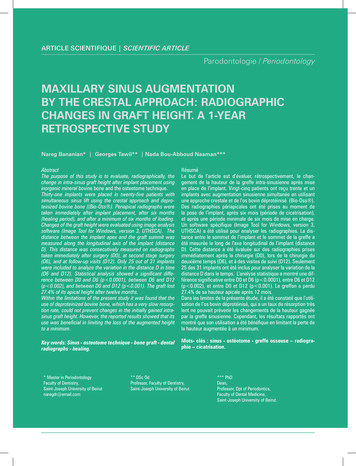
When describing a maxillary sinus that demonstrates no areas of mucosalthickening, one would see the maxillary sinus as a large black cavity (because it is filledwith air) which is surrounded by bony walls which appear radiopaque. In cases where nomucosal thickening is present, the Schniderian membrane cannot be demarcated easily asa space between the air in the cavity and surrounding bone (Figure 1). However, whenthere is some evidence of mucosal thickening, one will see the appearance of a grey massrepresenting the thickened tissue or fluid (Figure 2). This area of thickened tissue or fluidwill appear grey because its density is somewhere between that of air (black) and bone(white).Figure 1. Sagittal view of a CT scan demonstrating no mucosal thickening.8
Figure 2. Sagittal view of a CT scan demonstrating mucosal thickening.The purpose of this study was to determine the prevalence of maxillary sinuspathology in patients of the Misch International Implant Institute (MIII). Variables suchas age and gender were also studied to see if they had any influence on the results.9
CHAPTER 2METHODS AND MATERIALSDuring the course of this investigation, 275 scans (either CT or CBCT) wereanalyzed retrospectively. All of the patients were selected at random from the databaseof patients presenting to the MIII for maxillary sinus augmentation. The scans weretaken during the course of initial treatment planning. Information regarding the patients‟gender and age were recorded when available. All patients had agreed to the use of theirrecords for presentation and research purposes. In many cases, patients were referredfrom other dentists to the MIII and therefore had scans taken prior to becoming patientsof the institute. Therefore, the scans were taken from a wide number of different CT orCBCT scanning machines, and no attempt was made to accommodate for this. All thatwas required was for the scans to be available in a DICOM (digital imaging andcommunication in medicine) format which was then converted for use with the SimPlantPro treatment planning software (Materialise, Glen Burnie MD).The Simplant Pro treatment planning software allowed for the visualization of thescans in 3 different views: axial, sagittal, and panoramic. All scans were viewed by up-to3 different examiners. All examiners (2 periodontal residents – Drs. Aleem Manji andJoanie Faucher, and 1 prosthodontist – Dr. Randolph R. Resnik) were well versed in theutilization of CT scans for implant treatment planning. First the scans were reviewedindependently by Dr. Manji and Dr. Faucher. If there was a disagreement between thetwo in terms of the findings, Dr. Resnik cast the deciding vote. If there was not an10
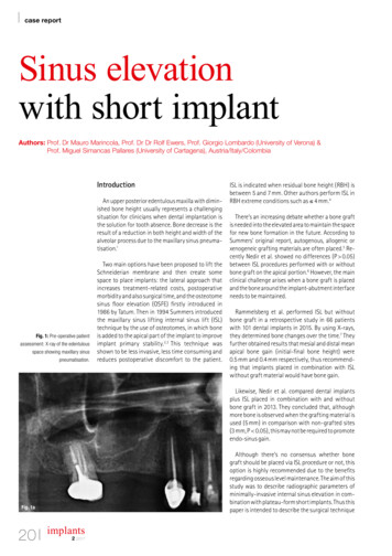
agreement between at least 2 of the reviewers the scan was eliminated fromconsideration. Furthermore, all reviewers had complete access to all features of thesoftware including the ability to change the color/contrast, zoom images and access allviews available in the scans.The mean age of the patients at the time of scan was 54.8 (range 13-91), and therewere 115 males and 160 females included in the study.The patients were alsocategorized based on age into one of 3 groups : 30, 30-59, and 60. There were 16aged 30, 112 aged 30-59 and 92 aged 60. Age data was only available for 220patients, as this information was missing from some of the scans reviewed.Patients were assigned to one of five different categories according to the leveland pattern of sinus inflammation: healthy, mucosal thickening ( 5mm), polypoidalmucosal thickening, partial opacification and/or air/fluid level or complete opacification.All 3 examiners were calibrated as to the 5 different categories and representativeexamples were discussed. Patients who were categorized as healthy had anywhere from0 to 5mm of mucosal thickening (Figure 3).When the mucosal thickening waspolypoidal in nature (as is the case when pseudocysts or nasal polyps are present) it wascategorized as polypoidal mucosal thickening (Figure 4). The next category includedmucosal thickening which displayed areas of partial opacification, or if an air/fluid levelwas apparent (Figure 5). The final category of complete opacification was noted whenthe sinus cavity was completely occupied with a grey or white appearance as opposed tothe normal black appearance (Figure 6).11
Figure 3. Panoramic (top), sagittal (middle) and axial (bottom) views of a patientcategorized as healthy.12
Figure 4. Panoramic (top), sagittal (middle) and axial (bottom) views of a patientcategorized as mucosal thickening 5mm.13
Figure 5. Panoramic (top), sagittal (middle) and axial (bottom) views of a patient withpolypoidal mucosal thickening.14
Figure 6. Panoramic (top), sagittal (middle) and axial (bottom) views of a patientcategorized as partial opacification and/or air/fluid.15
Figure 7. Panoramic (top), sagittal (middle) and axial (bottom) views of a patient with acompletely opacified sinus.16
Statistical calculations were performed using the Mann-Whitney U Test for nonparametric data to determine differences between groups, and subsequent p-values weregenerated.17
CHAPTER 3RESULTSOf the 275 CBCT scans which were reviewed, 151 patients were categorized ashealthy and the remaining 124 exhibited some evidence of maxillary sinus pathology(Figure 8). This translated to a prevalence of maxillary sinus pathology of 45.1% in thepopulation studied, with the remaining 54.9% not significant for any finding of maxillarysinus pathology.275250225200175151# of 150Patients 1251241007550250No PathologyPathologyFigure 8. Number of patients exhibiting no pathology and pathology respectively.When categorizing the results in terms of gender, 64 males and 60 femalespresented with evidence of sinus pathology. This translated to 55.6% of males and18
37.5% of females studied. There was a statistically significant difference in comparingthe incidence sinus pathology between males and females (p 0.01) (Figure Females30%20%10%0%PathologyNo PathologyFigure 9. Prevalence of sinus pathology based on gender.Out of the 151 patients who presented with evidence of sinus pathology, therewere 70 cases of mucosal thickening ( 5mm), 35 cases of polypoidal thickening, 11cases partial opacification and/or air/fluid level and 8 cases of complete opacification.This translated to 56.5%, 28.2%, 8.9% and 6.5% respectively for the 4 categories. (Figure10)19
Thickening nand/or air/fluidlevelCompleteOpacificationFigure 10. Percentage of each category of pathology of all cases exhibiting pathology.When categorizing the results in terms of age, the prevalence of sinus pathologywere 38%, 48% and 48% for the groups 30, 30-59 and 60 respectively. There was nostatistically significant difference between any of the age categories in terms of theincidence of sinus pathology (Figure 11).20
5%No PathologyPathology30%20%10%0% 3030-59 60Figure 11. Prevalence of sinus pathology and health by age group21
CHAPTER 4DISCUSSIONThe overall prevalence of 45.1% of cases showing some evidence of sinuspathology is consistent with other reports in the literature. However, in order to put thisnumber into perspective and how it differs from other studies, it is important to have anunderstanding of the history of imaging of the paranasal sinuses in both the medical anddental literature.A study by Havas in 1988 investigated the prevalence of radiographicabnormalities of the paranasal sinuses in 666 asymptomatic patients.They foundradiographic abnormalities in of 42% of scans, with 28.4% and 24.8% respectively withinthe ethmoid and maxillary sinuses. The authors also mentioned in their discussion thatmucosal thickening was the most common finding but that there may be a range ofmucosal thickening that can be considered normal and not evidence of pathology.17Moser in 1991 examined the use of MRI imaging to determine the prevalence ofparanasal sinus pathology in patients who did not present with symptoms indicative ofsinus pathology.The categories of pathology included mucoperiosteal thickening,retention cysts or polyps, air-fluid levels and complete opacification. Of the 263 imagesstudied, 24.7% demonstrated abnormalities in the paranasal sinuses.The authorsmentioned in their discussion that it was important to differentiate between „normalchanges‟ such as asymptomatic retention cysts or slightly thickened sinus mucosa fromtrue pathology. They stated that the 24.7% prevalence demonstrated changes that wouldbe interpreted as abnormalities in symptomatic patients.1822
A study by Rak in 1991 investigated mucosal thickening in the paranasal sinusesas a finding in patients undergoing MRI imaging.They concluded that mucosalthickening 3mm lacked clinical significance, and that mucosal thickening of 1-2mm inthe ethmoidal sinus was found in 63% of patients who were asymptomatic.19 Takentogether, the studies by Moser Havas and Rak provided a foundation for future study ofparanasal sinus pathology. Namely, that a large segment of the population may exhibitpathologic changes of the paranasal sinuses, and that an even larger number of patientsexhibit findings of the paranasal sinuses that are not considered pathologic or significant. A study by Tarp in 2000 investigated the prevalence of sinus pathology based onevaluation with MRI in patients presenting for intracranial neurological pathology.According to their study, 31.7% of patients exhibited pathology according to MRIimaging. It is important to note that similar to this study, mucosal thickening 5mmwas not considered pathology and was scored as normal. They found no correlationbetween positive findings of sinus pathology and either age or gender.20Studies published in the dental literature have also investigated the prevalence ofparanasal sinus pathology, but obviously they have focused mostly on the maxillarysinus. A study by Beaumont in 2005 studied 45 consecutively treated patients scheduledto undergo maxillary sinus augmentation and who were referred to an otolaryngologistfor consultation.After evaluation, 18 were diagnosed with sinus diseases and/orabnormalities for a prevalence of 40%. The categories of disease and/or abnormalitiesincluded chronic sinusitis, sinus cysts, nasal septum deviation and ostium stenosis.21 Astudy by Cha in 2007 examined 500 CBCT scans and found that 18.2% of incidentalfindings related to the “airway”. The findings of the airway category included sinusitis,23
sinus polyps, retention cysts, obstructed sinus, septum deviation, and thickened mucosa( 3mm). Interestingly, 22% of patients categorized as having „airway findings‟ hadrelated symptoms or medical histories.22A study by Pazera in 2011 examined the prevalence of incidental findings in themaxillary sinus of patients presenting to an orthodontic practice. They evaluated 134CBCT scans and the mean age of the patients was 17.5 years old. The six categories ofincidental findings included: acute inflammatory and/or allergic disease, chronicinflammatory and/or allergic disease, malformation and bone dysplasia, primary andsecondary neoplastic disease, metabolic disease and other. Their study found a 46.8%prevalence of incidental maxillary sinus findings, and all were suspected mucosalpathologies. They categorized the mucosal pathologies into 3 different categories: flatmucosal thickening ( 1mm) (23.7%), polypoid mucosal thickening (19.4%), and signsof acute sinusitis (3.6%). No differences were found based on patient‟s gender.23A study by Ritter in 2011 examined 1129 consecutively taken CBCT scans. Theycategorized maxillary sinus pathologic findings into mucosal thickening, partialopacification with liquid accumulation, total opacification and polypoidal mucosalthickening. They found a total prevalence of pathologies in the maxillary sinus of 56.3%.They found that patients 60 years of age had more pathology compared to younger agegroups, and males ha
implant dentistry it is imperative to have a comprehensive understanding of the maxillary sinus area. Both lateral wall and crestal approach sinus augmentation procedures have proven to be extremely successful for guided bone regeneration in the maxillary sinus. However, complications stemming from augmentation procedures are related to the