Transcription
Joerg WiltfangKarl Andreas SchlegelStefan Schultze-MosgauEmeka NkenkeRobert ZimmermannPeter KesslerAuthors’ affiliations:Joerg Wiltfang, Karl Andreas Schlegel, StefanSchultze-Mosgau, Emeka Nkenke, PeterKessler, Department of Oral and MaxillofacialSurgery, University of Erlangen-Nuremberg,Erlangen, Germany.Robert Zimmermann, Department ofTransfusion Medicine, University of ErlangenNuremberg, Erlangen, GermanyCorrespondence to:Jörg WiltfangDepartment of Oral and Maxillofacial SurgeryUniversity of Erlangen-NurembergGlückstr. 1191054 ErlangenGermanyTel: 49 9131 8534220Fax: 49 9131 8534219e-mail: joerg.wiltfang/mkg.imed.unierlangen.deDate:25 March 2002To cite this article:Wiltfang J, Schlegel KA, Schultze-Mosgau S, NkenkeE, Zimmermann R, Kessler P. Sinus flooraugmentation withb-tricalciumphosphate (b-TCP): does platelet-richplasma promote its osseous integration anddegradation?Clin. Oral Impl. Res, 14, 2003; 213–218Copyright C Blackwell Munksgaard 2003ISSN 0905-7161Sinus floor augmentation withb-tricalciumphosphate (b-TCP): doesplatelet-rich plasma promote itsosseous integration and degradation?Key words: b-tricalciumphosphate, foreign-body giant cell, platelet-rich plasma, sinus flooraugmentationAbstract: When dental implants are to be inserted, sinus floor augmentation is an effectivetreatment procedure to improve bone height in the posterior maxilla. In addition toautogenous bone material, allogenic materials, e.g. b-tricalciumphosphate (b-TCP), have beenused successfully. The purpose of this study was to investigate whether the combination of bTCP with platelet-rich plasma (PRP) enhances bony regeneration and resorption of thetricalciumphosphate material. In a randomized prospective trial, 45 sinus floor elevationswere performed in 39 patients. In 22 sites, PRP was added to the b-TCP granules, while in 23sites b-TCP without PRP was used. Six months later, bone specimens were harvested fromthe augmented region during the implant insertion procedure. The formation of new bonewas about 8–10% higher when PRP was applied. A faster degradation of the ceramic bonesubstitute was not observed. In conclusion, when PRP was added to b-TCP, bone regenerationwas supported to a small extent. However, the resorption of b-TCP was not accelerated andforeign-body giant cells and soft tissue surrounding the b-TCP granules were present.Sinus floor elevation in combination withdental implants is an accepted treatmentprocedure in the prosthetic restoration andesthetic rehabilitation of the severelyatrophic maxilla (Erbe et al. 1996; Engelke & Deckwer 1997; Wiltfang et al. 2000).In less severe cases, if the remaining alveolar bone has a minimum height of5mm, sinus floor elevation and implant insertion can be performed in one session. Incases of severe atrophy of the maxillary alveolar process, sinus floor elevation andimplant insertion are usually performed intwo stages (Boyne & James 1980; Wood &Moore 1988; Tatum et al. 1993). It is preferred to use autogenous bone from variousdonor sites for the augmentation procedure(Schenk et al. 2000; Nkenke et al. 2001). Ittakes approximately 4–6months followingaugmentation for the transplanted bone tobe integrated and substituted by osteoconduction (creeping substitution). Alternatively, autogenous bone transplants can bereplaced by bone substitutes, e.g. tricalciumphosphate ceramics, to avoid donor sitemorbidity (Leonardis & Pecora 1999; Piatelli et al. 1999). The degradation of thesematerials may take up to 12months if usedfor sinus floor augmentation.It would be beneficial for the patient toreduce this time interval by acceleratingthe process of integration of the transplanted bone or the bone substitute.The application of osteoinductive substances is a promising option as they areconstituents of platelet-rich plasma (PRP).Osteoinductive cytokines such as thetransforming growth factors TGF-b1 andTGF-b2, the platelet-derived growth factor(PDGF) and the insulin-like growth factor213
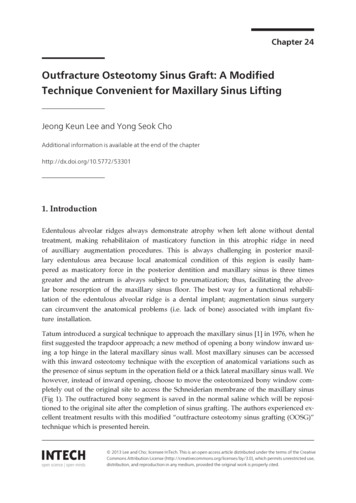
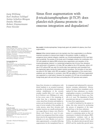
Wiltfang et al . Sinus floor augmentation with b-TCPFig.2. Harvesting of the bone biopsies 6months after sinus floor augmentation with a trephine bur of 3mmdiameter. The cranial 4mm of the biopsies was used for histological examination.Fig.1. (a) Access to the sinus by the lateral windowtechnique and sinus augmentation with 1–1.5g bTCP ceramic granules of 1000–2000mm diameter. (b)Careful mixing of 1ml of PRP with the b-TCP granules.IGF-I could be used for a faster integrationand substitution of autogenous bone transplants and bone substitutes (Canalis 1981;Cho et al. 1995; Giannobile 1996; Marxet al. 1998; Anitua 1999; Lynch et al.1999).The aim of this clinical investigationwas to determine whether the applicationof PRP in combination with tricalciumphophate ceramics could accelerate thedegradation and bony substitution of thisallogenic material in the case of sinus floorelevation (Merten et al. 2000).Material and methodsPatients and methodsIn the year 2000, 35 sinus floor elevationswere performed with b-tricalciumphosphate (b-TCP) in healthy subjects. The inclusion criteria were a blood concentrationof thrombocytes within the normal rangeand an absence of a history of maxillarysinus inflammations.Preoperatively, the residual bone heightswere assessed from panoramic radiographs.In the anterior maxillary sinus wall, a bone214 Clin. Oral Impl. Res. 14, 2003 / 213–218Fig.3. With PRP, light microscopy revealed that the remaining TCP ceramic bone substitute was surroundedby newly formed bone trabeculae. Direct bone-to-ceramic substitute contact could be observed (toluidine/alkaline fuchsine staining; a, magnification ¿2.8; b, magnification ¿7).lid was created using the window technique. Perforation of the maxillary sinusmucosal lining resulted in exclusion fromthe study. After careful elevation of themucosal layer, 1–1.5g CerasorbA (CurasanAG, Kleinostheim, Germany) containing
Wiltfang et al . Sinus floor augmentation with b-TCPb-TCP ceramic granules of 1000–2000mmdiameter were instilled for augmentationof the sinus floor. One milliliter of PRPwas administered in addition to the ceramic material in 17 sites in a randomizedprospective study approved by the ethicscommission of the University of ErlangenNuremberg (application no. 2075). Carewas taken to equally distribute the PRPover the b-TCP granules. In 18 sites noPRP was added (Figs1a and b). The averageage of the patients in the PRP group was45years (range 37–54years; 13 women andfour men). In the second group the averageage was 47years (range 32–64years; 14women and four men).PRP preparationUsing the tube technique ad modum CurasanA (Curasan AG, Kleinostheim, Germany), the patient’s blood plasma was prepared immediately prior to the operation inthe Institute for Transfusion Medicine ofthe University of Erlangen-Nuremberg.The PRP was required to contain plateletconcentrations at least triple the value fornormal plasma. Patients were excludedwhen the concentration factor was below3. The average platelet concentration was4.1 times higher. The PRP was then delivered to the surgical theatre so that itcould be applied within 2h or less.Harvesting of the specimensImplant insertion was performed 6monthsfollowing sinus floor augmentation. During this procedure, a bone biopsy from theaugmented site was harvested using a trephine bur of 3mm diameter. To guaranteethat the augmented region of interest wasexamined, only 4mm of the cranial end ofthe bone cylinder was taken for histological examination (Fig.2).Histological examinationUndecalcified thin sections of the bonespecimen were examined under the lightmicroscope. The specimens were sectionedlongitudinally in the axial plane. After fixation in 4% buffered formalin and dehydration through a series of decreasing concentrations of alcohol into de-ionizedwater, the test material was embedded inLR white hard-grade (Science Service,Munich, Germany) acrylic material. Thesamples were cut perpendicularly to thelongitudinal axis. Utilizing the ‘sawing andgrinding’ technique, the specimens wereFig.4. Direct bone-to-ceramic substitute contact could be observed as well as embedding of the ceramic granules in the soft tissue layer (toluidine/alkaline fuchsine staining; a, magnification ¿2.4; b, magnification ¿6.8).ground to 30–15mm (Exakt Apparatebau,Norderstedt, Germany). After toluidine/alkaline fuchsine staining, the thin sectionswere examined under the light microscope.The purpose of the light-microscope examination was to measure and evaluatethe bone regenerate as well as the relationbetween the ceramic substitute and bone.The images were assessed at magnifications of ¿1.25 to ¿100 and digitized(Duoscan T 2000 XL, Agfa, Mortsel, Belgium). NIH-Image software was used forimage analysis (National Institutes ofHealth, Bethesda, MD). The bone area ratiowas defined as the area of new bone insidean area of 9mm2/total area of 9mm2(¿100 to give a percentage).Statistical analysisMedian values were obtained with the interquartile range. For presentation of thedata, box-plots were used. To evaluate theinfluence of PRP on bony regeneration anddegradation of the ceramics, the Wilcoxontest was used. A P-value ⱕ 0.05% was considered statistically significant.ResultsHealing was uneventful in all patients. Alloperation sites showed primary woundhealing so that the sutures could be removed at term. Abnormal swelling orsigns of infection were absent.The preoperative bone height rangedfrom 2 to 7mm in the postoperative maxilla in the test and control groups. Directlyafter augmentation, the radioopacity variedin the augmented sinus floors. The cranialborder of the implanted material wascloudy. Radiographs taken 6months laterimmediately before implant insertionshowed a reduction of the vertical height215 Clin. Oral Impl. Res. 14, 2003 / 213–218
Wiltfang et al . Sinus floor augmentation with b-TCPFig.5. Without the addition of PRP, TCP ceramic remnants were surrounded by newly formed bone trabeculae.Direct bone-to-ceramic substitute contact could be observed (toluidine/alkaline fuchsine staining; a, magnification ¿2.72; b, magnification ¿6.8).by 10–20% in both groups. All implantsites allowed the installation of two tothree rough-surfaced implants of differentsystems with diameters up to 4.8mm inthe posterior maxillae. The structure of theaugmented material was now more homogenous. After implant insertion, all implants radiologically displayed full bony integration.All osseous biopsies of test and controlgroups could be included in the study. Thecranial 4mm never included bone of theresidual alveolar crest. Without PRP instillation osseous regeneration reached an average level of 29% compared to 38% in thePRP group. In the PRP group, bone formation was between 32 and 43%, whereas inthe non-PRP group values between 25 and37% were found. The wide range of scatterwas striking in both groups.Light microscopy revealed that the remaining TCP ceramic bone substitute wassurrounded by newly formed bone trabecu-216 Clin. Oral Impl. Res. 14, 2003 / 213–218lae (Kassolis et al. 2000). Direct bone-to-ceramic substitute contact could be observedas well as an embedding of the ceramicgranules in a soft tissue layer (Figs3, 4, 5aand 5b). Foreign-body giant cells could bedetected in these soft tissue layers thatwere more present in the bone specimensof patients that were treated with PRP (Figs6a and b). A faster degradation of the ceramic material (b-TCP) could not be observed in the group in which PRP had beenapplied. The relation between b-TCP granules and newly formed bone was 13.8% inthe group substituted with PRP comparedto 15% in the patient group without PRP(Figs7 and 8) (Donath 1985).DiscussionThrombocytic b-granules contain variousgrowth-stimulating factors, e.g. the plateletderived growth factor (PDGF) and the trans-forming growth factors TGF-b1 and -b2 (Harrison & Cramer 1993). These cytokines playan important role in osteogenetic and angiogenetic processes. Furthermore, PDGF isinvolved in almost all wound-healing reactions. Physiological wound healing ischaracteristically triple-phased and is supported by cellular and humoral factors.First, a catabolic latency period lasting approximately 1week can be observed. Thisperiod is succeeded by a proliferation phaseof about 2weeks. After the proliferationphase a period of reparation and reorganization follows, which may last for severalmonths. The proliferation phase is characterized by a high cellular and vasculardensity (Hosgood 1993). The intense cellmetabolism provides the necessary matrixfor cell differentiation, cell migration andthe initial steps for tissue repair. Thrombocytic growth factors on the whole have theirmain effect during the phase of proliferation. They support cell mitosis, neoangiogenesis and the activation and immigrationof macrophages. Cytokines of the TGF family in particular attract preosteoblastic cellsand fibroblasts by chemotaxis and supporttheir mitotic activity.The combined application of PRP andbone transplants leads to a higher cellularactivity in the bone-transforming cells, resulting in an accelerated integration of thetransplanted bone. The aim of this investigation was to determine whether the combination of b-TCP with PRP results in accelerated bone repair based on faster ceramic degradation and transformation. In arandomized prospective study, we tried tofind an answer to this question by clinically applying ceramic bone substitutes incombination with PRP during sinus flooraugmentation. In the two-stage procedure,there was no problem in harvesting relevant biopsy specimens during the implantinsertion 6months after sinus floor augmentation.Six months post-augmentation, our results showed a great intra- and interindividual range with respect to the amount ofnewly formed bone and the degree of ceramic degradation. With regard to the rateof bone regeneration, slightly increasednew bone formation of about 8–10% couldbe observed when PRP was applied. Afaster degradation of the ceramic bone substitute CerasorbA was not found. A striking result in the PRP group was the largenumber of foreign-body giant cells that
Wiltfang et al . Sinus floor augmentation with b-TCPre si des implants dentaires doivent y être insérés. A côtédu matériel osseux autogène, des matériaux allogènes telsque le phosphate tricalcique-b (b-TCP) ont été utilisésavec succès. Le but de cette étude a été de vérifier si l’association du b-TCP et du plasma riche en plaquettes(PRP) pouvait augmenter la régéneration osseuse et la résorption du matériel phosphate tricalcique. Dans un essaiprospectif au hasard, 45 épaississements du plancher sinusal ont été effectués chez 39 patients. Au niveau de 22sites, le PRP a été injecté aux granules b-TCP tandis que23 sites b-TCP se retrouvaient sans PRP. Six mois plustard, des échantillons osseux ont été prélevés des régionsépaissies durant le processus d’insertion des implants. Laformation du nouvel os était de 8 à 10% plus importantelosque le PRP avait été utilisé. Une dégradation plus rapide du substitut osseux en céramique n’s pas pû être misen évidence. Lorsque le PRP est ajouté au b-TCP la régénerátion osseuse est légérement favorisée. Cependant larésorption de b-TCP n’était pas accélérée et des cellulesgéantes de corps étrangers ainsi que du tissu mou entourant les granules TCP étaient présents en plus grandnombre.ZusammenfassungFig.6. Foreign-body giant cells could be detected in these soft tissue layers that were more present in the bonespecimens of patients that were treated with PRP (a, microtome section, Giemsa staining, magnification ¿68;b, microtome section, Giemsa staining, magnification ¿27.2).Ziel: Die Augmentation des Sinusbodens stellt eine effiziente Behandlung zur Verbesserung der Knochenhöhe inder posterioren Maxilla dar, wenn Implantate eingesetztwerden sollen. Neben autologem Knochenmaterial wurden auch allogene Materialien wie z.B. b-Trikalziumphosphat (b-TCP) erfolgreich verwendet. Es war das Ziel dieser Studie, zu untersuchen, ob die Kombination von bTCP mit plättchenreichem Plasma (PRP) die Knochenregeneration und die Resorption des Trikalziumphosphatmaterials fördern wird.Studienaufbau: Bei 39 Patienten wurden 45 ElevationenFig.7. Box-plot analysis for the bony regeneration.could be observed and the fact that not allceramic granules had direct bone contactbut were partially embedded in a softtissue shed. The latter can be explained byTGF, which is included in PRP and supports the chemotaxis of fibroblasts.In summary, this study suggests that theapplication of PRP will only result in accelerated new bone formation if target cellssuch as osteoblasts or osteocytes are present. A faster degradation of ceramic bonesubstitutes cannot be expected. Furtherstudies are necessary to investigate the influence of varying concentrations of PRPon wound-healing processes alone and incombination with other bone substitutesas tested here. Moreover, the patients operated on in the present study will be followed up once a year to obtain long-termdata on the success rate of the implants installed in the different sites.RésuméFig.8. Box-plot analysis for the degradation of the bTCP ceramics.L’épaississement du plancher sinusal est un processus efficace d’amélioration de la hauteur à l’arrière du maxillai-des Sinusbodens in einer randomisierten prospektivenUntersuchung durchgeführt. Bei 22 Stellen wurde dem bTCP-Granulat PRP zugefügt, während bei 23 Stellen bTCP ohne PRP verwendet wurde. Nach sechs Monatenwurden während des Eingriffs zum Setzen der ImplantateKnochenbiopsien von den augmentierten Regionen gewonnen.Resultate: Die Bildung von neuem Knochen war ca. 8 bis10% grösser wenn PRP appliziert wurde. Eine schnellereAuflösung des keramischen Knochenersatzmaterialskonnte nicht beobachtet werden.Schlussfolgerung: Wenn PRP dem b-TCP zugefügt wird,wird die Knochenregeneration leicht unterstützt. Jedochwird die Resorption von b-TCP nicht bescheunigt.Fremdkörperriesenzellen und Weichgewebe um die TCPGranulate sind mehr vorhanden und auffälliger.ResumenObjetivo: La elevación del suelo del seno es un procedimiento de tratamiento efectivo para mejorar la altura dehueso en el maxilar posterior, si hay que insertar implantes dentales. Aparte de material óseo autógeno, se hanusado con éxito materiales alogénicos como b-tricalciumphosphate (b-TCP). El propósito de este estudio fueinvestigar, si la combinación de b-TCP con plasma ricoen plaquetas (PRP) incrementaria la regeneración ósea yla reabsorción del material tricalciumphosphate.Diseño del estudio: En un ensayo aleatorio prospectivo sellevaron a cabo 45 elevaciones del suelo del seno en 39217 Clin. Oral Impl. Res. 14, 2003 / 213–218
Wiltfang et al . Sinus floor augmentation with b-TCPpacientes. En 22 lugares se añadió PRP a gránulos de bTCP, mientras que en 23 lugares se usó b-TCP sin PRP.Seis meses mas tarde se recolectaron especimenes de hueso de la zona aumentada durante el procedimiento de inserción del implante.Resultados: La formación de nuevo hueso fue un 8 a 10% mas alta cuando se aplicó PRP. No se observó unadegradación mas rápida del sustituto óseo cerámico.Conclusiones: Cuando se añade PRP a b-TCP, la regeneracion ósea es ligeramente ayudada. De todos modos, lareabsorción de b-TCP no se acelera y las células gigantesde cuerpo extraño y el tejido blando rodeando los gránulosde b-TCP fueron mas evidentes y estaban presentes.ReferencesAnitua, E. (1999) Plasma rich in growth factors: prelimi-Hosgood, G. (1993) Wound healing. The role of platelet-Piatelli, M., Favero, G.A., Scarano, A., Orsini, G. & Piatel-nary results of use in the treatment of future sites forimplants. International Journal of Oral and Maxillo-derived growth factor and transforming growth factorbeta. Veterinarian Surgery 22: 490–495.li, A. (1999) Bone reactions to anorganic bovine bone(Bio-Oss) used in sinus augmentation procedures. Inter-facial Implants 14: 529–535.Boyne, P.J. & James, R.A. (1980) Grafting of the maxillarysinus floor with autogenous marrow and bone. Journalof Oral and Maxillofacial Surgery 38: 613–616.Canalis, E. (1981) Effect on platelet-derived growth factorDANN and protein synthesis in cultered rats calvaria.Kassolis, J., Rosen, P.S. & Reynolds, M.A. (2000) Alveolarridge and sinus augmentation utilizing platelet-richplasma in combination with freeze-dried bone allograft:case series. Journal of Periodontology 71: 1654–1661.Leonardis, D. & Pecora, G.E. (1999) Augmentation of themaxillary sinus with calcium sulfate: one year clinicalreport from a prospective longitudinal study. International Journal of Oral and Maxillofacial Implants 14:869–878.Lynch, S.E., Genco, R.G. & Marx, R.E. (1999) Platelet-richplasma: a source for bone grafts. Tissue Engineering 4:71–78.Marx, R.E., Carlson, E.R., Eichstaedt, R.M., Schimmele,S.R., Strauss, J.E. & Georgeff, K.R. (1998) Platelet-richplasma, growth factor enhancement for bone grafts.Oral Surgery Oral Medicine Oral Pathology OralRadiology Endodontology 85: 638–646.national Journal of Oral and Maxillofacial Implants14: 835–840.Schenk, C., Schwarz, A., Kaupe, A.J.M., Wiltfang, J. &Neukam, F.W. (2000) Erfahrungen mit autogenenBeckenkammtransplantationen im Ober- und Unterkiefer. Deutsche Zahnärztliche Zeitschrift 55: 355–358.Tatum, O.J., Lebowitz, M.S., Tatum, C.A. & Borgner,R.A. (1993) Sinus augmentation: rationale, development, long term results. New York State DentalJournal 5: 43–48.Wiltfang, J., Schultze-Mosgau, S., Merten, H.A., Kessler,P., Ludwig, A. & Engelke, W. (2000) Endoscopic andultrasonographic evaluation of the maxillary sinusafter combined sinus floor augmentation and implantinsertion. Oral Surgery Oral Medicine Oral PathologyOral Radiology Endodontology 89: 288–291.Wood, R.M. & Moore, D.L. (1988) Grafting of the maxil-Merten, H.A., Wiltfang, J., Hönig, J.F., Funke, M. & Luhr,H.G. (2000) Intraindividueller Vergleich von Alphaund Beta-TCP-Keramik im Tierexperiment. Mund-Kiefer Gesichtschirurgie 4: 509–514.Nkenke, E., Schultze-Mosgau, S., Radespiel-Tröger, M.,Kloss, F. & Neukam, F.W. (2001) Morbidity of harvesting of chin grafts: a prospective study. ClinicalOral Implants Research 12: 495–502.lary sinus with intraorally harvested autogenous boneprior to implant placement. International Journal ofOral and Maxillofacial Implants 3: 209–214.Zimmermann, R., Jakubietz, R., Jakubietz, M., Strasser,E., Schlegel, K.A., Wiltfang, J. & Eckstein, R. (2001) Different preparation techniques to obtain platelet-richplasma as source of growth factors for local application.Transfusion, in press.Metabolism 30: 970–975.Cho, M., Lin, W.L. & Genes, R.J. (1995) Platelet-derivedgrowth factor modulated guided tissue regenerativetherapy. Journal of Periodontology 66: 522–528.Donath, K. (1985) The diagnostic value of the newmethod for the study of undecalcified bones and teethwith attached soft tissues (sawing and grinding). Pathology Research and Practice 179: 631–633.Engelke, W. & Deckwer, I. (1997) Endoscopic controlledsinus floor augmentation. A preliminary report. Clinical Oral Implants Research 8: 527–531.Erbe, M., Blijdorp, K., Hoppenreijs, T.J.M. & Stoelinga,P.J.W. (1996) Sinusbodenelevation und Onlayosteoplastik zur Verbreiterung des extrem schmalen Oberkieferkammes. Deutsche Zahnärztliche Zeitschrift 51: 49–55.Giannobile, D.G. (1996) Periodontal tissue engineering bygrowth factors. Bone 19: 235–237.Harrison, P. & Cramer, E.M. (1993) Platelet alpha-granules. Blood Review 7: 52–62.218 Clin. Oral Impl. Res. 14, 2003 / 213–218
5mm, sinus floor elevation and implant in-sertion can be performed in one session. In cases of severe atrophy of the maxillary al-veolar process, sinus floor elevation and implant insertion are usually performed in two stages (Boyne & James 1980;Wood& Moore 1988; Tatum et al. 1993). It is pre-ferred to use autogenous bone from various