Transcription
Article Scientifique Scientific ArticleParodontologie / PeriodontologyMAXILLARY SINUS AUGMENTATIONBY THE CRESTAL APPROACH: RADIOGRAPHICCHANGES IN GRAFT HEIGHT. A 1-YEARRETROSPECTIVE STUDYNareg Bananian* Georges Tawil** Nada Bou-Abboud Naaman***AbstractThe purpose of this study is to evaluate, radiographically, thechange in intra-sinus graft height after implant placement usinginorganic mineral bovine bone and the osteotome technique.Thirty-one implants were placed in twenty-five patients withsimultaneous sinus lift using the crestal approach and deproteinized bovine bone ((Bio-Oss ). Periapical radiographs weretaken immediately after implant placement, after six months(healing period), and after a minimum of six months of loading.Changes of the graft height were evaluated using image analysissoftware (Image Tool for Windows, version 3, UTHSCA). Thedistance between the implant apex and the graft summit wasmeasured along the longitudinal axis of the implant (distanceD). This distance was consecutively measured on radiographstaken immediately after surgery (D0), at second stage surgery(D6), and at follow-up visits (D12). Only 25 out of 31 implantswere included to analyze the variation in the distance D in time(D6 and D12). Statistical analysis showed a significant difference between D0 and D6 (p 0.0001), between D6 and D12(p 0.002), and between D0 and D12 (p 0.001). The graft lost27.4% of its apical height after twelve months.Within the limitations of the present study it was found that theuse of deproteinized bovine bone, which has a very slow resorption rate, could not prevent changes in the initially gained intrasinus graft height. However, the reported results showed that itsuse was beneficial in limiting the loss of the augmented heightto a minimum.RésuméLe but de l’article est d’évaluer, rétrospectivement, le changement de la hauteur de la greffe intra-sinusienne après miseen place de l’implant. Vingt-cinq patients ont reçu trente et unimplants avec augmentation sinusienne simultanée en utilisantune approche crestale et de l’os bovin déprotéinisé (Bio-Oss ).Des radiographies périapicales ont été prises au moment dela pose de l’implant, après six mois (période de cicatrisation),et après une période minimale de six mois de mise en charge.Un software spécifique (Image Tool for Windows, version 3,UTHSCA) a été utilisé pour analyser les radiographies. La distance entre le sommet de l’implant et le sommet de la greffe aété mesurée le long de l’axe longitudinal de l’implant (distanceD). Cette distance a été évaluée sur des radiographies prisesimmédiatement après la chirurgie (D0), lors de la chirurgie dudeuxième temps (D6), et à des visites de suivi (D12). Seulement25 des 31 implants ont été inclus pour analyser la variation de ladistance D dans le temps. L’analyse statistique a montré une différence significative entre D0 et D6 (p 0.0001), entre D6 et D12(p 0.002), et entre D0 et D12 (p 0.001). Le greffon a perdu27.4% de sa hauteur apicale après 12 mois.Dans les limites de la présente étude, il a été constaté que l’utilisation de l’os bovin déprotéinisé, qui a un taux de résorption trèslent ne pouvait prévenir les changements de la hauteur gagnéepar la greffe sinusienne. Cependant, les résultats rapportés ontmontré que son utilisation a été bénéfique en limitant la perte dela hauteur augmentée à un minimum.Key words: Sinus - osteotome technique - bone graft - dentalradiographs - healing.Mots- clés : sinus - ostéotome - greffe osseuse – radiographie – cicatrisation.* Master in PeriodontologyFaculty of Dentistry,Saint-Joseph University of Beirutnaregdr@email.com** DSc OdProfessor, Faculty of Dentistry,Saint-Joseph University of Beirut*** PhDDean,Professor, Dpt of Periodontics,Faculty of Dental Medicine,Saint-Joseph University of Beirut.
48IAJD Vol. 3 – Issue 2Article Scientifique Scientific ArticleIntroductionImplant TypeThe edentulous posterior segments of the maxilla are often characterized by severe bone resorptionand pneumatization of the maxillaryantrum, compromising rehabilitationwith implant supported prostheses[1]. Sinus floor elevation restores theheight of available bone in the posterior maxilla, thus allowing the placement and integration of endosseousimplants. The conventional procedureprovides access through the lateralwall of the antrum [2]. Tatum in 1986[3] proposed a different technique, thecrestal approach. However, it was notuntil 1994 that Summers describedthe use of specifically designed instruments called osteotomes, hence the“osteotome technique”, to compressbone and elevate the sinus floor undermalleting forces [4]. Clinical studiesreported favorable long-term outcomesusing this technique [5-8]. However,the discrepancy in the reported success/survival rates (between 88.6 %and 100 %) were attributed to someconfounder factors such as the residual bone height, the type of implantused and the surgical technique.Long-term stability of the initiallyregenerated bone is a concern regardless the site and the type of surgical procedure. Pneumatization of themaxillary sinus continues even aftersinus floor elevation; this can lead toloss in the intra-sinus graft height [9].Using the conventional technique, fewstudies observed these changes andreported that the amount of loss in theinitially gained height was related tothe type of grafting material used [10-12]. With the osteotome technique,Bragger et al. [13] used a combinationof autogenous bone and deproteinizedbovine bone (Bio-Oss ); they reporteda considerable loss in the apical graftheight after twelve months [13].Since Bio-Oss was reported tohave a very slow resorption rate [14, 15],the aim of this study was to observe,radiographically, the changes in theapical graft height in sinuses graftedwith deproteinized bovine bone aloneFrequencyPercent3I 825.8Brånemark 1341.9Xive 412.9Replace 26.5Screw Vent 26.5ITI 26.5Total31100.0Table 1: Types of implants used.with simultaneous implant insertionusing the osteotome technique.Materials and MethodsThe files of patients who underwent osteotome sinus floor elevationin three specialized periodontal clinics were screened. Cases were selectedbased on the following criteria:- Osteotome sinus floor augmentation was performed using deproteinized bovine bone as the grafting material.- Implants had been loaded for aminimum of six months.- Standardized periapical radiographs were taken at the following intervals: preoperative,immediately after implant insertion, at the second stage surgeryfor submerged implants, or at 4to 6 months for trans-mucosalimplants, and at follow-up visitsbetween 6 to 12 months.- The following landmarks could beclearly identified on consecutiveradiographs: the entire length ofthe implant, at least two to threemillimeters past the cranial end(or summit) of the intra-sinusgraft.- Implants should fulfill the success criteria described in 1986 byAlbrektsson et al. [16] during theperiod covered by the study.Twenty-five patients fulfilled theselection criteria. They received thirtyone implants, each inserted withsimultaneous sinus augmentationusing the crestal approach. The meanage of the patients was 47.96 10.350(range: 28 to 65 years). Ten were males(40%), 15 were females (60%). Only twopatients (8%) were smokers.Surgical treatmentThe surgical procedure was performed using either the modifiedosteotome sinus floor elevation technique as described by Davarpanah[17], or a modified technique similarto that described by Ferrigno et al. [5].All sites were grafted with deproteinized bovine bone, namely BioOss (Geistlich Sons Ltd, Wolhusen,Switzerland). Six different types oftextured surface implants were used(Table 1). Implant length variedbetween 8.5- to 12-mm, the majority (71.0%) were 10-mm long. Implantdiameter varied between 3.75 and 5mm, most were either 4 or 5 mm. 23implants (74.2%) were inserted at firstand second molar sites, the remainder8 implants (25.8%) at the second premolar site.Healing periods extended from 4to 9 months, the majority of implants(83.9%) were allowed to heal for 6months, and 80.6% in a submergedmanner, the rest (19.4%) had a transmucosal healing. Restorative treatment was either a single crown (35.5%),or two- to three-unit implant supported fixed partial denture (64.5%).Radiographic analysisAll periapical radiographs weretaken using extension cone paralleling technique (XCP instruments, RinnCorporation). Some were taken using
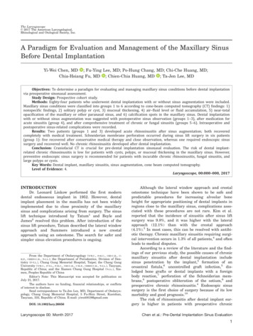
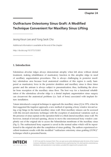
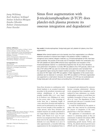
49Parodontologie / Periodontologya digital sensor, others were conventional. Each conventional radiographwas digitized by placing it against anilluminated X-ray viewer using a digital camera (Canon Digital Rebel XT Clinical – Sigma EM-140DG); a pictureof the radiograph was taken and transferred to the computer.Analysis was performed using acomputerized measuring techniquewith image analysis software (ImageTool for Windows, version 3, UTHSCSA).The software allows for internal calibration performed for each radiographusing either the known implant lengthas a reference, or the distance betweenthree consecutive threads (based onthe distance between two consecutivethreads as detailed by the manufacturer). The precision of the measuringsystem is 0.01 mm. In order to improvethe quality of the images, and henceanalysis, image-enhancement procedures were performed. These includedsharpening, brightness, contrast andcolor to gray-scale.Figure 1 details the distances measured on each radiograph:Distance A: The residual boneheight (RBH): The distance betweenthe most coronal bone to implant contact to the original sinus floor.Distance B: The length of the partof the implant protruding into thesinus, measured form the most apicalbone to implant contact (original sinusfloor) to the implant apex parallel tothe long axis of the implant.Distance C: The height of the intrasinus graft immediately after surgery.This distance was measured along theaxis of the implant from the most apical bone to implant contact (originalsinus floor) to the graft summit.Distance D: The distance betweenthe implant apex and the graft summit as measured along the longitudinal axis of the implant. This distancewas consecutively measured on radiographs taken immediately after surgery, at the second stage surgery, andat follow-up visits. The change in thisdistance reflects change in the intrasinus bone height. It had a positivevalue when the graft summit was api-Fig. 1: Diagram illustrating the distancesmeasured on each radiograph.Distance A: The residual bone height(RBH); distance B: The length of the partof the implant protruding into the sinus;distance C: The height of the intra-sinus graftimmediately after surgery; distance D: thedistance between the implant apex and thegraft summit.cal to the implant apex, and a negative value when the implant apex washigher than the graft.Statistical analysisThe statistical analysis was performed using a software program(SPSS for Windows version 11.0). Eachoutcome (distance) was scored twotimes by the same evaluator. For statistical analysis, the average of the twomeasures was obtained. The reproducibility among measures was evaluatedusing Pearson product-moment correlation coefficient. The normal distribution of the data was tested using theKolmogorov Smirnov test.The statistical analysis includeddescriptive statistics and non parametric analysis of variance (Friedmantest) followed by the Wilcoxon signrank tests with the Bonferroni correction, for determination of differencesin distance between reference pointsmeasured after implant insertion,after 6 and 12 months. Relationshipsbetween measurements and timewere evaluated using linear regressionanalysis. The relationship between distances was investigated using Pearsoncorrelation coefficient. Paired t-testwas used to compare two distancesin the same group. The “implant” wasdefined as the statistical unit. The testwas two-sided and a significance of5% or less was considered statisticallysignificant.ResultsThe quality of bone encounteredwas type III in the majority of the sites(80.6%), only few sites (19.4%) exhibited type II bone.Radiographic analysisAll measurements were performedby one operator (N.B.) at two different time points. The coefficient of correlation “rho” demonstrated a strongreproducibility between measurements towards 1.The mean preoperative residual bone height (distance A) was6.44 1.62mm ranging from 2.17mm to9.34mm. In 27 out of 31 sites ( 85%),this distance was more than 5 mm.Around 35.4% of all sites had less than6mm preoperative bone height (Fig. 2).Figure 3 depicts the measured values for distance B. The mean length ofthe part of the implant protruding intothe sinus was 3.47 1.28mm rangingfrom 0.79 to 5.51mm. In twenty-oneout of 31 sites ( 67.7%), the implantsprotruded more than 3mm past theoriginal sinus boundaries, and morethan 4 mm in around 38.7% of the sites.The post-operative intra-sinus graftheight (distance C) was 4.91 1.52mm.It ranged from 1.33 to 7.16mm.Distance C was greater than 5mm in17 out of 31 sites ( 54.5%), and morethan 6mm in 25.8% (8 out of 31 sites) ofthe sites (Fig. 4).In all thirty-one sites, the sinusgraft covered the implant apex as evident in periapical radiographs takenimmediatelypostoperatively.Atimplant insertion, the mean distancebetween the graft summit and implantapex (distance D0) was 1.70 0.85mm,ranging from 0.47 to 4.48mm (Fig. 5).In fourteen out of 31 sites (45.4%),distance D0 was greater than 1.5mm,
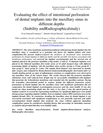
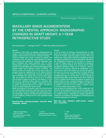
50IAJD Vol. 3 – Issue 2Article Scientifique Scientific ArticleFig. 2: Residual bone height (distance A).Fig. 3: Length of implant protruding into the sinus (distance B).Fig. 4: Distance between graft summit and first apical BIC atbaseline (distance C).Fig. 5: Distance between graft summit and implant apex atbaseline (distance D).while it was greater than 2mm in only25.8% of the sites (8 out of 31 sites).Changes of distance D in timeOnly 25 out of 31 implants wereincluded for this analysis due to lackof radiographs taken at one of the twofollow-up periods (either during thefirst 6 months or after 12 months).Figure 6 presents the changes indistance D from implant insertionthrough the follow-up periods. Theapical graft height was reduced from1.7 0.85mm at implant insertion(D0) to 1.49 0.77mm during the first6 months (D6). Further reduction to1.11 0.74mm was observed after 12months (D12).Using the Wilcoxon Signed Rankstests with the Bonferroni correction,statistically significant differenceswere found between baseline (D0) and(D6) (p 0.0001), between (D6) and(D12) (p 0.002), and between (D0) and(D12) (p 0.001).Table 2 shows the mean percentageof loss in apical graft height observedbetween the observation periods.As illustrated in table 2, the graftlost 27.4% of its apical height aftertwelve months. Moreover, althoughnot statistically significant, the loss ofapical graft height was greater between(D6) and (D12), as compared to thatobserved between (D0) and (D6).Using the Pearson productmoment correlation coefficient, astrong, positive correlation was foundbetween distance B (length of implantprotruding into the sinus at baseline)and distance C (the post-operativeintra-sinus graft height) [r 0.882,n 31, p 0.0001], with high levels ofdistance B associated with high levelsof distance C. In addition, there was amedium positive correlation betweendistance C and distance D0 (apicalgraft height at baseline) [r 0.443,n 31, p 0.013], with high levels of distance C associated with high levels ofdistance D0. However, no correlationcould be found between distance Band distance D0, between distance Band distance D12, or between distanceC and distance D12.Figure 7 shows the radiographicchanges observed during the one yearobservation period. In most cases thenew sinus floor is demarcated withwell defined boundaries.DiscussionThe crestal approach for sinus floorelevation offers a less invasive technique when compared to the lateral
51Parodontologie / PeriodontologyFig. 6: Changes of distance D in time.Between(D0) & (D6)Between (D6) & (D12)Between (D0) & (D12)% loss in apical graft height13.7%14.7%27.4%The 95% confidence able 2: Mean percentage of loss in apical graftheight observed between the observation periods.Fig. 7: Consecutive radiographs showing graft consolidation and the formation of new sinus floor.a: pre-operative; b: after surgery; c: at 2nd stage surgery (6 months); d: at twelve months (D0, D6 and D12).window approach, to compensate forthe vertical inadequacy of the residualbone height in the posterior segmentsof the maxilla. However, it is not theaim of the present study to addressthe predictability of this technique.Rather, it describes the changes of theapical graft height within a one yearperiod, after functional loading of theimplants for a minimum of 6 months.Consequently, only cases which fulfilled previously described successrates for implants along with adequateclinical and radiographic documentations were included in the study.Residual bone height is consideredthe most important factor in determining the success of the osteotometechnique. Few studies evaluated success/survival rates as a function ofRBH. Rosen et al. [18], using the boneadded osteotome sinus floor elevation technique, reported 96% or highersurvival rate when pretreatment boneheight was 5 mm or more. These valuesdropped to 85.7% when pretreatmentbone height was 4 mm or less. Morerecently, Toffler [19] described a similar trend in a clinical report. An overall survival rate of 93.5% was reportedfor 276 implants. However, when siteswith a residual bone height of 4mm orless were only considered, the survivalrate dropped to 73.3%. In the presentstudy, the mean RBH was 6.44 1.62mm which might explain the good per-formance of implants included in thisstudy.In a human cadaver study, Reiseret al. [20] reported that the incidenceof class II membrane perforationsincreased whenever the sinus membrane was elevated 6 mm or moreusing osteotomes. In the presentstudy, the mean value of distance Cwas 4.91 1.52 mm. But even in siteswhere the sinus membrane was elevated up to 7.16 mm, periapical radiographs showed a clearly visible domeshaped area beyond the apex of theimplant and no collapse of the graftedarea was observed on the consecutiveradiographs.
52IAJD Vol. 3 – Issue 2Article Scientifique Scientific ArticleFew in vitro studies evaluated stressdistribution around implants placedin augmented sinuses. Fanuscu et al.[21] designed composite photoelasticmodels to simulate conditions beforesinus grafting and at various stages ofgraft maturation. The stress analysisindicated that before placement of thesimulated graft, loading on the implanttransferred the highest stresses tocortical bone. As the model simulated more advanced stages of graftmaturation, proportionately increasedstresses developed around the apicalportion of the implant and the graftassumed a greater proportion of theload, with concomitant stress reduction in the native bone simulants.In a three-dimensional finite element analysis, Tepper et al. [22]designed several models which mimicked different sinus graft conditionsto evaluate the surgical procedureand the amount of peri-implant packing that would yield the best bonysupport for dental implants. Clinicalconditions were simulated with only5.3 mm of sub-antral bone height.The results showed that in modelswith adequately regenerated intrasinus bone, interfacial stresses were30-50% lower than in the referencemodels with no or inadequate (1 mm)packing. Moreover, complete packingaround the implant reduced 30-45%of the cortical stresses as comparedto no grafting or to a thin peri-implantbone sheath (1 mm). Based on theseresults, the authors recommended thatas much as of the space available forsinus floor elevation should be packedwith grafting material up to the apicalimplant bed. With the osteotome technique the amount of grafting material packed around the implant tip isgenerally limited. Moreover, maxillarysinus pneumatization is a continuousprocess that could lead to bone resorption even after sinus floor augmentation. Consequently, in time, the reduction in apical graft volume increasesthe stress values generated in thebone surrounding the implant, even ifthe prosthetic loads are the same. Thiscombined with the often poor qualityof bone encountered in the posteriormaxilla, and the increased occlusalloads in the posterior segments of thejaws would compromise the bone toimplant integration. Hence, the physical stability of the graft is important forthe mechanical stability of the implant.For the conventional sinus floorelevation, few studies investigatedthe changes in the height and volumeof the sinus graft in time. These studies showed that regardless the type ofgraft, there was a gradual decrease ingraft height over the observation periods. In the 1996 sinus floor elevationconsensus conference [10], changes insinus graft height of 9 different graftmaterials and various graft combinations were evaluated. In the 3-year timeframe, the data showed that the leastchange in graft height was observed forthe combination of an autograft (intraoral bone) and an alloplast (mostlyporous Hydroxyapatite) with a meanbone loss of 0.79 mm. The greatestloss in graft height was observed forthe freeze-dried demineralized bone(DFDBA), with a mean change of 2.09mm. In a prospective study, Froumet al. [11] evaluated the stability ofthe volume of the augmented boneover 2 to 3 years in 13 sinuses graftedwith inorganic bovine bone mineral(Osteograf/N , Ceramed ). Whilethe authors reported minimal changes(1.4%) in graft height in sites graftedwith OsteoGraf/N , in two other caseswhere the sinuses were grafted withDFDBA the radiographic follow-updemonstrated 15.5% and 16.5% loss ingraft height, respectively. On the otherhand, Hatano et al. [12] performed along-term radiographic evaluation ofgraft height changes for up to 10 years.Maxillary sinuses were augmentedwith a 2: 1 autogenous bone/xenograft mixture along with simultaneousplacement of dental implants. Afteraugmentation, the grafted sinus floorwas consistently located above theimplant apex. However, the sinus graftheight decreased significantly in timeand approached the original sinusfloor. After 2 to 3 years, the graftedsinus floor was at the same level of orslightly below the implant apex. Thesedifferences in results have been mainlyattributed to the use of different grafting materials.In the study of Bragger et al. [13],sinuses were grafted with a combination of Bio-Oss and autogenousbone using the osteotome technique.The apical portion of the graft showeda statistically significant loss of morethan 50 % of its initial height, droppingfrom a mean of 1.52 mm at baselineto 0.29 mm at 12 months at mesialaspects. Similar values were reportedat the distal aspects.Space maintenance has beenreported to influence the amount ofnewly formed bone during regeneration. In this regard, one of the drawbacks of using autogenous bone is itsfast resorption. It would be advantageous to use a material which resiststhe air pressure encountered withinthe sinus, promotes bone formation,and persists for a long period of time.Deproteinized bovine bone has beenreported to be biocompatible, osteoconductive, and more importantly tohave a very slow resorption rate whichgrants it long-lasting and space-maintaining characteristics [23, 24]. Still,the apical portion of the graft in thepresent study did lose around 27.4 %of its initial height at the end of the12 months observation period whichwas found to be statistically significant. However, the new sinus floor wasstill located above the implant apex inall of the evaluated cases, confirmingthe ability of this material to supportthe elevated membrane and, hence,reduce the loss in the initially gainedheight.In conclusion, within the limitations of this retrospective study itwas found that the use of deproteinized bovine bone alone along with theosteotome technique for sinus floorelevation and simultaneous implantplacement could not prevent changesin the initially gained intra-sinus graftheight. However, the reported resultsshow its benefit in limiting the loss ofthe augmented height to a minimum.
53Parodontologie / PeriodontologyReferences1. Smiler DG, Johnson PW, Lozada JL, Misch C, Rosenlicht JL,Tatum OH, Wagner JR. Sinus lift grafts and endosseousimplants. Dent Clin North Am 1986;30: 207-229.2. Boyne PJ, James RA. Grafting of the maxillary sinus floor withautogenous marrow and bone. J Oral Surg 1980;38:613-616.3. Tatum H. Maxillary and sinus implant reconstructions. Dent ClinNorth Am 1986;30:207229.4. Summers RB. The osteotome technique; Part 3. Less invasivemethods of elevating the sinus floor. Compend Contin EducDent 1994;15(6):698–708.5. Ferrigno N, Laureti M, Fanali S. Dental implants placement inconjunction with osteotome sinus floor elevation: A 12-year lifetable analysis from a prospective study on 588 ITI implants. ClinOral Impl Res 2006;17:194-205.6. Deporter D, Todescan R, Caudry S. Simplifying managementof the posterior maxilla using short, porous-surfaced dentalimplants and simultaneous indirect sinus elevation. Int JPeriodontics Restorative Dent 2000;20:477-485.7. Fugazzotto P. Immediate implant placement following amodified trephine/osteotome approach: Success rates of 116Implants to 4 years in function. Int J Oral Maxillofac Implants2002;17:113-120.8. Cavicchia F, Bravi F, Petrelli G. Localized augmentation ofthe maxillary sinus floor through a coronal approach for theplacement of implants. Int J Periodontics Restorative Dent2001;21:475–485.9. Hürzeler MB, Kirsch A, Ackermann KL, Quinones CR.Reconstruction of the severely resorbed maxilla with dentalimplants in the augmented maxillary sinus: a 5-year clinicalinvestigation. Int J Oral Maxillofac Implants 1996;11: 466–475.10. Jensen OT, Shulman LB, Block MS & Iacone VJ. Report of thesinus consensus conference of 1996. Int J Oral MaxillofacImplants 1998;13 (Special Suppl): 11– 32.11. Froum SJ, Tarnow DP, Wallace SS, Rohrer MD, Cho SC.Sinus floor elevation using anorganic bovine bone matrix(OsteoGraf/N) with and without autogenous bone: A clinical,histologic, radiographic, and histomorphometric analysis-Part 2of an ongoing prospective study. Int J Periodontics RestorativeDent 1998;18:529-543.12. Hatano N, Shimizu Y, Ooya K. A clinical long-term radiographicevaluation of graft height changes after maxillary sinus flooraugmentation with a 2:1 autogenous bone/xenograft mixtureand simultaneous placement of dental implants. Clin Oral ImplRes 2004;15: 339–345.13. Brägger U, Gerber C, Joss A, Haenni S, Meier A, Hashorva E,Lang NP. Patterns of tissue remodeling after placement of ITIdental implants using an osteotome technique: a longitudinalradiographic case cohort study. Clin Oral Impl Res 2004;15:158–166.14. Wetzel AC, Stich H & Caffesse RG. Bone apposition ontooral implants in the sinus area filled with different graftingmaterials. A histological study in beagle dogs. Clin Oral ImplRes 1995;6:155–163.15. Hämmerle CH, Olah AJ, Schmid J, Flückiger L, Gogolewski S,Winkler JR & Lang NP. The biological effect of natural bonemineral on bone formation on the rabbit skull. Clin Oral ImplRes 1997;8: 198–207.16. Albrektsson T, Zarb G, Worthington P, Eriksson AR. The longterm efficacy of currently used dental implants: A review andproposed criteria of success. Int J Oral Maxillofac Implants1986;1:11-25.17. Davarpanah M, Martinez H, Tecucianu JF, Hage G, Lazzara R. Themodified osteotome technique. Int J Periodontics RestorativeDent 2001;21:599-607.18. Rosen PS, Summers R, Mellado JR et al. The bone addedosteotome sinus floor elevation technique: Multicenterretrospective report of consecutively treated patients. Int J OralMaxillofac Implants 1999;14:853-858.19. Toffler M. Osteotome-mediated sinus floor elevation: A clinicalreport. Int J Oral Maxillaofac Implants 2004;19:266-273.20. Reiser GM, Rabinovitz Z, Bruno J, Damoulis PD, Griffin TJ.Evaluation of maxillary sinus membrane response followingelevation with the crestal osteotome technique in humancadavers. Int J Oral Maxillofac Implants 2001;16:833-840.21. Fanuscu MI, Iida K, Caputo AA, Nishimura RD. Load transferby an implant in a sinus-grafted maxillary model. Int J OralMaxillofac Implants 2003;18:667–674.22. Tepper G, Haas R, Zechner W, Krach W, Watzek G. Threedimensional finite element analysis of implant stability in theatrophic posterior maxilla. Clin Oral Impl Res 2002;13:657-665.23. Xu H, Shimizu Y, Asai S, Ooya K. Grafting of deproteinized boneparticles inhibits bone resorption after maxillary sinus floorelevation. Clin Oral Impl Res 2004;15:126–133.24. Yildirim M, Spiekermann H, Biesterfeld S & Edelhoff D. Maxillarysinus augmentation using xenogenic bone substitute materialBio-Oss in combination with venous blood: A histologicand histomorphometric study in humans. Clin Oral Impl Res2000;11: 217–229.
the long axis of the implant. Distance C: The height of the intra-sinus graft immediately after surgery. This distance was measured along the axis of the implant from the most api-cal bone to implant contact (original sinus floor) to the graft summit. Distance D: The distance between the implant apex and the graft sum-