Transcription
Treatment and Managementof Venous Sinus ThrombosisSebastian Pollandt, MDNeurocritical Care/EpilepsyRush University Medical Center04/29/2016
Disclosures No actual or potential conflict of interest in regards to thispresentation The planners, editors, faculty and reviewers of this activity haveno relevant financial relationships to disclose. This presentation was created without any commercial support.
Learning ObjectivesAt the conclusion of this course participants will be able to Identify the epidemiology, pathophysiology and clinicalfeatures of cerebral venous sinus thrombosis (CVST) Analyze diagnostic modalities of CVST Evaluate principles of treatment of CVST
Case 35 year old healthy Female delivered a healthy infant viaC-section 3-4 days later, she developed headaches Another 3 days later, friends found her confused on thefloor with her baby next to her Later in the day had a witnessed episode of generalizedshaking
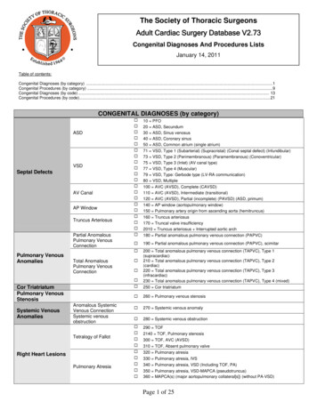
Case Head CT at OSH shows cerebral sinus venous thrombosis Admitted, heparin drip started, transitioned tosubcutaneous enoxaparin Had several odd episodes of generalized shaking whilemaintaining consciousness “Screaming in pain” with one of those episodes Repeat head imaging showed progression of CVSTthrombosis despite anticoagulation Transfer to Rush
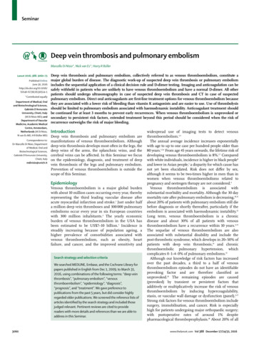
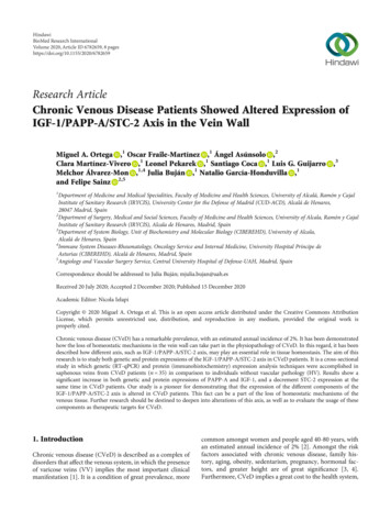
Extensive dural venous sinus thrombosisinvolving the entire superior sagittal sinus,right transverse and sigmoid sinuses Partial thrombosis of theleft transverse/sigmoid sinuses and severalparasagittal cortical veins
CaseUpon arrival to Rush: Somnolent, but easily arousable Complains of severe headache Fully oriented Intact cranial nerves Fundoscopy shows blurred disc margins Full motor strength throughout No sensory deficit Heparin gtt restarted
CaseNext day: More somnolent, difficult to arouse, inattentive, non-verbal CN intact All 4 extremities drift to bed when she becomes inattentive,improves when stimulated Consulted endovascular team– Severe thrombosis of all the intracranial sinuses– Undergoes mechanical thrombectomy and partialrecanalization of all sinuses
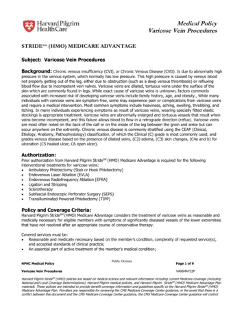
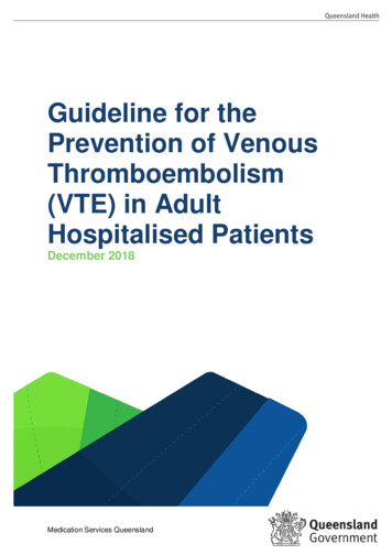
Case – MRI brain/ HCT
Case - Hospital Course Mental status and speech slowly improve over days Transitioned to coumadin Discharged to acute rehab3 months later: Doing very well Neurologic exam is normal Headaches subsided 2-3 weeks after discharge Continues on warfarin, INR is therapeutic Levetiracetam for seizures, which have not recurred
Learning ObjectivesEpidemiology, pathophysiologyand clinical features of CVST
CVST - Epidemiology Cerebral venous thrombosis– Venous sinus thrombosis– Cortical vein thrombosis Relatively rare cause of stroke ( 1%) Annual incidence estimated 3 - 7 cases per million Comparable incidence to acute bacterial meningitis in adults More common among young women and children Can cause devastating injury to the brain, but most patientshave a good prognosis if it is recognized and treated earlySamuels & Webb, NCS Practice Update 2013Ferro & Canhao, Curr Cardiol Rep (2014) 16:523
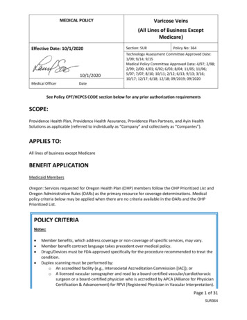
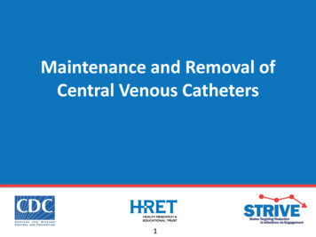
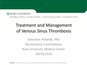
CVST - Pathophysiology% location of CVST(International Study on CerebralVenous and Dural SinusesThrombosis, n 624)Saposnik et al., Stroke. 2011;42:1158-1192
CVST - PathophysiologyThrombosis of the cortical veins:– Localized vasogenic edema– Venous infarction with cytotoxic edema– Hemorrhage Symptoms:– Seizures– focal neurologic symptomsSamuels & Webb, NCS Practice Update 2013
CVST - PathophysiologyThrombosis of the large venous sinuses:– Obstructs venous drainage– Impaired CSF absorption through arachnoid villi– Intracranial hypertension without hydrocephalus Symptoms:– Elevated intracranial pressure– Bi-hemispheric symptoms (stupor, coma)Samuels & Webb, NCS Practice Update 2013
CVST - Pathophysiology Multiple etiologic factors Usually one or more predisposing risk factors plus oneinciting factor Thrombosis develops through common pathways of: Hypercoagulability Hemoconcentration Direct injury or inflammation of the vessel Venous stasis Transient and/or permanent risk factors raise suspicion forCVST and influence treatment durationSamuels & Webb, NCS Practice Update 2013
CVST - Transient Risk Factors Infections– Central nervous system (empyema, meningitis)– Ear, sinus, mouth, face and neck (otitis, mastoiditis, tonsillitis,stomatitis, sinusitis, cellulitis)– Systemic infections (sepsis, endocarditis, tuberculosis, HIV, malaria) Pregnancy and puerperium Physical precipitants– Head trauma– Lumbar puncture, myelography, intrathecal medications, spinalanesthesia– Radical neck surgery– Neurosurgical procedures– Jugular and subclavian cathetersFerro & Canhao, Curr Cardiol Rep (2014) 16:523
CVST - Transient Risk Factors Drugs with ral contraceptivesHormone replacement therapyAndrogensMedroxyprogesterone acetateL- alidomide– Ecstasy– Sildenafil Other conditions– Dehydration– Diabetic ketoacidosisFerro & Canhao, Curr Cardiol Rep (2014) 16:523
CVST - Permanent Risk FactorsProthrombotic conditionsGenetic Protein S, C, antithrombindeficiencies Factor V Leiden Prothrombin mutationsAcquired Antiphospholipid AB syndrome Nephrotic syndrome Cyanotic congenital heartdiseaseFerro & Canhao, Curr Cardiol Rep (2014) 16:523
CVST - Permanent Risk Factors Malignancy– Central nervous system (meningioma)– Solid tumors outside central nervous system– Hematological (leukemias, lymphomas) Hematological condition– Anemias (sickle cell disease and trait, iron deficiency, folic aciddeficiency)– Paroxysmal nocturnal hemoglobinuria– Polycythemia (primary or secondary)– Thrombocythemia (primary or secondary)Ferro & Canhao, Curr Cardiol Rep (2014) 16:523
CVST - Permanent Risk Factors CNS disorders– Dural fistulae Inflammatory diseases– Behçet’s disease– Systemic lupuserythematosus– Sjögren’s syndrome– Wegener’s granulomatosis– Temporal arteritis– Thromboangiitis obliterans– Inflammatory bowel disease– Sarcoidosis Other disorders– Thyroid disease Hyperthyroidism HypothyroidismFerro & Canhao, Curr Cardiol Rep (2014) 16:523
CVST - Clinical Features Onset acute, subacute or chronic Headache is most common, nearly 90% of patients Other common presenting symptoms:– Focal or generalized seizure (40%)– Focal motor weakness (37%)– Encephalopathy or change in mental status (22%)– Vision loss (13%)– Diplopia (13%)– Stupor or coma (13%)Samuels & Webb, NCS Practice Update 2013
CVST - Clinical Features Papilledema in 25-30% of patients Thrombosis of the cavernous sinus produces acharacteristic syndrome:– Orbital pain– Proptosis– Chemosis– Variable dysfunction of cranial nerves III, IV, V, and VISamuels & Webb, NCS Practice Update 2013
Learning ObjectivesDiagnosis of CVST
CVST - DiagnosisHigh degree of clinical suspicion is key to the diagnosis– Head CT– CT Venography (CTV)– MRI/MRV– Catheter Angiography (DSA)Samuels & Webb, NCS Practice Update 2013
CVST DiagnosisHead CT Non-contrast head CT may be normal Cannot exclude a diagnosis of CVST Suspicious findings include:– Cerebral edema– Bilateral infarction– Infarction in a non-arterial distribution– Lobar intracerebral or subarachnoid hemorrhage– Hyperdense thrombosed cortical veins– Hyperdensities within the venous sinusesSamuels & Webb, NCS Practice Update 2013
CVST - DiagnosisCT Venography (CTV) Sensitivity of 95% compared with digital subtractionangiography, widely available, quick Less expensive than MRI Less invasive than conventional angiography Provides good visualization of the major venous sinuses Suboptimal for thrombosis in deep venous structures andcortical veins Radiation exposure and administration of intravenouscontrastSamuels & Webb, NCS Practice Update 2013
CVST - DiagnosisMRI/MRV MRI in combination with time-of-flight or contrastenhanced MR venography (MRV) highly sensitive forthe diagnosis CVST Abnormal T1 and/T2 signal within the venous sinus andabsence of normal flow through the venous sinus on MRVconfirms the diagnosis Age of the thrombus determines T1 and T2 signalcharacteristicsSamuels & Webb, NCS Practice Update 2013
CVST - DiagnosisCatheter Angiography (DSA) CTV or MRI/MRV is usually adequate for the diagnosis orexclusion of CSVT DSA may be necessary for:– Identification of an isolated cortical vein thrombosiswithout venous sinus involvement– Diagnosis and characterization of dural arteriovenousfistula associated with a CVSTSamuels & Webb, NCS Practice Update 2013
Learning ObjectivesTreatment of CVST
CVST - TreatmentAnticoagulation Cornerstone of treatment for CVST Prevent extension of the thrombosis and support spontaneousthrombus resolution Indicated even in the presence of intracranial hemorrhageSamuels & Webb, NCS Practice Update 2013
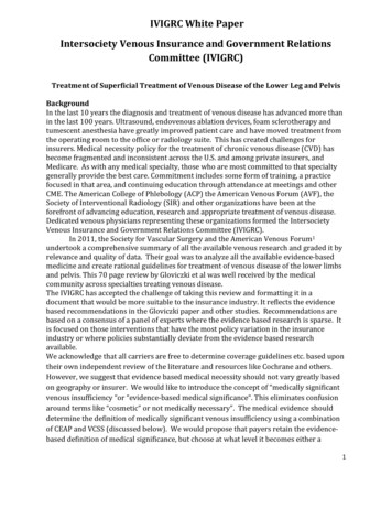
CVST - Treatment“no new symptomaticcerebral hemorrhages.Anticoagulation proved safe,even in patients with cerebralhemorrhage”Patients with ICH and CVST:27 treated with IV heparin, 4 died (mortality 15%)13 not treated with heparin, 9 died (mortality 69%)“ICH is not a contraindication to heparin treatment ”de Bruijn & Stam, Stroke 1999;30:484-8.Einhäupl et al., Lancet. 1991 Sep 7;338(8767):597-600.
CVST - Treatment American Heart Association recommendations:“ initial anticoagulation with adjusted-dose UFH or weight-based LMWH in fullanticoagulant doses is reasonable, followed by vitamin K antagonists,regardless of the presence of ICH”(Class IIa; Level of Evidence B)“Continuation of oral anticoagulation with vitamin-K antagonists is reasonablefor 3-6 months followed by antiplatelet therapy“(Class IIa, Level B) Essentially identical recommendations from (now defunct)European Federation of Neurological SocietiesSaposnik et al., Stroke. 2011;42:1158-1192
CVST - TreatmentThrombolytics and Endovascular Treatment Options Numerous case reports using localized thrombolytics and mechanical clotdisruption No controlled trials to establish efficacy or safety of these therapies Appropriate agent, dose, route of administration and clinical situation haveyet to be defined Increased risk of intracranial hemorrhage is most commonly reportedcomplication Thrombolytic and endovascular treatment should be limited to selectpatients who decline despite anticoagulation Should be performed only in centers with sufficient expertise in neuroendovascular interventionsSamuels & Webb, NCS Practice Update 2013
CVST - TreatmentSeizures Most common in patients with– Focal edema– Venous infarcts– Intracranial hemorrhage Prophylactic anticonvulsants may be considered Duration of treatment depends on:– Seizure recurrence (unprovoked, 5% to 32% of patients)– EEG findings in follow-up– Tolerability of antiseizure drugsSamuels & Webb, NCS Practice Update 2013
CVST - TreatmentIntracranial Pressure Intracranial hemorrhage, edema and infarction lead to localized mass effect Venous outflow impairment causes decreased CSF reabsorption,communicating hydrocephalus and intracranial hypertension Hyperosmolar therapy (mannitol, hypertonic saline) should be administeredto patients at risk for cerebral herniation Acetazolamide is reasonable to reduce CSF production CSF diversion (lumbar puncture, ventriculostomy) or optic nervedecompression can be effective if there is progressive visual loss Resection of hemorrhagic infarction or decompressive craniectomy may berequired Anticoagulation should be resumed as soon as possible following surgicalinterventionSaposnik et al., Stroke. 2011;42:1158-1192Samuels & Webb, NCS Practice Update 2013
CVST - Outcome Recanalization– At 3 months 84%– At 1 year 85%– Highest recanalization rates in deep cerebral veins and cavernoussinus thrombosis, lowest in lateral sinus thrombosis In adults, recanalization of the occluded sinus is notrelated to outcomeSaposnik et al., Stroke. 2011;42:1158-1192
CVST - Outcome 3% to 15% of patients die in the acute phase Patients at risk:–––––Depressed consciousnessAltered mental statusThrombosis of the deep venous systemRight hemisphere hemorrhagePosterior fossa lesions. Main cause of acute death with CVT is transtentorial herniation dueto large hemorrhagic lesion Second is herniation due to multiple lesions or to diffuse brainedema. Status epilepticus, medical complications, and PE are other causesSaposnik et al., Stroke. 2011;42:1158-1192
CVST - Outcome 79% of patients will have complete recovery 9.7% are functionally dependent (mRS 3 or greater) 50% of survivors feel depressed or anxious, minor cognitive orlanguage deficits may preclude them from resuming previousjobs Abulia, executive deficits, and amnesia result fromthrombosis of the deep venous system, with bilateralpanthalamic infarcts Memory deficits, behavioral problems, or executive deficitsmay persistSaposnik et al., Stroke. 2011;42:1158-1192
CVSTQuestions?
intervention Samuels & Webb, NCS Practice Update 2013 Saposnik et al., Stroke. 2011;42:1158-1192 . CVST - Outcome Recanalization -At 3 months 84% -At 1 year 85% -Highest recanalization rates in deep cerebral veins and cavernous sinus thrombosis, lowest in lateral sinus thrombosis