Transcription
Marquez et al. World Journal of Surgical Oncology (2018) ARCHOpen AccessImmediate implant replacement with DIEPflap: a single-stage salvage option in failedimplant-based breast reconstructionMiguel De La Parra Marquez1*, Ricardo Fernandez-Riera2, Hector Vela Cardona1 and Jesus María Rangel Flores1AbstractBackground: Implant-based immediate breast reconstruction after skin-sparing mastectomy has shown asignificant improvement in patients’ quality of life, making the procedure steadily more popular year afteryear. However, this technique has a high morbidity rate, including skin necrosis and implant exposure.Methods: A retrospective review of a prospectively held database for autologous breast reconstruction inour institution of the last 5 years found eight cases with exposed implants after nipple-sparing mastectomyand immediate reconstruction. A single-stage procedure consisting on implant removal and immediatereplacement with a deepithelialized DIEP flap was performed in all cases (10 DIEP flaps).Results: All flaps were successful. Patients’ mean age was 45 years old. Three patients developed seroma(5, 7, and 14 days after surgery, respectively). No infections were detected in up to 24 months of follow-up.Conclusions: Nipple-sparing mastectomy with immediate implant-based reconstruction is consideredoncologically safe. However, it has a high rate of complications that could require implant removal.Immediate free flap reconstruction is a feasible and safe option to replace the missing volume with low riskof complications that result in a soft and natural-shaped breast.Keywords: Tertiary breast reconstruction, Autologous breast reconstruction, Implant failure, ReconstructionfailureBackgroundProven improvement in most patients’ quality of life hasmade the frequency of immediate breast reconstructionafter skin-sparing mastectomy to steadily rise in the lastyears. Women are also increasingly demanding nipplesparing and skin-sparing procedures from their surgicaloncologists [1].To date, there is no universally accepted criteria forpatient selection for skin-sparing mastectomy; however,the most accepted are tumor size less than 3 cm, tumorlocation greater than 2 cm from the nipple-sparingmastectomy, clinically negative axillary nodes, absenceof skin involvement or inflammatory cancer, and cleanmargins beneath the nipple [2–5].* Correspondence: drdelaparra@yahoo.com.mx1Division of Plastic and Reconstructive Surgery, Mexican Institute of SocialSecurity (IMSS), No. 21 Pino Suárez y 15 de Mayo, Av. Hidalgo 2480 pte, col.Obispado. C.p.64060. Cons. 212, Monterrey Nuevo León, MexicoFull list of author information is available at the end of the articleTraditionally, immediate breast reconstruction withimplants requires a two-stage procedure includingsubpectoral placement of a tissue expander at thetime of mastectomy followed by replacement of thetissue expander with the definitive breast implant as asecond stage once breast expansion has beenachieved. Acellular dermal matrix use for pocket creation and total implant coverage after nipple-sparingmastectomy is booming as a one-stage implant-basedreconstruction option and has been widely studiedand discussed in medical literature [6–15].Implant-based reconstruction is associated with risksand complications, which may lead to complete implantloss due to infection, implant exposure, or capsular contracture [16–18], the latter with reported incidencehigher than 50% of all implant-based reconstructions[19]. Breast infection or implant exposure increases therate of failure by 30%, and implant loss has been The Author(s). 2018 Open Access This article is distributed under the terms of the Creative Commons Attribution 4.0International License (http://creativecommons.org/licenses/by/4.0/), which permits unrestricted use, distribution, andreproduction in any medium, provided you give appropriate credit to the original author(s) and the source, provide a link tothe Creative Commons license, and indicate if changes were made. The Creative Commons Public Domain Dedication o/1.0/) applies to the data made available in this article, unless otherwise stated.
Marquez et al. World Journal of Surgical Oncology (2018) 16:80reported to occur in 4 to 18% of all prosthetic breast reconstructions [16]. Failure of primary or secondarybreast reconstruction creates a stressful situation forboth patient and surgeon, and decisions should be takento adjust the strategy and eliminate potential causes ofrecurrent failure [18].Hamdi et al. coined the term “tertiary breast reconstruction” for those “redo” reconstructions of cases withunsatisfactory results or failure of previous immediate ordelayed procedures [17]; several authors have used thatterm afterwards [20–23].The goal of tertiary reconstruction is the completerestoration of the breast after a failed previous reconstruction; it can be achieved with an implant, autologous tissue, or a combination of both. Long-termimplant-based reconstruction complications and improvement on microvascular techniques are makingpatients ask for autologous reconstruction more often[16, 24]. In 1994, Feng et al. reported the use of autogenous tissue for breast reconstruction followingimplant failure [25]; nowadays, there are several autologous flaps used for this purpose, such as deep inferior epigastric artery perforator flap (DIEP), superiorgluteal artery perforator flap (SGAP), transverse musculocutaneous gracilis flap (TMG), superficial inferiorepigastric artery perforator flap (SIEA), and manyothers [15]. These procedures have the advantage ofresulting in a breast that responds to weight changes,that has a natural texture, and that eliminates the riskof capsular contracture, but they require certainmicrosurgical expertise, lead to additional scars in thedonor site, and need longer surgeries than theirprosthetic-based counterparts [18, 26, 27].The purpose of this study was to prove the safety andfeasibility of the management of implant exposure afternipple-sparing breast mastectomy and implant-based reconstruction with implant removal and immediate replacement with DIEP flap.MethodsFor a period of 5 years (starting June 2012), we haveprospectively held an independent database for patients admitted at our institution for breast reconstruction. We performed a retrospective review of thisdatabase looking for all patients referred to the plasticsurgery department of our hospital who had previously been managed with nipple-sparing mastectomyand immediate prosthetic-based reconstruction anddeveloped implant exposure. A one-stage procedure withimplant removal and immediate replacement with DIEPflap was performed in all cases. Demographic data andcomplications were studied to assess the safety and feasibility of our single-stage tertiary breast reconstructiontechnique.Page 2 of 6Operative techniquePreoperative markings are performed with the patient inthe standing and supine positions. The superior marginof the flap is shifted slightly above the umbilicus to include periumbilical perforators. Perforators are identifiedwith a handheld 8 MHz Doppler. No additional imagestudies are routinely performed in our institution.The procedure is approached by two teams. In the abdomen, DIEP flap is elevated from lateral to medial in asuprafascial plane until adequate perforators are found.The superficial inferior epigastric vein (SIEV) is preserved. The largest perforators are dissected, and the anterior rectus sheath is opened around the perforatingvascular bundle, allowing the perforators to be traced tothe deep inferior epigastric vessels. Intercostal nervesshould be left intact to avoid denervating the musclesmedially. The rectus sheath and muscle are separated toallow isolation of the pedicle until desired pedicle lengthand diameter is obtained.Simultaneously, the second team proceeds to removethe exposed implant, debride the borders of the skin defect, and wash the pocket with iodine and saline solutions, hypochlorous acid solution also being a goodalternative. After pocket irrigation, we perform multiplecapsulotomy incisions to attain good compliance of thechest flap. The internal mammary artery and vein aredissected as the recipient vessels of choice; when thearea of implant exposure is too close to the inframammary fold, the fourth rib is selected; otherwise, the thirdrib level is preferred. We approach the vessels by resecting one rib-cartilage level to improve visibility and facilitate the microvascular anastomosis in these difficultcases. The flap is placed in the same pocket where theprevious implant was; no new pocket or change of planeis advocated by our team.After dividing the pedicle, the flap is transferred to thethorax for anastomosis to the internal mammary vesselswith 9-0 interrupted nylon sutures under surgical microscope augmentation. The flap is deepithelialized, and askin paddle is left in place to monitor the flap, usuallywhere the implant was exposed to make up for the losttissue in that area. The flap is inserted in the pocket andthe wound closed in two layers with 3-0 Monocryl ontop of a suction drain.The rectus sheath is closed with no tension, and theabdominal flap is advanced and closed in three layersover suction drains (Figs. 1, 2, 3, 4, 5, and 6).ResultsFrom June 2012 through May 2017, 140 DIEP flaps wereperformed for breast reconstruction in the Departmentof Microsurgery in our institution. Eight of these patients were included in our study as they were previouslytreated with nipple-sparing mastectomy and immediate
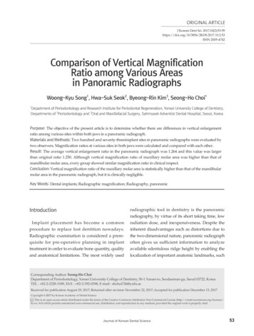
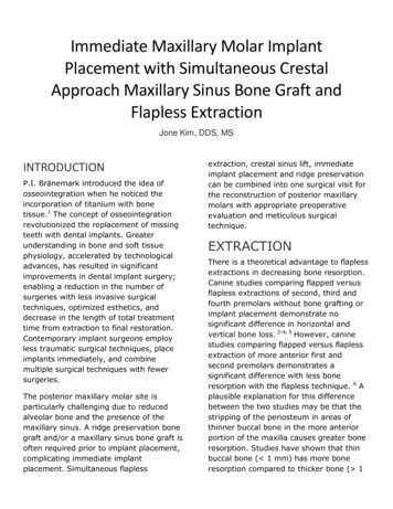
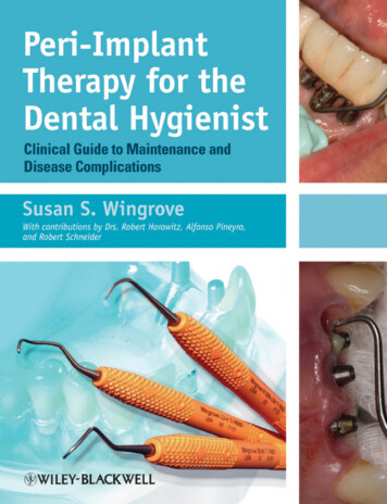
Marquez et al. World Journal of Surgical Oncology (2018) 16:80Page 3 of 6Fig. 2 Case 1. After failed reconstruction with implant exposure onthe left breast, the patient was recruited in our study. The leftimplant was removed. Trans-operative view before DIEP flap harvestFig. 1 Case 1. Preoperative view before right prophylactic nipplesparing mastectomy and left therapeutic nipple-sparing mastectomywith immediate bilateral implant-based reconstructionreconstruction with implant and developed partialskin necrosis and implant exposure. Two of these patients suffered bilateral implant exposure. Immediatedeepithelialized DIEP flap was performed in all patients. The two patients with bilateral implant exposure underwent tertiary reconstruction with immediatebilateral DIEP flaps.Patients’ mean age was 45 years old, (42–50 years; SD 3.30), and the average implant volume removed was463 cm3 (410–525 cm3; SD 47.14 cm3). The mean timefrom implant exposure to tertiary DIEP flap-based reconstruction was 8 days (7–14 days). Exposed implants were seven silicones, two polyurethanes, andone saline. All patients were referred from differentinstitutions, so we ignored the frequency with whichthey use each kind of implant to learn the rate offailure according to the implant type. Mean hospitalstay was 6 days after surgery.Three of the reconstructed breasts (30%) presentedseroma that needed re-exploration and placement of anew drain 5, 7, and 14 days after surgery, respectively.Of these cases, one had had silicone, one saline, and onepolyurethane implants. No partial or total flap necrosisor infection was present in these patients.Discussion.Nipple-sparing mastectomy and immediate implantbased reconstruction is increasingly accepted as a desirable option for patients with breast cancer or hereditaryrisk of breast cancer. The oncological safety of the procedure has been well demonstrated [7, 28]. However, ensuring total resection of glandular tissue might lead toextremely thin skin envelopes with compromised vasculature. Skin flap ischemia is reported to happen in 5 to15% of cases, and short-term complication rate is about33 to 55% with implant loss in 8 to 18% of patients [7,14, 29, 30]. In our study, all eight patients presentednipple-areola or skin necrosis and implant exposure thatwarranted prosthesis explantation.Our technique for flap harvest does not differ fromthat previously described [17, 31–35]; however, flap insetvaries in some aspects. First, if the third costal cartilageis far away from the skin defect, the fourth rib cartilage
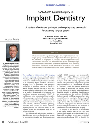
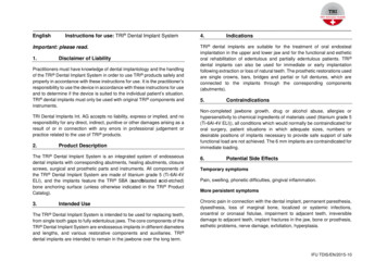
Marquez et al. World Journal of Surgical Oncology (2018) 16:80Page 4 of 6Fig. 4 Case 2. Forty-five years old female after right nipple sparingtherapeutic mastectomy and left prophylactic nipple-sparingmastectomy and immediate bilateral reconstruction with polyurethaneround implants. Exposure of both polyurethane implants 5 weeks afterthe surgeryFig. 3 Case 1. Postoperative view after 8 weeks of single-stageimplant removal and replacement with DIEP flapis preferred. In case of peri-areolar exposure, thethird cartilage is used. Next, our experience is thatcapsulotomy is sufficient to provide chest flap compliance, allowing to create enough space to fit in theDIEP flap. Also, avoiding capsulectomy which, for us,has been unnecessary decreases bleeding and reducessurgery time. Fear about infection when using thesame pocket is understandable, but it seems that thenew, well-vascularized tissue and postoperative antibiotic coverage are enough to avoid it as we had nocase of infection after surgery.Changing the plane of dissection by creating a newpocket as suggested by Bramhall on his recent publication [36] would also lead to increased bleeding and timein the operating room. On the downside, three patients(30%) in our study presented seroma which neededdraining. It is possible that our capsulotomy-only approach could be related to this complication, but furthercomparative studies are needed to objectively supportthis theory. All three seroma cases were resolved by placing a new drain without further complications.Positioning the skin monitor of the DIEP flap wherethe implant was exposed serves two purposes: adequateflap monitoring and to avoid further breast deformity byreplacing missing tissue lost due to skin necrosis. Therest of the flap needs only to be deepithelialized as it isproven that the dermis plays a significant role inFig. 5 Removed implant and deepithelialized left DIEP flap
Marquez et al. World Journal of Surgical Oncology (2018) 16:80Page 5 of 6However, it has a high rate of complications that couldnecessitate implant removal. Immediate tertiary DIEPflap reconstruction with our technique replaces the exposed implant with low risk of infection (no cases in thisstudy) and approximately 30% rate of seroma that canbe resolved by appropriate draining with no furthercomplications. It results in a soft and natural final breastshape.Availability of data and materialsPlease contact the author for data requests.Authors’ contributionsMPM is the main author, attending physician, and lead surgeon in all casesand contributed to the manuscript drafting, critical revision, and finalapproval. RFR contributed to the manuscript drafting and proofreading andis the surgical assistant. HVC contributed to the manuscript design anddrafting and is the surgical assistant. JMRF contributed to the dataacquisition and data analysis. All authors read and approved the finalmanuscript.Ethics approval and consent to participateThis study complies with the Helsinki Declaration and has been reviewedand authorized by the research and bioethics committee of the MexicanInstitute of Social Security 21, Monterrey NL, with the reference number R2017-1903-36.Consent for publicationWritten informed consent was obtained from the patient for the publicationof this report and any accompanying images.Competing interestThe authors declare that they have no competing interests.Fig. 6 Case 2. Postoperative view after single-stage salvagereconstruction with bilateral implant replacement with bilateral deepithelialized DIEP flapenhancing the overall DIEP flap perfusion through thepreservation of indirect linking vessels in the subdermalplexus [37].Tertiary breast reconstruction after implant failure orcapsular contracture has been widely discussed [17, 20,38, 39]. However, to our knowledge, there is only one recent publication in English literature addressing the possibility of one-stage implant removal and immediateautologous reconstruction [36]. This technique addresses some problems found in delayed tertiary reconstruction such as skin retraction and added fibrosis, notto mention the cost and risks associated with an additional surgery. We believe that, on the psychologicalstandpoint, the immediate implant replacement withDIEP flap is also beneficial to the patient just as the impact of immediate breast reconstruction is better thanthat of the delayed one [40, 41].ConclusionNipple-sparing mastectomy and immediate implantbased reconstruction is an oncologically safe technique.Publisher’s NoteSpringer Nature remains neutral with regard to jurisdictional claims inpublished maps and institutional affiliations.Author details1Division of Plastic and Reconstructive Surgery, Mexican Institute of SocialSecurity (IMSS), No. 21 Pino Suárez y 15 de Mayo, Av. Hidalgo 2480 pte, col.Obispado. C.p.64060. Cons. 212, Monterrey Nuevo León, Mexico.2Department of Plastic and Reconstructive Surgery, Hospital General Dr.Ruben Leñero, Plan de San Luis esq Salvador Díaz Mirón SN, Col. SantoTomás. Deleg. Miguel Hidalgo. Cp. 11340. CDMX, Mexico City, Mexico.Received: 17 October 2017 Accepted: 10 April 2018References1. Cassileth L, Kohanzadeh S, Amersi F. One-stage immediate breastreconstruction with implants: a new option for immediate reconstruction.Ann Plast Surg. 2012;69:134–8.2. Endara M, Chen D, Verma K, Nahabedian MY, Spear SL. Breast reconstructionfollowing nipple-sparing mastectomy: A systematic review of the literaturewith pooled analysis. Plast Reconstr Surg. 2013;132:1043–54.3. Spear SL, Willey SC, Feldman ED, Cocilovo C, Sidawy M, Al-Attar A, et al.Nipple-sparing mastectomy for prophylactic and therapeutic indications.Plast Reconstr Surg. 2011;128:1005–14.4. Benediktsson KP, Perbeck L. Survival in breast cancer after nipple-sparingsubcutaneous mastectomy and immediate reconstruction with implants: aprospective trial with 13 years median follow-up in 216 patients. Eur J SurgOncol. 2008;34:143–8.5. Mosahebi A, Ramakrishnan V, Gittos M, Collier SJ. Aesthetic outcome ofdifferent techniques of reconstruction following nipple-areola-preservingenvelope mastectomy with immediate reconstruction. Plast Reconstr Surg.2007;119:796–803.
Marquez et al. World Journal of Surgical Oncology (2018) 2.23.24.25.26.27.28.Sbitany H, Sandeen SN, Amalfi AN, Davenport MS, Langstein HN. Acellulardermis-assisted prosthetic breast reconstruction versus completesubmuscular coverage: a head-to-head comparison of outcomes. PlastReconstr Surg. 2009;124:1735–40.Woerdeman LAE, Hage JJ, Smeulders MJC, Rutgers EJT, van der HorstCMAM. Skin-sparing mastectomy and immediate breast reconstruction byuse of implants: an assessment of risk factors for complications and cancercontrol in 120 patients. Plast Reconstr Surg. 2006;118:321–30.Kroll SS, Schusterman MA, Tadjalli HE, Singletary SE, Ames FC. Risk ofrecurrence after treatment of early breast cancer with skin-sparingmastectomy. Ann Surg Oncol. 1997;4:193–7.Kroll SS, Ames F, Singletary SE, Schusterman MA. The oncologic risks of skinpreservation at mastectomy when combined with immediatereconstruction of the breast. Surg Gynecol Obstet. 1991;172:17–20.Hidalgo DA, Borgen PJ, Petrek JA, Heerdt AH, Cody HS, Disa JJ. Immediatereconstruction after complete skin-sparing mastectomy with autologoustissue. J Am Coll Surg. 1998;187:17–21.Gherardini G, Thomas R, Basoccu G, Zaccheddu R, Fortunato L, Cortino P, et al.Immediate breast reconstruction with the transverse rectus abdominismusculocutaneous flap after skin-sparing mastectomy. Int Surg. 2001;86:246–51.Disa JJ, Cordeiro PG, Heerdt AH, Petrek JA, Borgen PJ, Hidalgo DA. Skinsparing mastectomy and immediate autologous tissue reconstruction afterwhole-breast irradiation. Plast Reconstr Surg. 2003;111:118–24.Sufi PA, Gittos M, Collier DSJ. Envelope mastectomy with immediatereconstruction (EMIR). Eur J Surg Oncol. 2000;26:367–70.Contant CME, Van Geel AN, Van Der Holt B, Griep C, Tjong Joe Wai R,Wiggers T. Morbidity of immediate breast reconstruction (IBR) aftermastectomy by a subpectorally placed silicone prosthesis: the adverse effectof radiotherapy. Eur J Surg Oncol. 2000;26:344–50.Toth BA, Lappert P. Modified skin incisions for mastectomy: the needfor plastic surgical input in preoperative planning. Plast Reconstr Surg.1991;87:1048–53.Spear SL, Masden D, Rao SS, Nahabedian MY. Long-term outcomes of failedprosthetic breast reconstruction. Ann Plast Surg. 2013;71(3):286–91.Hamdi M, Casaer B, Andrades P, Thiessen F, Dancey A, D’Arpa S, et al.Salvage (tertiary) breast reconstruction after implant failure. J Plast ReconstrAesthetic Surg. 2011;64:353–9.Visser NJ, Damen THC, Timman R, Hofer SOP, Mureau MAM. Surgical results,aesthetic outcome, and patient satisfaction after microsurgical autologousbreast reconstruction following failed implant reconstruction. Plast ReconstrSurg. 2010;126:26–36.Tarantino I, Banic A, Fischer T. Evaluation of late results in breastreconstruction by latissimus dorsi flap and prosthesis implantation. PlastReconstr Surg. 2006;117:1387–94.Mohan AT, Al-Ajam Y, Mosahebi A. Trends in tertiary breast reconstruction:literature review and single centre experience. Breast. 2013;22:173–8.Pülzl P, Huemer GM, Schoeller T. Transverse musculocutaneous gracilis flapfor treatment of capsular contracture in tertiary breast reconstruction. AnnPlast Surg. 2015;74:167–72.Munhoz AM, Montag E, Arruda EG, Okada A, Fonseca A, Filassi JR, et al. Theuse of contralateral free extended latissimus dorsi myocutaneous flap for atertiary failed breast reconstruction: is it still an option? J Plast ReconstrAesthetic Surg. 2016;69:1087–91.Endara MR, Verma K, Nahabedian MY. Tertiary breast reconstruction using afree contralateral latissimus dorsi musculocutaneous flap. J ReconstrMicrosurg. 2014;30:141–3.Hamdi M, Andrades P, Thiessen F, Stillaert F, Roche N, Van Landuyt K, et al.Is a second free flap still an option in a failed free flap breastreconstruction? Plast Reconstr Surg. 2010;126:375–84.Feng LJ, Mauceri K, Berger BE. Autogenous tissue breast reconstruction inthe silicone-intolerant patient. Cancer. 1994;74:440–9.Schoeller T, Huemer GM, Wechselberger G. The transversemusculocutaneous gracilis flap for breast reconstruction: guidelines for flapand patient selection. Plast Reconstr Surg. 2008;122:29–38.Rabey NG, Lie KH, Kumiponjera D, Erel E, Simcock JW, Malata CM. Salvage offailed prosthetic breast reconstructions by autologous conversion with freetissue transfers. Eplasty. 2013;13:e32.Albornoz CR, Matros E, McCarthy CM, Klassen A, Cano SJ, Alderman AK,et al. Implant breast reconstruction and radiation: a multicenter analysis oflong-term health-related quality of life and satisfaction. Ann Surg Oncol.2014;21:2159–64.Page 6 of 629. Hoffman JP, Kusiak J, Boraas M, Genter B, Steuber K, Weese JL, et al. Riskfactors for immediate prosthetic postmastectomy reconstruction. Am Surg.1991;57:514–21. discussion 52230. Nahabedian MY, Tsangaris T, Momen B, Manson PN. Infectiouscomplications following breast reconstruction with expanders and implants.Plast Reconstr Surg. 2003;112:467–76.31. Healy C, Allen RJ. The evolution of perforator flap breast reconstruction:twenty years after the first DIEP flap. J Reconstr Microsurg. 2014;30:121–6.32. Koshima I, Soeda S. Inferior epigastric artery skin flaps without rectusabdominis muscle. Br J Plast Surg. 1989;42:645–8.33. Van Waes C, Seidenstuecker K, Mahajan A, Roche N, Stillaert F, Andree C,et al. The safety of the deep inferior epigastric artery perforator flapprocedure. Plast Reconstr Surg. 2011;128:57.34. Scheflan M, Dinner MI. The transverse abdominal island flap: part I. Indications,contraindications, results, and complications. Ann Plast Surg. 1983;10:24–35.35. Scheflan M, Dinner MI. The transverse abdominal island flap: part II. Surgicaltechnique Ann Plast Surg. 1983;10:120–9.36. Bramhall RJ, Hernan I, Harris PA. A single-centre, retrospective proof-ofconcept review of salvage of infected or exposed implant breastreconstructions with explantation and one-stage free flap replacement. JPlast Reconstr Aesthet Surg. 2018;71:194–200.37. Laungani A, Christner JA, Lachman N, Ritman EL, Pawlina W, Saint-Cyr M.Impact and role of subdermal plexus in DIEP flap perfusion. Plast ReconstrSurg. 2013;132:128.38. Bennett SPH, Fitoussi AD, Berry MG, Couturaud B, Salmon RJ. Managementof exposed, infected implant-based breast reconstruction and strategies forsalvage. J Plast Reconstr Aesthetic Surg. 2011;64:1270–7.39. Levine SM, Lester ME, Fontenot B, Allen RJ. Perforator flap breast reconstructionafter unsatisfactory implant reconstruction. Ann Plast Surg. 2011;66(5):513–7.40. Stevens LA, McGrath MH, Druss RG, Kister SJ, Gump FE, Forde KA. Thepsychological impact of immediate breast reconstruction for women withearly breast cancer. Plast Reconstr Surg. 1984;73:619–28.41. Zhong T, Hu J, Bagher S, Vo A, AC ON, Butler K, et al. A comparison ofpsychological response, body image, sexuality, and quality of life betweenimmediate and delayed autologous tissue breast reconstruction: aprospective long-term outcome study. Plast Reconstr Surg. 2016;138:772–80.
culocutaneous gracilis flap (TMG), superficial inferior epigastric artery perforator flap (SIEA), and many others [15]. These procedures have the advantage of resulting in a breast that responds to weight changes, that has a natural texture, and that eliminates the risk of capsular contracture, but they require certain