Transcription
SCIENTIFIC ARTICLE CAD/CAM Guided Surgery inImplant DentistryA review of software packages and step-by-step protocolsfor planning surgical guidesBy Michael D. Scherer, DMD, MS;Author ProfileMathew T. Kattadiyil, DDS, MDS, MS;Ewa Parciak, DDS;Shweta Puri, BDSABSTRACTDr. Michael Scherer, DMD,MS, is currently an assistantprofessor in residence at University of Nevada – Las Vegas,an assistant clinical professorat Loma Linda University, anda fellow of the AmericanCollege of Prosthodontists.He has published articlesrelated to clinical prosthodontics and implant dentistryand completed a master’sdegree related to implantoverdentures with multiplepublications pending. As anavid technology and computer hobbyist, Dr. Scherer’sinvolvement in digital implantdentistry has led him to develop and utilize new technology with CAD/CAM surgicalsystems and implement student facilitated CBCT implantplanning. Dr. Scherer servesas the director of the implantdentistry curriculum at UNLVand is actively engaged inguided surgical placementand prosthetic restoration ofimplants.32Three-dimensional radiographic imaging for dental implant treatment planning is gaining widespread interest and popularity. However, application ofthe data from 3D imaging can be a complex and daunting process initially.The purpose of this article is to describe features of three software packagesand the respective computerized guided surgical templates (GST) fabricatedfrom them. A step-by-step method of interpreting and ordering a GST to simplify the process of the surgical planning and implant placement is discussed.The paradigm of 2-dimensional (2D) imaging,treatment planning with model-based surgicalplanning, and surgical guide fabrication for thepurposes of dental implant placement has shiftedto a completely digital workflow. Traditionalimaging, such as panoramic and periapicalradiographs is inherently difficult to utilize fordental implant planning because it does notrepresent all dimensions of the bone volume.1-2As a result, 3-dimensional (3D) imaging withcone-beam computed tomography (CBCT) incombination with computerized interpretationsoftware packages enable the clinician to visualizea greater amount of diagnostic information.3 Thisimproved visualization improves assessment ofbone volumes, enhances treatment planning,allows for more accurate and precise control ofimplant position, and offers the ability to linktogether guides and restorations with the 3Dradiograph image.4Three-dimensional radiographic imaging fordental implant treatment planning, however, canbe a seemingly complex and daunting process.Alpha Omegan Spring 2014Multiple CBCT machines are commerciallyavailable and utilize different controls andscan settings. In addition, numerous softwarepackages exist with varying capabilities andfeatures with proprietary computerized guidedsurgical templates (GST), all of which may leadto confusion. Dental and medical radiologistshave strived to standardize the complex worldof medical imaging by setting a standard formatfor digital imaging in radiology: digital imagingand communications in medicine (DICOM).Engineering and modeling standards have alsobeen established for enabling the sharing of opticalimaging data and exporting stereolithographyimages (STL). This standardization allows fora common language to exist between softwarepackages and greatly enhances the ability for acollaborative workflow with digital imaging.The purpose of this article is to review threeof the more popular software packages andcomputerized guided surgical templates (GST).This article provides a step-by-step method ofinterpreting and ordering GSTs to simplify thewww.ao.org
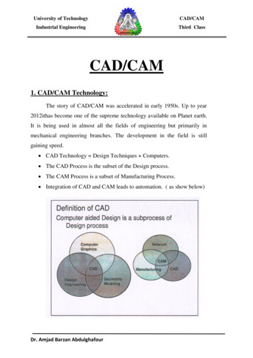
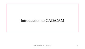
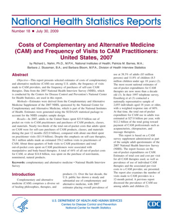
CAD/CAM GUIDED SURGERY IN IMPLANT DENTISTRY SCIENTIFIC ARTICLEprocess of the surgical planning andplacement of implants.Software Package FeaturesMultiple software packages areavailable for the clinician to choose fromfor analysis, treatment planning, andfabrication of guided surgical templates.In order to simplify the complexlandscape of digital implant treatmentplanning, three major software packagesare reviewed: Invivo (Anatomage; SanJose, Calif.), Simplant (Materialise NV;Leuven, Belgium), and NobelClinician(NobelBiocare; Yorba Linda, Calif.).While each software has unique featuresand specific protocols, they sharecommon features (Table 1).Invivo and Anatomage GuideAnatomage Inc. is a medical imagingcompany that produces softwarefor medical and dental specialties.Invivo Dental is a dental software thatallows clinicians to visualize DICOMdatasets in 3D volumetric renderingsof hard and soft tissues, measureairway volumes, trace orthodonticlandmarks, and plan dental implants.The dental implant module allows fullcontrol of 3D renderings allowing theclinician to section, slice, identify, andmark anatomical landmarks, and thendetermine placement of dental implants.In the implant module, the cliniciancan manipulate implant position,angulation, trajectory, and measurerelative bone density. Unique featuresof the Invivo software are instantaneous3D rendering / image processing(thresholding) (Figure 1), a dynamicclipping mode (virtual slicing), allowingthe clinician to visualize 3D restorativespace (Figure 2), and creation of virtualwax patterns based upon a digitalrestorative library (Figure 3).Anatomage Guide is the surgicalguide fabricated by Anatomage uponcompletion of the implant planning inthe Invivo software. The following typesof templates are currently available forAnatomage Guide: Tooth-supported Mucosa-supported Bone-supported reduction andimplant surgical guides (mandibleonly)Table 1.Figure 1. Invivo software features automatic thresholding and volumetric rendering.Figure 2. Clipping mode allows the clinician to visualize prosthetic space between implantplatform and the occlusal surface of the planned restoration.Alpha Omegan Spring 201433
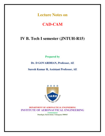
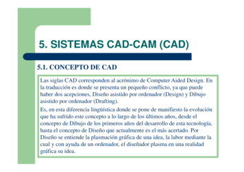
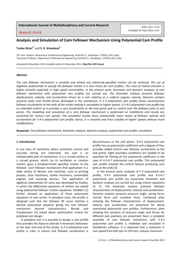
SCIENTIFIC ARTICLE CAD/CAM GUIDED SURGERY IN IMPLANT DENTISTRYFigure 4. Zest Locator overdenture implant placed using an Anatomageguide with universal master sleeves and drills.Figure 3. Virtual restorations added to dental implants to finely tune the implant angulationand position.Universal master sleevesAuthor ProfileDr. Ewa Parciak, DDS, is athird year graduate student inthe Advanced Specialty Education Program in Prosthodontics at Loma Linda University,School of Dentistry (LLUSD).She earned her dental degreein her home country of Polandin 2005. She completed theInternational Dentist Program(LLUSD) in 2008 and aGeneral Practice Residency atVAH in Loma Linda in 2010.She enjoys traveling andphotography.Anatomage Guide controls the trajectoryand osteotomy depth through the master sleeveintegrated into the acrylic drill guide template.A unique feature of the Anatomage Guide is theuniversal master sleeve that allows a clinician toinsert any implant that uses a parallel drillingprotocol such as Straumann Bone Level, NobelActive/Speedy, Dentsply AstraTech, and ZestLocator Overdenture Implants (LODI). (Figure 4)The handle and drill kits accommodate narrowsleeves (3.1mm), regular sleeves (4.1mm), andwide sleeves (5.1mm), and the drill kit includes21mm and 26mm universal drills. (Figure 5)The universal kit allows full osteotomypreparation except for drill tapping and final implantinsertion (partially-guided); these procedures shouldbe performed without the guide in the mouth.If a tapered implant drilling protocol is required,universal drills should be used up until the last drillsize and the final tapered drill in the manufacturer’skit used to prepare the final osteotomy. Implanttrajectory is controlled by tilting the master sleevesrelative to teeth, mucosa, or bone; implant depth iscontrolled by raising or lower the sleeve position inrelation to the above. The clinician needs to be awareof the surgical drilling protocol for the respectiveimplant when using a universal kit. For example, ifa 3.5mm x 11.5mm Nobel Replace implant were tobe placed, the universal drilling protocol would be aguided universal 2.0mm drill, followed by a guideduniversal 2.8mm drill, and finally by non-guidedNobel tapered 3.5mm x 13mm drill.Manufacturer-specific master sleevesIf preferred, Anatomage can also fabricate aguide with manufacturer-specific master sleevesfor the following systems:34Alpha Omegan Spring 2014Figure 5. Universal Anatomage Guide drills and handles allow forosteotomy preparation for multiple systems.Figure 6. Manufacturer specific master sleeve with a universalguide allows for a fully-guided implant surgical procedure.Nobel GuideZimmer3i NavigatorStraumannCamlogImplant DirectManufacturer-specific master sleeves allow forfull trajectory and depth control, guided drill tap,and guided implant insertion (Figure 6). Fullyguided implant insertion capability with Anatomageguide, however, is currently available only forNobelBiocare, 3i, and Camlog implant systems.Full implant guidance may be less of an issue withclinicians who routinely use guided surgery to placeimplants without immediate provisional restorationspre-fabricated in the laboratory. If a clinician wishesto have a provisional restoration pre-fabricated todeliver the day of implant insertion, fully guidedimplant placement may be important.www.ao.org
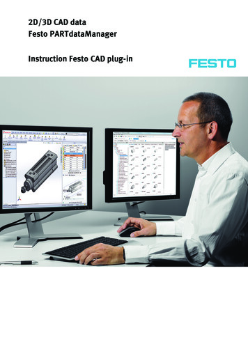
CAD/CAM GUIDED SURGERY IN IMPLANT DENTISTRY SCIENTIFIC ARTICLEFigure 7. Soft tissue separation performed with cotton rolls held inplace during the CBCT scan. Checklist for Anatomage Guide Partially Dentate Teeth-Supported Guide Patient CBCT scan with occlusal and tissueseparation (Figure 7) Patient cast optical or CBCT scan Diagnostic wax pattern optical scan (optional)Fully Edentulous Bone-Supported Guide Patient CBCT scanFully Edentulous Soft Tissue-Supported Guide Patient CBCT scan with denture relined withradiopaque PVS material (Green-Mousse,Parkell, USA). (Figure 8) Prosthesis CBCT Scan Patient cast optical or CBCT scanSubmit for Surgical Guide FabricationFor either partially or fully edentulous patients,it is recommended that the clinician initially planimplant positions according to proposed implantsites. Importing the DICOM files into the Invivosoftware will create an initial planning file (.inv) tofacilitate upload to Anatomage’s uploading service(Anatomodel). For fully edentulous templates,the clinician must import the prosthesis scanand convert the file to an initial planning file.Anchor pin placement is recommended for fullyedentulous templates, and unless specified, 2 to4 pins should be placed around the arch. Onceapproved, the surgical guide takes from 3 to 5 daysto manufacture.VERSIONCOSTFigure 8. Radiopaque PVS placed prior to making a CBCT allowingthe software to recognize the soft tissue profile.SimPlant and SurgiGuideMaterialise NV is a worldwide leader in medicaladditive manufacturing and biomedical researchand produces SimPlant, one of the pioneeringdental implant imaging software. SimPlant is aninteractive dental implant software that allowsclinicians to visualize DICOM datasets in 3D surfacerenderings with transparency controls. While mostclinicians will typically use SimPlant Planner fordental implant planning, Materialise offers severaldifferent versions of the software package (Table 2).In SimPlant, the dental implant module allowsfull control of 3D renderings, allowing the user tosection, identify, and mark anatomical landmarks,and then determine placement of dental implants.In the implant module, the clinician can manipulateimplant position, angulation, trajectory, and measurerelative bone density. Unique features of SimPlantare the ability to section layers of scans based upondensity value image processing and view in 3Dlayers, create virtual diagnostic wax-patterns, utilizea comprehensive implant and abutment library, anddirect visualization of a virtual surgical guide oneither bone, soft tissue, or on teeth (Figure 9).SurgiGuide is the surgical guide fabricated byMaterialise upon completion of the implant planningin the SimPlant software. The following types oftemplates are currently available for SurgiGuide: Tooth-supported Mucosa-supported Bone-supported reduction and surgical guidesIn SimPlant,the dentalimplant moduleallows fullcontrol of3D renderings.DESCRIPTIONSimPlant ViewFree3D viewer for sharing previously planned surgical cases but does not allowthe user to make modifications to a planSimPlant Planner Full 3D planning software with virtual implant library. Requires a DICOMconversion fee (per scan)SimPlant Pro Same as SimPlant Planner but DICOM conversion built inSimPlant Master Enterprise/Laboratory version that allows all of the above but enables you toconvert DICOMs for other usersTable 2. SimPlant versionsAlpha Omegan Spring 201435
SCIENTIFIC ARTICLE CAD/CAM GUIDED SURGERY IN IMPLANT DENTISTRYAuthor ProfileDr. Mathew T. Kattadiyil,DDS, MDS, MS, received hisBDS from the College ofDental Surgery, KasturbaMedical College (KMC),Manipal, India in 1989. Hecompleted his masters in dental science (MDS) in prosthodontics from the College ofDental Surgery, KMC, Manipalin 1992. He began full-timeteaching in the department ofprosthodontics at the Collegeof Dental Surgery, KMC,Manipal, India in 1992.In 1997, he received the professional certificate in prosthodontics from the School ofDentistry and, in 1999, earnedhis masters of science (MS)degree from Loma LindaUniversity,Loma Linda, Calif.Dr. Kattadiyil is a diplomate of the American Boardof Prosthodontics and aFellow of the American College of Prosthodontists. He iscurrently the director of theAdvanced Specialty EducationProgram in Prosthodontics atLoma Linda University.He has been a full-timefaculty member at Loma LindaUniversity since 1999 andlimits his practice toprosthodontics.Universal templates (Pilot SurgiGuide,Universal SurgiGuide)Pilot SurgiTemplates control initial trajectoryand osteotomy depth through the master sleeveintegrated into the acrylic drill guide template. Thepilot surgical guide provides drill guidance anddepth control for the pilot drill only, which typicallyis 2.0mm in diameter. Once the pilot osteotomy isperformed, the guide is removed and the remainderof the drilling and implant placement is performedwithout the assistance of the guide. This guide isrecommended for experienced users who would liketo have guidance during the initial drilling step.Universal SurgiTemplates control full trajectoryand osteotomy depth through the master sleeve. Theuniversal guide allows for comprehensive drillingguidance from pilot to final drills, however, thisdepends upon the implant system being used. If animplant is being placed that requires a tapered drillingprotocol (Nobel Replace or 3i Tapered Certain), thelast drill must be performed without the assistanceof the guide. Complete osteotomy can be performedentirely through the guide for cylindrical drillingprotocol implants (Nobel Active, 3i Parallel, ImplantDirect Legacy). After the completion of the drillingprotocol, the guide is removed and the implant isplaced without the assistance of the guide.Either the pilot or universal SurgiGuide can utilizea drill and handle kit provided by the manufacturer.Available in three diameters (1.95, 2.75, 3.15 mm) & 6lengths (15, 18, 20, 23, 25, 28 mm), drills are availablefrom the manufacturer (SurgiGuideLongStop Drills,Materialise NV) for use in the pilot and universaltemplates (Figure 10).Figure 10. Universal drills for the Simplant SurgiGuide system allowsfor the initial osteotomy preparation.Manufacturer-specific guide (SAFESurgiGuide)If preferred, Materialise can also fabricate aguide with manufacturer-specific master sleevesfor the following systems:AstraTech FacilitateCamlog3i NavigatorStraumann SafeNobel GuideFriadent ExperteaseZimmerSAFE SurgiTemplates allow for full trajectoryand depth control, guided drill tap, and guidedimplant insertion. This method allows for a fullyguided implant insertion, however, fully guidedimplant insertion capability is currently availableonly for Nobel, 3i, Camlog, and AstraTech. Zimmer,Straummann, and Friadent implants can be placedthrough the guide but the guide will not be fullycontrolling the guidance depth and turn of theimplant. Bone-supported templates are available36Figure 9. Simplant allows for visualization of a bone level surgicalguide and implant surgical planAlpha Omegan Spring 2014Figure 11. Stereolithographic mandibles illustrating a bone reduction and an implant surgical guide.with the option of a stereolithographic maxilla ormandible (Figure 11).Checklist for Materialise GuidePartially Dentate Teeth-Supported Guide Patient CBCT scan with radiolucent PVSocclusal occlusal spacer and a radiographictemplate with 6-8 gutta percha markers Patient cast optical scanFully Edentulous Bone-Supported Guide Patient CBCT scanFully Edentulous Soft Tissue Supported Guide Patient CBCT scan with barium-sulfateradiographic template Prosthesis CBCT Scanwww.ao.org
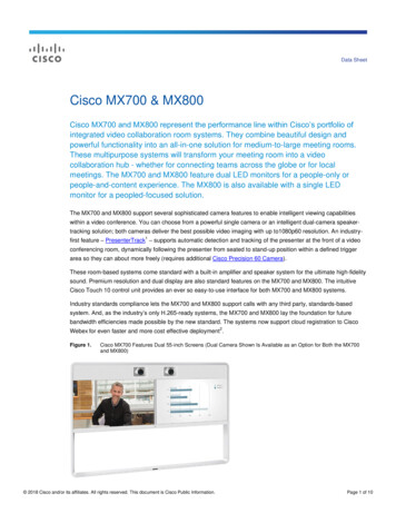
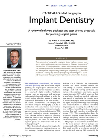
CAD/CAM GUIDED SURGERY IN IMPLANT DENTISTRY SCIENTIFIC ARTICLEFigure 12. Virtual maxillary surgical guide with implant trajectoriesillustrating surgical planFigure 14. NobelClinician allows for full control and visualization of Nobel implants and abutments. (Reproduced with permission from NobelBiocare)Figure 13. Materialise guide fixated to maxillary alveolar bone priorto implant placementSubmit for SAFE Surgiguide FabricationIf using the SimPlant Pro or Master versions,importing the DICOM files will require noconversion; however, if using the SimPlant Plannersoftware, it is necessary to convert the files prior toopening. Many conversion services are available fora fee, costing between 50 to 100 per scan. Oncethe files have been converted, they are opened intothe software package and then thresholding limitsfor bone, soft tissue, and teeth are completed.Additionally, sectioning of prosthesis and/or teethcan be completed to create virtual layers by allowingthe user to toggle (turning on and off the individuallayers). A clinician can also input a second DICOMfile dataset scan and/or an optical STL scan of thepatient’s cast to perform a dual scan digital registrationusing the dual-scan module. Alternatively, DICOMfiles can be sent to Materialise’s planning service(DentalPlanit) to have the files converted and digitalregistration completed.Implants are placed according to bone volumesand the restorative plan, while being cognizant ofvital structures. System-specific abutments canbe visualized for most major systems and genericabutment options are available to control thelocation of the implant platform’s interface withthe abutment to achieve a predictable result. Oncesatisfied with the dental implant plan, the softwarewill allow visualization of the surgical guide priorFigure 15. Guided surgical kit for Nobel Replace tapered implantsto ordering (Figure 12). After confirming the guidedesign and shape compatibility with the surgical plan,the guide is ordered via a web-portal and is fabricatedby a stereolithographic printing process (Figure 13).Surgical templates typically take 4 to 7 business daysto fabricate and ship and rush services are available.Nobel Clinician/Nobel GuideNobel Biocare is one of the largest manufacturersof dental implants and CAD/CAM-basedindividualized prosthetics with its own scannersand software. Nobel Biocare also developed asoftware program for clinicians (Nobel Clinician)for proper diagnostic studies and implant treatmentplanning, with production of customized, patientspecific, guided-surgery templates to assist inimplant placement surgeries. Nobel Clinician allowsfull control of 3D renderings allowing the user tosection, identify, and mark anatomical landmarksand to place virtual dental implants (Figure 14). Inthe implant module, the clinician can manipulateimplant position, angulation/trajectory, andmeasure relative bone density. A unique feature ofthe software is the calibration procedure, allowingThe combinationof restorativebased digitalplanning andcomputerizedguided surgicaldeliveryallows for atremendousintegrativeworkflow.Alpha Omegan Spring 201437
SCIENTIFIC ARTICLE CAD/CAM GUIDED SURGERY IN IMPLANT DENTISTRYFigure 17. Implant with fixture mount assembly allows for animplant to be precisely placed according to plan. (Courtesy NakulRathi)Figure 16. Mucosa-supported guided surgical template for Nobel implantsAuthor Profilethe clinician to separate 3D renderings of theradiographic guide from the CBCT based onIsovalue. Nobel Guide is the surgical guide fabricatedby Nobel upon completion of the implant planningin the Nobel Clinician software.The following types of templates are currentlyavailable for Nobel Guide: Tooth-supported Mucosa-supportedGuided surgery kitsDr. Shweta Puri, BDS, is athird year graduate student inthe Advanced Specialty Education Program in Prosthodontics at Loma Linda University,School of Dentistry (LLUSD).She received her BDS fromthe Christian Dental College,Ludhiana, India in 2004.After graduating, she workedfor three years as a generaldentist in a specialty dentalclinic in India. Her researchinterests brought her to theCenter for Dental Researchat LLUSD, Loma Linda, Calif.Prior to being accepted tothe prosthodontics program,Dr. Puri had been involvedin multiple projects includingsponsored trials, clinical trials,and animal studies. She enjoysexploring new places whiletraveling.38Nobel Biocare has surgical kits compatiblewith the following implant systems: Branemark System Guided Surgery Kit Nobel Active Guided Surgery Kit Nobel Replace Straight Guided Surgery kit Nobel Replace Tapered Guided Surgery Kit(Figure 15)Checklist for Nobel Guide(for either Partially Dentate Teeth-Supportedor Fully Edentulous Tissue-Supported Guide) Patient CBCT scan with radiolucent PVSocclusal registration spacer and a clearradiographic template replacing the teeth tobe restored with 6-8 gutta percha markers Prosthesis CBCT ScanSubmit for Surgical Guide FabricationDICOM files are automatically converted bythe Nobel Clinician software. Once opened intothe software package, image processing guidelines(limits) for bone, soft tissue, and teeth are completed.Nerve mapping and sectioning of prosthesis and/or teeth can be completed to create virtual layersto allow the user to switch (toggle) on and off. Aclinician can also input a second DICOM file datasetscan to perform a dual scan digital registration usingthe dual-scan module.Alpha Omegan Spring 2014Implants are placed according to bone volumes,restorative plan, and with respect to vital structures.System-specific abutments can be visualized formost major systems and generic abutment optionsare available. Once satisfied with the dental implantplan, the software will allow visualization of thevirtual surgical guide prior to ordering. Afterconfirming the guide design and shape compatibilitywith the surgical plan, the guide is ordered via a webportal and is fabricated by a STL printing process(Figure 16). Most clinicians will typically workwith a NobelClinician certified laboratory to ensurethat the pre-surgical and surgical requirementsare properly coordinated to reduce surgical error(Figure 17).ConclusionNumerous implant software packages exist withvarying capabilities and features that will allowfor the design and manufacture of proprietarycomputerized guided surgical templates (GST).While these systems are diverse, many sharesimilar features and methodology in how theyinterpret DICOM data and facilitate assessmentof potential sites for placement of dental implants.The combination of restorative-based digitalplanning and computerized guided surgicaldelivery allows for a tremendous integrativeworkflow that facilitates the team approach todental implantology. AOREFERENCES1. Angelopoulos C, Aghaloo T.Imaging technology in implantdiagnosis. Dent Clin North Am. 2011; 55:141-58.2. Dreiseidler T, Mischkowski RA, Neugebauer J, RitterL, et al. Comparison of cone-beam imaging withorthopantomography and computerized tomographyfor assessment in presurgical implant dentistry. Int J OralMaxillofac Implants 2009; 24:216-25.3. Orentlicher G, Abboud M. Guided surgery for implanttherapy. Dent Clin North Am. 2011; 55:715-44.4. Spector L. Computer-aided dental implant planning. DentClin North Am. 2008; 52:761-75.All authors of this article work and study at Loma LindaUniversity under a research grant from NobelBiocare.www.ao.org
Camlog Implant Direct Manufacturer-specific master sleeves allow for full trajectory and depth control, guided drill tap, and guided implant insertion (Figure 6). Fully guided implant insertion capability with Anatomage guide, however, is currently available only for NobelBiocare, 3i, and Camlog implant systems. Full implant guidance may be less of an issue with clinicians who routinely use .