Transcription
Physiol. Res. 69 (Suppl. 3): S403-S419, EWTestosterone and the Brain: From Cognition to AutismDaniela OSTATNÍKOVÁ1, Silvia LAKATOŠOVÁ1, Jaroslava BABKOVÁ1, JúliusHODOSY1,2,3, Peter CELEC1,21Institute of Physiology, Academic Research Centre for Autism, Faculty of Medicine, ComeniusUniversity, Bratislava, Slovak Republic, 2Institute of Molecular Biomedicine, Faculty of Medicine,Comenius University, Bratislava, Slovak Republic, 3University Hospital, Bratislava, SlovakRepublicReceived March 30, 2020Accepted October 6, 2020SummaryComenius University, Sasinkova 2, 811 08 Bratislava, SlovakSex and gender matter in all aspects of life. Humans exhibitRepublic. E-mail: daniela.ostatnikova@fmed.uniba.sksexual dimorphism in anatomy, physiology, but also pathology.Many of the differences are due to sex chromosomes and, thus,Introductiongenetics, other due to endocrine factors such as sex hormones,some are of social origin. Over the past decades, huge number ofscientific studies have revealed striking sex differences of onsequences. Prenatal and postnatal testosterone influencebrain structures and functions, respectively. Cognitive sexdifferences include especially certain spatial and language tasks,but they also affect many other aspects of the neurotypical brain.Sex differences of the brain are also relevant for thepathogenesis of neuropsychiatric disorders such as autismspectrum disorders, which are much more prevalent in the malepopulation. Structural dimorphism in the human brain was welldescribed, but recent controversies now question its importance.On the other hand, solid evidence exists regarding genderdifferences in several brain functions. This review tries tosummarize the current understanding of the complexity of theeffects of testosterone on brain with special focus on their role inthe known sex differences in healthy individuals and people inthe autism spectrum.Key wordsAndrogen Intelligence Psychoneuroendocrinology Sexsteroids Mental rotationCorresponding authorD. Ostatníková, Institute of Physiology, Faculty of Medicine,Humans as sexually reproducing living beingsexhibit sexual dimorphism with well-known sex orgender differences in anatomy, physiology andpathophysiology. While sex refers to biologicalcharacteristics assigned at birth based on reproductiveorgans and main sex hormones they produce, genderrepresents person’s social and cultural role based onone’s identity. Over the past decades research hasrevealed a number of sex differences in the human brainwith remarkable functional behavioral and cognitiveconsequences (Collaer and Hines 1995, Hedges andNowell 1995, Gur et al. 1999). Androgens are keyregulators of male sexual differentiation and developmentof a normal male phenotype. The main human androgentestosterone plays a dominant role in sexual dimorphism(Durdiakova et al. 2011). Genetic and environmentaleffects modulating gene expression of enzymes forsteroid metabolism in the steroidogenic cascade inconcordance with the modulation of expression ofrespective receptors imply complex and sophisticatedmechanisms of androgen effects. In addition, the nongenomic effects of testosterone on behavior bypassing thenuclear receptors have attracted the interest of researchersand might complicate the general understanding (Filovaet al. 2015a, Filova et al. 2015b). But they might explainPHYSIOLOGICAL RESEARCH ISSN 1802-9973 (online) 2020 Institute of Physiology of the Czech Academy of Sciences, Prague, Czech RepublicFax 420 241 062 164, e-mail: physres@fgu.cas.cz, www.biomed.cas.cz/physiolres
S404Ostatníková et al.some of the puzzling observations of non-linearassociations between testosterone and brain functionsfrom the past (Celec et al. 2015). Testosterone and othersex steroids are not the only cause for sex differences.Testosterone itself is under genetic control, although thisis complex and indirect (Harden et al. 2014). The role ofsex-different androgen receptor gene dose due to itslocation on the X chromosome might be involved. Theoverall testosterone/androgen signaling is determined bynumerous factors from genes, over their usage to variousenvironmental factors including physical exercise andphytoestrogen intake (Celec et al. 2007a, Hodosy et al.2012a). It should be noted that sex differences and theirmechanisms might vary during the development and becompletely different in aging (Domonkos et al. 2017a).Effects of testosterone on brain during thelife spanThe effect of testosterone on sexualdifferentiation of the reproductive system depends on theconcentrations of androgens during the critical periods ofprenatal life (Wilson and Davies 2007). The actions ofsex steroids on brain and behavior traditionally have beendivided into organizational and activational effects.Organizational effects are permanent and occur early indevelopment; activational effects are transient and occurthroughout life (Arnold and Breedlove 1985). In humanas early as in the 7th week of gestation, testes begin tosecrete testosterone and determine male typical genitalia(Ivell et al. 2017). Testosterone is involved also in thesexual differentiation of the behavior (Auyeung et al.2009b). The surge of this hormone in male fetusesbetween 16th and 24th week of gestation mediates the socalled organizational effect (Auyeung et al. 2009c). Highconcentrations of testosterone during this period arecrucial for the differentiation of androgen dependenttissues including specific brain areas (Connolly andResko 1994, Hiort 2013). In humans, the prepubertalperiod lasts approximately from year 1 to year 8, as theendogenous gonadal steroids remain in relatively stablelow concentrations. A wide spectrum of endocrinechanges is associated with the maturation of thereproduction function during the adolescent period –testosterone production peaks again and remainsrelatively stable during the fertile period of life respectingbiological fluctuations and rhythms (Oerter et al. 1990,Celec et al. 2002, Celec et al. 2009a). The critical periodfor cyclicity induction in girls is likely the pubertalVol. 69transition (Barbieri, 2014). But although it is assumedthat this is valid only for women, the true cyclicity oftestosterone was found to be even higher in men (Celecet al. 2003). So, the general knowledge on the acyclicityof men might be challenged in the near future. Thecritical period for cyclicity induction in girls is likely thepubertal transition (Barbieri 2014). But although it isassumed that this is valid only for women, the truecyclicity of testosterone was found to be even higher inmen (Celec et al. 2003). So, the general knowledge on theacyclicity of men might be challenged i the near future.Both, prenatal and postnatal exposure lead to structuraland functional consequences that are both, time and dosedependent (Hines 2006). Particular attention is given tothe role of prenatal hormone exposure, which appears tobe vital for early organization of the brain (Collaer andHines 1995, Chura et al. 2010). In later life, testosteroneis found to influence the behavior, but the effect is a kindof activation or fine-tuning of the already establishedearly organization of the brain (Romeo et al. 2002,Schulz et al. 2009). Organizational effects producepermanent changes in the wiring and sensitivity of thebrain areas and are largely irreversible. Activationaleffects occur later in life and are associated with changesof circulating testosterone levels, which activate neuralsystems organized early in life (Celec et al. 2015).However, as shown in our experiment, prenataltestosterone affects postnatal hormonal status and, thus,a clear experimental division of organizational andactivational effects is nearly impossible (Domonkos et al.2017b).Organizational effects of testosterone may primethe brain structures by changing its responsivity to thehormonal fluctuations later in life (Cohen-Bendahan et al.2005). Due to ethical constraints it is extremely difficultto analyze prenatal testosterone concentrations. It hasbeen proposed that the ratio of the second to fourth digitlengths (2D:4D) may be a proxy of prenatal androgenexposure. Low 2D:4D ratio is associated with highprenatal androgen exposure (Manning et al. 1998,Manning et al. 2000, Manning and Robinson 2003,Manning and Fink 2008, Beaton et al. 2011).Activational effect reflects the influence of actualhormonal concentrations on preprogrammed brainstructures with behavioral and cognitive consequences(Goel and Bale 2008). We have previously shown thatlow 2D:4D associated with higher prenatal testosteronewas also associated with lower empathy in intellectuallygifted boys (Durdiakova et al. 2015). On the other hand,
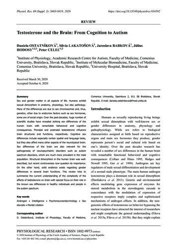
2020experimentally we have not proved that exogenoustestosterone lowers the 2D:4D in mice (Suchonova et pothesis is old, but fascinating concept on how sexdifferences in mating behavior are induced by prenataltestosterone (Phoenix et al. 1959). Later the hypothesishas been widen to include all sex differences in brainfunctions and even to all other organs and tissues (Arnold2009). The mechanisms underlying the organizationaleffects have been studied extensively, but the picture isstill not complete. It is clear that it includes theconversion of testosterone to estradiol by aromatase, atleast for the effects on the brain (McCarthy et al. 2009).The hypothesis, however, is not generally accepted.Testosterone has been shown to affect plasticity of thebrain even in the adulthood (Losecaat Vermeer et al.2016). This means that the organizational effects are notlimited to the prenatal period, but in contrast to theactivational effects might rather be mediated by otherreceptors and signaling pathways.Metabolism of testosterone contributes to theTestosterone and the BrainS405complexity of its actions. Testosterone does not only actper se, but also via the products of its metabolism.Reduction to dihydrotestosterone by 5-alpha reductaseincreases the androgen activity, conversion to estradiol byaromatase converts the androgen to estrogen activity(Filova et al. 2013). The biological effects of testosteronedepend on the sensitivity of androgen receptor, that isaffected by the genetic variation of the androgen receptorgene (Hillmer et al. 2005). Genetic variation in steroidreceptors and enzymes related to steroid metabolism iscrucial for understanding of endocrine phenotypicvariability. Several studies examined the associationbetween (CAG)n polymorphism and cognitive abilities(Yaffe et al. 2003, Lee et al. 2010). In the range ofnormal variation low number of (CAG)n repeats causehigher transactivational activity (more intense activationof transcription of the regulated genes) of receptor and,thus, higher sensitivity to androgens (Irvine et al. 2000,Greenland et al. 2004). Genetic and environmentalfactors determine the effects and the role of testosteronein physiology and pathogenesis of neurodevelopmentaldisorders (Fig. 1).Fig. 1. Causes and consequences of testosterone/androgen signaling variability on the brain. Genetic variants, sex differences,endocrine biorhythms, phytoestrogens in the nutrition, treatments affect the concentration of testosterone as well as its metabolism andrecognition by the corresponding receptors. Activational and organizational effects on the brain and its functions such as spatial abilitiesare physiological. Testosterone, however, also increases the risk of developmental disorders such as autism.
S406Ostatníková et al.Testosterone in relation to anatomical andfunctional brain differencesStructural sex differences in the human brain areof interest for the understanding of both, differences inphysiological psychological traits and psychiatricdisorders. It is suggested that the presence, magnitude,and direction of observed sex differences strongly dependon (1) the brain structure examined (cerebral cortex,corpus callosum, etc.), (2) on the specific brain featureassessed (cortical thickness, cortical convolution, etc.),(3) on the degree of regional specificity (global graymatter volume, voxel-wise gray matter volume, etc.), and(4) whether measurements are adjusted for individualbrain size or not (Luders and Toga 2010). There are fewstudies providing the insight into the adult brain structureand function. One of the biggest studies reported on sexdifferences studied in 2750 female and 2466 maleparticipants from a UK biobank (Ritchie et al. 2018).Males showed higher raw volumes, raw surface areas andwhite matter fractional anisotropy; females had on theother side higher raw cortical thickness and higher whitematter tract complexity. This implies that the averagemale brain is better adapted for connecting sensoryperception with coordinated motor activity, while averagefemale brain is structured for enhanced communicationbetween analytic and intuitive processing (Ingalhalikaret al. 2014). Most of the differences were observed onadults. But the question remains when the sexuallydimorphic brains develop. Studies in newborns andinfants have shown that already at this stage males andfemales vary in brain volume and cortical thickness(Knickmeyer et al. 2005, Choe et al. 2013) implicatinga likely emergence of sexual dimorphism in the brainbefore birth. Gao and colleagues assessed brainfunctional connectivity at birth, at the age of 1 and2 years and observed greater age-dependent increase infrontoparietal connectivity in males than females (Gaoet al. 2015b). However, no other sex differences innetwork connectivity were observed. In another study nodifferences were observed in developmental trajectoriesfor cortical growth or myelination of brain regionsbetween male and female brains at the ages of 1-6 years(Deoni et al. 2015).Sexual differentiation was supposed to occur inthe first weeks of life (Hines 2010). One of the mostconsistent dimorphic findings is that male infants,children and adults have greater whole-brain volume thanfemales (Gilmore et al. 2007, Koolschijn and CroneVol. 692013). A number of structural elements in the humanbrain differ between males and females. “Structural”refers to actual parts of the brain and the way they arebuilt, including their size and/or mass. Specifically, maleshave been documented to have larger prefrontal cortex,amygdala, and hippocampi than females (Ruigrok et al.2014). However, it is important not only to describeregional differences in structure, but also the connectivitybetween regions and how these connections differ early indevelopment between males and females in utero.Females often have a higher density of neuralconnections into the hippocampus. Regional differencesin brain function developed in utero may predisposeindividuals to specific abilities, and also to healthoutcomes later in life (DiPietro and Voegtline 2017).Male and female brains process the sameneurochemicals but in different concentrations andthrough sex-specific connections. One dominantneurochemical is testosterone mostly known as sex andaggression hormone that has a profound influence onbrain structures and functions. Individual variations oftestosterone in relation to other hormones within the brainlead to a fluctuating neuroendocrine milieu that hasan impact on typical male behavior using differentstrategies in problem solving and decision making(Gouchie and Kimura 1991). Testosterone influencescognitive styles based on the principles of theempathizing-systemizing theory of typical sex differrences, proposing that females on average havea stronger drive to empathize – to identify thoughts andfeelings of other persons and to respond to these withan appropriate emotion, while males on average havea stronger drive to systemize – to analyze or constructrule-based systems (Baron-Cohen 2002). Possiblebiological mechanisms include the effect of fetal sexsteroids or the role of sex chromosomes. Sex steroidhormones have the capacity for epigenetic modulation ofneuronal functions and fetal brain development duringearly critical periods (Auyeung et al. 2009b, Lombardoet al. 2012).According to the callosal theory, prenataltestosterone mediates early axon pruning in callosaltissue, and thus the higher the prenatal testosterone, themore lateralization occurs in the brain (Witelson andNowakowski 1991, Ypsilanti et al. 2008). Additionally,before boys or girls are born, their brains develope withdifferent divisions of labor between hemispheres. Themale brain is set up for better connecting sensoryinformation with coordinated activity, while female brain
2020is predisposed for communication between analytic andintuitive processing (Ingalhalikar et al. 2014). As a result,girls and women tend to receive more sensorial andemotive information than males do. Both, girls andwomen tend to sense a lot more of what is going onaround and they retain sensorial information longer thanmen (Chura et al. 2010).Testosterone effect on intelligenceIndividual differences in general cognitiveability can be measured by the intelligence quotient (IQ),which assesses abilities including planning, reasoning,comprehension, abstraction and learning (Deary 2013).IQ is also strongly predictive of various important tional attainment, social mobility and jobperformance (Deary et al. 2007). Compared with females,males produce much more testosterone, which triggersa cascade of reactions that culminate in themasculinization of genital tissue and of the developingnervous system. There are several unique neurobiologicalfeatures present in the majority of academically giftedpopulation supporting the connection betweenextraordinary intelligence and brain pattern development.Although it has been not entirely understood how thedevelopment of the central nervous system can beaffected by testosterone, suggestive data haveaccumulated to prove that testosterone act on the brainstructure and architecture (de Lacoste et al. 1991, Hillekeet al. 2006, Beking et al. 2018). While no difference wasfound in mean IQ between boys and girls, there wasa difference in the proportion of boys and girls in theintellectually gifted group and also in the mentallydisabled group: both groups contained more males thanfemales (Ostatnikova et al. 2000). This was in accordancewith findings of other researchers who observed greatervariability in intellectual performance among boys (Hydeet al. 1990, Johnson et al. 2008). Researchers have foundmore male than female pre-adolescents and adolescents ingroups of above-average ability (Benbow 1988, Hydeet al. 1990). Sex differences in the occurrence ofneurodevelopmental disorders have been reported withmales being more often affected, including severe mentalretardation, language disorders, learning difficulties andothers, irrespective of race and severity of disability(Flannery et al. 2000, Liederman et al. 2005). Intellectualgiftedness defined as an IQ score of 130 or higher isassociated with prenatal testosterone (Mrazik andTestosterone and the BrainS407Dombrowski 2010). The hypothesis is that similarly tophysical and cognitive deficiencies that may develop inutero, in utero exposure to testosterone might result inintellectual giftedness. In the attempt to find thebiological correlate for intellectual ability, our researchgroup correlated salivary testosterone levels with generalintelligence in preadolescent academically giftedparticipants and general population controls (Ostatnikovaet al. 2000). Salivary testosterone levels in the controlgroup of children attending regular primary school werecompared with those in the group of intellectually giftedchildren scoring 130 or more in the independentstandardized intelligence tests. The results provedsignificant differences in all ages – from the age of 5 tillthe age of 8. Lower testosterone concentrations werefound in academically gifted children. Interestingly,intellectual ability measured as IQ was negativelyassociated with salivary testosterone in both sexes.Similar results were found in our follow-up studyshowing significantly lower testosterone in gifted boysthan controls (Ostatnikova et al. 2007).Without access to prenatal testosterone it can bespeculated that being exposed to higher prenataltestosterone in utero may lead to a lower postnatal set-upof the hypothalamo-pituitary-gonadal axis. This findingcould also indicate later maturation of intellectually giftedchildren. It has been proposed that the hormonal changesresponsible for the timing of puberty reflect thedifferences in cognitive abilities (Waber 1976). The latematurers were reported to score better in spatial tasks thatindividuals who matured early. This finding wasconfirmed later by others (Ray et al. 1981, Sanders andSoares 1986). There is a solid evidence that intellectuallygifted children are born with atypical brains as a productof genes and their messengers – hormones in criticalperiods. There are several other unique neurobiologicalfeatures present in the gifted children. For example, thereis a trend toward increased right-hemisphere involvementin this population. Geschwind-Behan-Galaburda theoryof cerebral dominance (1987) argued that higher thannormal concentrations of testosterone in utero may inhibitthe development of the left hemisphere while enhancingother areas, such as the right hemisphere (Geschwind andGalaburda 1985). The proposed consequences of thisdevelopmental disbalance is the ability to visualizeproblems individuals are working on and to translatethose visual images into the abstract language ofmathematics. According to this well-known theory, giftedchildren with exposure to higher testosterone in utero will
S408Ostatníková et al.also have non-standard hemispheric specialization andhandedness but also higher risk of allergy. Our findingsof higher allergy prevalence and lower percentage ofright handedness in intellectually gifted childrensupported this hypothesis (Ostatnikova et al. 2002).Studies on mathematically gifted individuals confirmedthe unique functional characteristic with biggerinvolvement of the right hemisphere in mental processingthat would also indicate an effect of intrauterinetestosterone on the right hemispheric development(O'Boyle et al. 1991). Testosterone concentration is notthe only determinant of androgen signaling. Thesensitivity of the androgen receptor has to be consideredas well. Several studies examined the association betweenthe CAG polymorphism in exon 1 of the androgenreceptor gene and cognitive abilities. Lee with colleaguesfound no association between CAG repeat length andfluid intelligence in older men (Lee et al. 2010). Ourstudy revealed a significantly lower number of CAGrepeats in the AR gene in gifted boys in comparison withcontrols indicating stronger androgen signaling in thispopulation. This could also explain why lowertestosterone is found in gifted boys, for the overallandrogen signaling it might be sufficient (Celec et al.2009b, Celec et al. 2013).The neurobiological basis of variation inintelligence remains unresolved. According to Catell’sintelligence theory (Cattel, 1943) fluid intelligence is thepure general ability to discriminate and perceive relationsbetween any fundaments. It is predominant in childhoodand increases until adolescence. Crystalized intelligencerefers to discriminatory habits long established ina particular field and culminates in adulthood. It is knownthat fluid intelligence depends on cognitive processes thatare mediated most prominently by frontal lobes (Duncanet al. 2000, Duncan 2005). Frontal cortical regions arepart of those neural networks that are under the influenceof androgens including testosterone (Bramen et al. 2012).Gonadal steroids induce morphological changes in theneuronal system such as growth, morphologicaldifferentiation or programmed apoptosis of particulargroups of neurons in topographically specific regions thatcontain androgen and/or estrogen receptors (MacLuskyet al. 1997, MacLusky et al. 2006).The effects of testosterone on brain structure andfunction are complex. Besides abovementionedmetabolism of testosterone that might end in activation ofestrogen rather than androgen receptors, the associationbetween outcomes and testosterone concentrations is notVol. 69linear. Tan and Tan (1998) proposed a curvilinearcorrelation between total testosterone and fluidintelligence in men and women. They studied therelationship between serum total testosterone and fluidintelligence in young men and women. As expected, therewas no significant difference between average IQ of menand women. IQ tended to increase with testosteronelevels in men, except at very high levels. The authorsconcluded that too low or too high testosteroneconcentrations may be disadvantageous for fluidintelligence. The non-linear nature of the associationcould explain the difficulties to reproduce the associationas many scientists only look for linear correlations andnot for complex interactions.Testosterone and specific cognitive abilitiesAdult men generally outperform women inspatial abilities (Linn and Petersen 1985). Especially,mental rotation shows a sex difference in favor of men.Even though some authors argue that contribution ofandrogens to human performance on mental rotation tasksmay be limited to early organizational periods (Puts et al.2010, Courvoisier et al. 2013), there is some evidencesupporting the existence of the association between actualtestosterone and mental rotation performance (O'Connoret al. 2001, Alexander and Son 2007). Some studies havereported a positive relationship – the higher thetestosterone concentration, the higher the performance(Hausmann et al. 2009), whereas other studies havefound that the association follows an inverted U-shapewith best performance at average testosteroneconcentrations (O'Connor et al. 2001). Again, this nonlinear association could lead to bias and misleadinginterpretations of results depending on the analysis used.So, it is unsurprisingly that some authors report alsoa negative correlation between mental rotation andtestosterone (Vuoksimaa et al. 2012). An additionalfactor that increases the complexity of the issue is thegenetic variability of the androgen sensitivity asdescribed for mental rotation and androgen receptor genein our study (Durdiakova et al. 2013).Activational effects of gonadal hormones werestudied by relying on naturally occurring biorhythms.Although not free from bias, the menstrual cycle providesa convenient method for studying specific cognitiveperformance in relation to fluctuation of actualtestosterone concentrations. The mentioned bias mightarise from fluctuating well-being of women during the
2020menstrual cycle that might affect the performance incognitive tests (Celec et al. 2011). Some studiesconfirmed curvilinear or U-shaped relationship betweentestosterone and spatial abilities (O'Connor et al. 2001).Shute (1983) found that females with the highestphysiological androgen levels performed better in spatialcognition test than females with the lowest androgenlevels, whereas males who had the highest androgenlevels performed worse than males with the lowestandrogen levels. Our research group has tested healthymen and women in reproductive age for mental rotationand spatial visualization. Performance in these twostandardized tests were related to actual salivarytestosterone concentrations, which reflected thebioavailable free fraction of this hormone (Celec et al.2002, Ostatnikova et al. 2010). A negative relationshipbetween testosterone and spatial performance was foundin male subjects while a positive though only marginallysignificant relationship was observed in women. The bestspatial performance was proposed in hormonallyandrogynous subjects. This was a proof of the U-shapedassociation between testosterone and specific cognitiveabilities on an interindividual level. However, in the samestudy we have also shown association of the short termchanges, as part of the infradian fluctuations inendogenous salivary testosterone, with spatial cognitionin humans (Celec et al. 2009a). In men, an intraindividualincrease of testosterone was associated with worse spatialperformance in mental rotation test, a decrease oftestosterone was accompanied by an improved spatialperformance. In women, the opposite associations werefound. These results support the hypothesis ofactivational effects of testosterone fluctuations during themenstrual cycle in women or the proposed infradiancircatrigintan (e.g. with a period of approximately30 days) cycle in men on spatial abilities and likely alsoother cognitive functions (Celec et al. 2003, Ostatnikovaet al. 2010). Whether other biological rhythms oftestosterone affect cognitive abilities or other brainfunctions is surprisingly unexplored. Even the widelyknown and accepted daily rhythm of testosterone has notbeen analyzed yet in relation to cognition (Reinberg et al.1975, Guignard et al. 1980). Less surprisingly, neither theyearly, nor other less known infradian rhythms oftestosterone were tested as determinants of cognitiveabilities (Celec et al. 2007b, Smith et al. 2013). Thereason might be simply the demanding design of theresearch studies. As the rhythms can be detected only byanalyzing testosterone in long time series, sampling isTestosterone and the BrainS409one major issue. The other is the need for repeated testingof cognitive abilities which might bias the outcome asmost of the tests are affected by learning (Tao et al.2019).Despite contradictory results, partially due todifferent analysis used, the majority of studies indicatethat the association between testosterone and cognitive,especially spatial abilities is curvilinear and sexdependent. In women, higher testosterone is associatedwith better mental rotation; in men, lower testosterone isassociated with better performance. This seems to be truefor both, actual and prenatal te
early organization of the brain (Romeo et al. 2002, Schulz et al. 2009). Organizational effects produce permanent changes in the wiring and sensitivity of the brain areas and are largely irreversible. Activational effects occur later in life and are associated with changes