Transcription
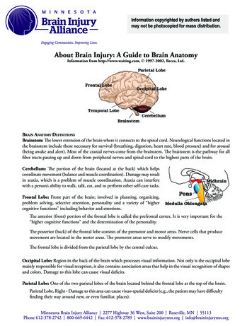
Information copyrighted by authors listed andmay not be photocopied for mass distribution.About Brain Injury: A Guide to Brain AnatomyInformation from http://www.waiting.com, 1997-2002, Becca, Ltd.Brain Anatomy DefinitionsBrainstem: The lower extension of the brain where it connects to the spinal cord. Neurological functions located inthe brainstem include those necessary for survival (breathing, digestion, heart rate, blood pressure) and for arousal(being awake and alert). Most of the cranial nerves come from the brainstem. The brainstem is the pathway for allfiber tracts passing up and down from peripheral nerves and spinal cord to the highest parts of the brain.Cerebellum: The portion of the brain (located at the back) which helpscoordinate movement (balance and muscle coordination). Damage may resultin ataxia, which is a problem of muscle coordination. Ataxia can interferewith a person’s ability to walk, talk, eat, and to perform other self-care tasks.Frontal Lobe: Front part of the brain; involved in planning, organizing,problem solving, selective attention, personality and a variety of “highercognitive functions” including behavior and emotions.The anterior (front) portion of the frontal lobe is called the prefrontal cortex. It is very important for the“higher cognitive functions” and the determination of the personality.The posterior (back) of the frontal lobe consists of the premotor and motor areas. Nerve cells that producemovement are located in the motor areas. The premotor areas serve to modify movements.The frontal lobe is divided from the parietal lobe by the central culcus.Occipital Lobe: Region in the back of the brain which processes visual information. Not only is the occipital lobemainly responsible for visual reception, it also contains association areas that help in the visual recognition of shapesand colors. Damage to this lobe can cause visual deficits.Parietal Lobe: One of the two parietal lobes of the brain located behind the frontal lobe at the top of the brain.Parietal Lobe, Right - Damage to this area can cause visuo-spatial deficits (e.g., the patient may have difficultyfinding their way around new, or even familiar, places).
Information copyrighted by authors listed andmay not be photocopied for mass distribution.Parietal Lobe, Left - Damage to this area may disrupt a person’s ability to understand spoken and/or writtenlanguage.The parietal lobes contain the primary sensory cortex which controls sensation (touch, pressure). Behind theprimary sensory cortex is a large association area that controls fine sensation (judgment of texture, weight,size, and shape).Temporal Lobe: There are two temporal lobes, one on each side of the brain located at about the level of the ears.These lobes allow a person to tell one smell from another and one sound from another. They also help in sortingnew information and are believed to be responsible for short-term memory.Right Lobe - Mainly involved in visual memory (i.e., memory for pictures and faces).Left Lobe - Mainly involved in verbal memory (i.e., memory for words and names).About Brain Injury:The Areas of the Brain, Their Function, & Associated Signs & SymptomsBrain StructureFunctionCerebral CortexThe outermost layer of the cerebralhemisphere which is composed of graymatter. Cortices are asymmetrical.Both hemispheres are able to analyzesensory data, perform memoryfunctions, learn new information,form thoughts and make decisions.Left HemisphereSequential Analysis: systematic,logical interpretation of information.Interpretation and production ofsymbolic information: language,mathematics, abstraction and reasoning.Memory stored in a language format.Associated Signs andSymptoms
Information copyrighted by authors listed andmay not be photocopied for mass distribution.Holistic Functioning:processing multi-sensory inputsimultaneously to provide“holistic” picture of one’senvironment. Visual spatial skills.Holistic functions such as dancingand gymnastics are coordinatedby the right hemisphere. Memoryis stored in auditory, visual andspatial modalities.Corpus CallosumConnects right and left hemisphereto allow for communicationbetween the hemispheres. Formsroof of the lateral and thirdventricles.Damage to the CorpusCallosum may result in “SplitBrain” syndrome.Frontal LobeCognition and memory. Prefrontalarea: The ability to concentrateand attend, elaboration ofthought. The “Gatekeeper”;(judgment, inhibition).Personality and emotionaltraits.Movement:Motor Cortex(Brodman’s): voluntary motoractivity.Premotor Cortex: storageof motor patterns and voluntaryactivities. Language: motorspeech. Impairment of recentmemory, inattentiveness,inability to concentrate,behavior disorders, difficultyin learning new information.Lack of inhibition(inappropriate social and/orsexual behavior). Emotionallability. “Flat” affect. Contralateral plegia, paresis. Expressive/motor aphasia.Parietal LobeProcessing of sensory input,sensory discrimination. Inability to discriminatebetween sensory stimuli. Inability to locate andrecognize parts of the body(Neglect). Severe Injury: Inability torecognize self. Disorientation ofenvironment space. Inability to write.Body orientation.Primary/ secondary somaticarea.
Information copyrighted by authors listed andmay not be photocopied for mass distribution.Occipital LobePrimary visual reception area.Primary visual association area:Allows for visual interpretation. Primary Visual Cortex: loss ofvision opposite field. Visual Association Cortex:loss of ability to recognizeobject seen in opposite field ofvision, “flash of light”, “stars”.Temporal LobeAuditory receptive area andassociation areas. Hearing deficits. Agitation, irritability, childishbehavior. Receptive/ sensory aphasia.Expressed behavior.Language: Receptive speech.Limbic SystemMemory: Information retrieval.Olfactory pathways:Amygdala and their differentpathways. Loss of sense of smell. Agitation, loss of controlof emotion. Loss of recentmemory.Hippocampi and their differentpathways.Limbic lobes: Sex, rage, fear, andemotions. Integration of recentmemory, biological rhythms.Hypothalamus.Basal GangliaThalamusSubcortical gray matter nuclei.Processing link between thalamusand motor cortex. Initiation anddirection of voluntary movement.Balance (inhibitory), Posturalreflexes. Movement disorders:chorea, tremors at rest andwith initiation of movement,abnormal increase in muscletone, difficulty initiatingmovement.Part of extrapyramidal system:regulation of automatic movement. Parkinson’s.Processing center of the cerebralcortex. Coordinates and regulates allfunctional activity of the cortex viathe integration of the afferent inputto the cortex (except olfaction).Contributes to affectual expression. Altered level ofconsciousness. Loss of perception. Thalamic syndrome spontaneous pain oppositeside of body.
Information copyrighted by authors listed andmay not be photocopied for mass distribution.HypothalamusIntegration center of Autonomic Nervous Hormonal imbalances.System (ANS): Regulation of body Malignant hypothermia.temperature and endocrine function. Inability to controltemperature. Diabetes Insipidus (DI).Anterior Hypothalamus: parasympathetic Inappropriate ADHactivity (maintenance function). (SIADH). Diencephalicdysfunction: “neurogenicPosterior Hypothalamus: sympathetic storms”.activity (“Fight” or “Flight”, stressresponse.Behavioral patterns: Physical expressionof behavior.Appestat: Feeding center.Pleasure center.Internal CapsuleMotor tracts.Reticular ActivatingSystem (RAS)Responsible for arousal fromsleep, wakefulness, attention.CerebellumCoordination and control ofvoluntary movement.Contralateral plegia (Paralysisof the opposite side of thebody).Altered level ofconsciousness. Tremors. Nystagmus (Involuntarymovement of the eye). Ataxia, lack of coordination.Brain Stem:Ner ve pathway of cerebralhemispheres.Auditory and Visual reflex centers.Cranial Nerves: CN III - Oculomotor (Related toeye movement), [motor]. CN IV - Trochlear (Superior obliquemuscle of the eye which rotates theeye down and out), [motor]. Weber’s: CN III palsy andptosis (drooping) ipsalateral(same side of body). Pupils: Size: Midposition todilated. Reactivity: Sluggish tofixed. LOC (Loss of consciousness):Varies Movement: Abnormalextensor ( muscle that extendsa part). Respiratory: Hyperventilating. CN (Cranial Nerve) Deficits:CN III, CN IV.
Information copyrighted by authors listed andmay not be photocopied for mass distribution.PonsMedulla OblongataRespiratory Center.Cranial Nerves: CN V - Trigeminal (Skinof face, tongue, teeth;muscle of mastication),[motor and sensory]. CN VI - Abducens (Lateralrectus muscle of eye whichrotates eye outward),[motor]. CN VII - Facial (Muscles ofexpression), [motor andsensory]. CN VIII - Acoustic (Internalauditory passage),[sensory].Crossing of motor tracts.Cardiac Center.Respiratory Center.Vasomotor (nerves havingmuscular control of the bloodvessel walls) Center Centers forcough, gag, swallow, and vomit.Cranial Nerves: CN IX - Glossopharyneal(Muscles and mucousmembranes of pharynx, theconstricted openings from themouth and the oral pharynxand the posterior third oftongue.), [mixed]. CN X - Vagus (Pharynx,larynx, heart, lungs, stomach),[mixed]. CN XI – Accessory (Rotationof the head and shoulder),[motor]. CN XII – Hypoglossal(Intrinsic muscles of thetongue), [motor]. Pupils: Size: Pinpoint LOC:Semi-coma“Akinetic Mute”“Locked In” Syndrome. Movement:Abnormal extensor.Withdrawal. Respiratory:Apneustic (Abnormalrespiration marked bysustained inhalation). Hyperventilation. CN Deficits: CN VI, CNVII. Movement: Ipsilateral(same side) plegia (paralysis). Pupils:Size: DilatedReactivity: Fixed. LOC: Comatose. Respiratory:Abnormal breathingpatterns.Ataxic.Clustered.Hiccups. CN Palsies (Inability tocontrol movement): Absent Cough. Gag.
Information copyrighted by authors listed andmay not be photocopied for mass distribution.About Brain Injury: Intracraniel PressureDoes the brain always swell? How do you know if the brain is swelling? Doesn’t the CT scan show swelling?Is it possible that the person’s brain did not swell because of the use of the drug manitol (protocol treatmentin all ICU’s)? Is the chemical released if there is no swelling? If a person didn’t need a shunt, can we assumethere was no swelling?Pretty much all tissues in the body swell when traumatized. They also require more oxygen to heal. The brainis unique in that it rests inside a bone case, so when it swells, it experiences more trauma.The more damage the brain receives, the more it swells. This is caused by leakage from blood vessels. Whenthe brain swells, because it is housed inside the skull, it has no room to expand. This leads to a rise inpressure within the brain. This rise in pressure rapidly equals the arterial pressure thereby affecting the bloodflow to the brain. This diffuse pressure which decreases blood flow affects the ability of the cells within thebrain to metabolize properly; the cells are unable to eliminate toxins which then accumulate.This phenomenon leads to a spiral effect which is what kills brain injured people who don’t get promptattention. One of the big breakthroughs that lead to the survival rate we have now for brain injury todaywas learning to break this cycle. We are still very much in the stage of learning to break this cycle. Themost significant factor has been the use of monitoring devices to inform treatments to prevent furtherdamage.The brain requires both oxygen and glucose. In response to the trauma, changes occur in the brain whichrequire monitoring to prevent further damage. The brain’s size frequently increases after a severe head injury.This is called brain swelling and occurs when there is an increase in the amount of blood to the brain.Water may collect in the brain which is called Brain Edema. Both Brain swelling and Brain Edema result inexcessive pressure in the brain called Intracranial Pressure (ICP). Around-the-clock monitoring during thistime is essential in order that ICP can be immediately treated. Treatment of brain swelling can be difficult.Very strong medications are administered. Medications which help to draw fluid back out of the brain andinto blood vessels may be useful. Some medications help by decreasing the metabolic requirements of thebrain. Other medications increase blood flow into the brain which can help diminish the spiral effect causedby brain swelling.In some cases, removal of small amounts of fluids or from the brain or surgery may be beneficial. And insome cases, removal of damaged tissue may prevent further damage.
In response to the trauma, changes occur in the brain which require monitoring to prevent further damage. The brain’s size frequently increases after a severe head injury. This is called brain swelling and occurs when there is an increase in the amount of blood to the brain. Water may collect in the bra