Transcription
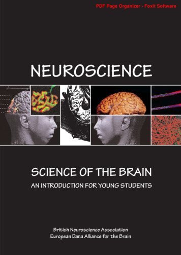
PDF Page Organizer - Foxit SoftwareNEUROSCIENCESCIENCE OF THE BRAINAN INTRODUCTION FOR YOUNG STUDENTSBritish Neuroscience AssociationEuropean Dana Alliance for the Brain
PDF Page Organizer - Foxit SoftwareNeuroscience: the Science of the Brain1The Nervous SystemP22Neurons and theAction PotentialP43Chemical MessengersP74Drugs and the BrainP95Touch and PainP116VisionP147MovementP198The DevelopingNervous SystemP229DyslexiaP2510PlasticityP2711Learning and MemoryP3012StressP3513The Immune SystemP3714SleepP3915Brain ImagingP4116Artificial Brains andNeural NetworksP4417When things go wrongP4718NeuroethicsP5219Training and CareersP5420Further Reading andAcknowledgementsP56Inside our heads, weighing about 1.5 kg, is an astonishing living organ consisting ofbillions of tiny cells. It enables us to sense the world around us, to think and to talk.The human brain is the most complex organ of the body, and arguably the mostcomplex thing on earth. This booklet is an introduction for young students.In this booklet, we describe what we know about how the brain works and how muchthere still is to learn. Its study involves scientists and medical doctors from manydisciplines, ranging from molecular biology through to experimental psychology, aswell as the disciplines of anatomy, physiology and pharmacology. Their sharedinterest has led to a new discipline called neuroscience - the science of the brain.The brain described in our booklet can do a lot but not everything. It has nerve cells- its building blocks - and these are connected together in networks. Thesenetworks are in a constant state of electrical and chemical activity. The brain wedescribe can see and feel. It can sense pain and its chemical tricks help control theuncomfortable effects of pain. It has several areas devoted to co-ordinating ourmovements to carry out sophisticated actions. A brain that can do these and manyother things doesn’t come fully formed: it develops gradually and we describe someof the key genes involved. When one or more of these genes goes wrong, variousconditions develop, such as dyslexia. There are similarities between how the braindevelops and the mechanisms responsible for altering the connections betweennerve cells later on - a process called neuronal plasticity. Plasticity is thought tounderlie learning and remembering. Our booklet’s brain can remember telephonenumbers and what you did last Christmas. Regrettably, particularly for a brainthat remembers family holidays, it doesn’t eat or drink. So it’s all a bit limited.But it does get stressed, as we all do, and we touch on some of the hormonal andmolecular mechanisms that can lead to extreme anxiety - such as many of us feel inthe run-up to examinations. That’s a time when sleep is important, so we let it havethe rest it needs. Sadly, it can also become diseased and injured.New techniques, such as special electrodes that can touch the surface of cells,optical imaging, human brain scanning machines, and silicon chips containingartificial brain circuits are all changing the face of modern neuroscience.We introduce these to you and touch on some of the ethical issues and socialimplications emerging from brain research.The Neuroscience Communityat the University of EdinburghThe EuropeanDana Alliancefor the BrainTo order additional copies: Online ordering: www.bna.org.uk/publicationsPostal: The British Neuroscience Association, c/o: The Sherrington Buildings, Ashton Street, Liverpool L68 3GETelephone: 44 (0) 151 794 4943/5449 Fax: 44 (0) 794 5516/5517
PDF Page Organizer - Foxit SoftwareThe NervousSystemNeurons have an architecture that consists of a cell bodyand two sets of additional compartments called‘processes’. One of these sets are called axons; their job isto transmit information from the neuron on to others towhich it is connected. The other set are called dendrites their job is to receive the information being transmitted bythe axons of other neurons. Both of these processesparticipate in the specialised contacts called synapses(see the Chapters 2&3 on Action Potential and ChemicalMessengers). Neurons are organised into complex chainsand networks that are the pathways through whichinformation in the nervous system is transmitted.Human central nervous system showing the brain andspinal cordBasic structureThe nervous system consists of the brain, spinal cord andperipheral nerves. It is made up of nerve cells, calledneurons, and supporting cells called glial cells.There are three main kinds of neurons. Sensory neurons arecoupled to receptors specialised to detect andrespond to different attributes of the internal and externalenvironment. The receptors sensitive to changes in light,sound, mechanical and chemical stimuli subserve the sensorymodalities of vision, hearing, touch, smell and taste.When mechanical, thermal or chemical stimuli to the skinexceed a certain intensity, they can cause tissue damageand a special set of receptors called nociceptors areactivated; these give rise both to protective reflexes and tothe sensation of pain (see chapter 5 on Touch and Pain).Motor neurons, which control the activity of muscles, areresponsible for all forms of behaviour including speech.Interposed between sensory and motor neurons areInterneurones. These are by far the most numerous (in thehuman brain). Interneurons mediate simple reflexes as wellas being responsible for the highest functions ofthe brain. Glial cells, long thought to have a purelysupporting function to the neurons, are now known to makean important contribution to the development of thenervous system and to its function in the adult brain.While much more numerous, they do not transmitinformation in the way that neurons do.2The brain and spinal cord are connected to sensoryreceptors and muscles through long axons thatmake up the peripheral nerves. The spinal cord has twofunctions: it is the seat of simple reflexes such as the kneejerk and the rapid withdrawal of a limb from a hot object or apinprick, as well as more complex reflexes, and it forms ahighway between the body and the brain for informationtravelling in both directions.These basic structures of the nervous system are the samein all vertebrates. What distinguishes the human brain is itslarge size in relation to body size. This is due to an enormousincrease in the number of interneurons over the course ofevolution, providing humans with an immeasurably wide choiceof reactions to the environment.Anatomy of the BrainThe brain consists of the brain stem and the cerebralhemispheres.The brain stem is divided into hind-brain, mid-brain and a‘between-brain’ called the diencephalon. The hind-brain is anextension of the spinal cord. It contains networks ofneurons that constitute centres for the control of vitalfunctions such as breathing and blood pressure. Withinthese are networks of neurons whose activity controls thesefunctions. Arising from the roof of the hind-brain is thecerebellum, which plays an absolutely central role in thecontrol and timing of movements (See Chapters onMovement and Dyslexia).The midbrain contains groups of neurons, each of which seemto use predominantly a particular type of chemicalmessenger, but all of which project up to cerebralhemispheres. It is thought that these can modulate theactivity of neurons in the higher centres of the brain
PDF Page Organizer - Foxit SoftwareThe human brain seen from above, below and the side.Side view of the brainshowing division betweenthe cerebral hemisphereand brain stem, anextension of which is thecerebellumto mediate such functions as sleep, attention or reward.The diencephalon is divided into two very different areascalled the thalamus and the hypothalamus: The thalamusrelays impulses from all sensory systems to the cerebralcortex, which in turn sends messages back to the thalamus.This back-and-forward aspect of connectivity in the brain isintriguing - information doesn’t just travel one way.The hypothalamus controls functions such as eating anddrinking, and it also regulates the release of hormonesinvolved in sexual functions.The cerebral hemispheres consist of a core, the basalganglia, and an extensive but thin surrounding sheet ofneurons making up the grey matter of the cerebral cortex.The basal ganglia play a central role in the initiation andcontrol of movement. (See Chapter 7 on Movement).Packed into the limited space of the skull, the cerebral cortexis thrown into folds that weave in and out to enable a muchlarger surface area for the sheet of neurons than wouldotherwise be possible. This cortical tissue is the most highlydeveloped area of the brain in humans - four times biggerthan in gorillas. It is divided into a large number of discreteareas, each distinguishable in terms of its layers andconnections. The functions of many of these areas areknown - such as the visual, auditory, and olfactory areas, thesensory areas receiving from the skin (called thesomaesthetic areas) and various motor areas.The pathways from the sensory receptors to the cortex andfrom cortex to the muscles cross over from one side to theother. Thus movements of the right side of the body arecontrolled by the left side of the cortex (and vice versa).Similarly, the left half of the body sends sensory signals tothe right hemisphere such that, for example, sounds in theleft ear mainly reach the right cortex. However, the twohalves of the brain do not work in isolation - for the left andright cerebral cortex are connected by a large fibre tractcalled the corpus callosum.The cerebral cortex is required for voluntary actions,language, speech and higher functions such as thinking andremembering. Many of these functions are carried out byboth sides of the brain, but some are largely lateralised toone cerebral hemisphere or the other. Areas concerned withsome of these higher functions, such as speech (which islateralised in the left hemisphere in most people), have beenidentified. However there is much still to be learned,particularly about such fascinating issues as consciousness,and so the study of the functions of the cerebral cortex isone of the most exciting and active areas of researchin Neuroscience.Cerebral HemisphereCerebellumBrain StemCross section throughthe brain showing thethalamus andhypothalamusThalamusHypothalamusCross section throughthe brain showing thebasal ganglia and corpuscallosumCerebral HemisphereCorpus CallosumBasai GangliaThe father of modernneuroscience, Ramon yCajal, at his microscopein 1890.Cajal’s first picturesof neurons and theirdendrites.Cajal’s exquisiteneuron drawings these are of thecerebellum.gInternet Links: aculty.washington.edu/chudler/neurok.html http://psych.hanover.edu/Krantz/neurotut.html3
PDF Page Organizer - Foxit SoftwareNeurons and theAction PotentialWhether neurons are sensory or motor, big or small, theyall have in common that their activity is both electrical andchemical. Neurons both cooperate and compete with eachother in regulating the overall state of the nervoussystem, rather in the same way that individuals in asociety cooperate and compete in decision-makingprocesses. Chemical signals received in the dendrites fromthe axons that contact them are transformed intoelectrical signals, which add to or subtract from electricalsignals from all the other synapses, thus making a decisionabout whether to pass on the signal elsewhere. Electricalpotentials then travel down axons to synapses on thedendrites of the next neuron and the process repeats.The dynamic neuronCell BodyReceivingIntegratingPyramidal cellPurkinje cell of cerebellumCell BodyCell BodyCell BodyAxonAxonAxon3 different types of NeuronsAs we described in the last chapter, a neuron consists ofdendrites, a cell body, an axon and synaptic terminals.This structure reflects its functional subdivision intoreceiving, integrating and transmitting compartments.Roughly speaking, the dendrite receives, the cell-bodyintegrates and the axons transmit - a concept calledpolarization because the information they processsupposedly goes in only one direction.DendritesSpinal motor neuronAxonSynapseTransmittingInside neurons are many inner compartments. Theseconsist of proteins, mostly manufactured in the cell body,that are transported along the cytoskeleton. Tinyprotuberances that stick out from the dendrites calleddendritic spines. These are where incoming axons makemost of their connections. Proteins transported to thespines are important for creating and maintaining neuronalconnectivity. These proteins are constantly turning over,being replaced by new ones when they’ve done their job.All this activity needs fuel and there are energy factories(mitochondria) inside the cell that keep it all working. Theend-points of the axons also respond to molecules calledgrowth factors. These factors are taken up inside and thentransported to the cell body where they influence theexpression of neuronal genes and hence the manufacture ofnew proteins. These enable the neuron to grow longerdendrites or make yet other dynamic changes to its shapeor function. Information, nutrients and messengers flow toand from the cell body all the time.The key concepts of a neuronLike any structure, it has to hold together. The outermembranes of neurons, made of fatty substances, aredraped around a cytoskeleton that is built up of rods oftubular and filamentous proteins that extend out intodendrites and axons alike. The structure is a bit like a canvasstretched over the tubular skeleton of a frame tent.The different parts of a neuron are in constant motion, aprocess of rearrangement that reflects its own activity andthat of its neighbours. The dendrites change shape,sprouting new connections and withdrawing others, and theaxons grow new endings as the neuron struggles to talk a bitmore loudly, or a bit more softly, to others.4Dendritic spines are the tiny green protuberances stickingout from the green dendrites of a neuron. This is wheresynapses are located.
PDF Page Organizer - Foxit SoftwareReceiving and decidingThe action-potentialOn the receiving side of the cell, the dendrites have closecontacts with incoming axons of other cells, each of which isseparated by a miniscule gap of about 20 billionths of metre.A dendrite may receive contacts from one, a few, or eventhousands of other neurons. These junctional spots arenamed synapses, from classical Greek words that mean “toclasp together”. Most of the synapses on cells in theTo communicate from one neuron to another, the neuronalsignal has first to travel along the axon. How do neuronsdo this?cerebral cortex are located on the dendritic spines thatstick out like little microphones searching for faint signals.Communication between nerve cells at these contact pointsis referred to as synaptic transmission and it involves achemical process that we will describe in the next Chapter.When the dendrite receives one of the chemical messengersthat has been fired across the gap separating it from thesending axon, miniature electrical currents are set up insidethe receiving dendritic spine. These are usually currentsthat come into the cell, called excitation, or they may becurrents that move out of the cell, called inhibition. All thesepositive and negative waves of current are accumulated inthe dendrites and they spread down to the cell body. If theydon’t add up to very much activity, the currents soon diedown and nothing further happens. However, if the currentsadd up to a value that crosses a threshold, the neuron willsend a message on to other neurons.pulses called action potentials. These travel along nervefibres rather like a wave travelling down a skipping rope.This works because the axonal membrane contains ionchannels, that can open and close to let through electricallycharged ions. Some channels let through sodium ions (Na ),while others let through potassium ions (K ). When channelsopen, the Na or K ions flow down opposing chemical andelectrical gradients, in and out of the cell, in response toelectrical depolarisation of the membrane.The answer hinges on harnessing energy locked in physicaland chemical gradients, and coupling together these forcesin an efficient way. The axons of neurons transmit electricalSo a neuron is kind of miniature calculator - constantlyadding and subtracting. What it adds and subtracts are themessages it receives from other neurons. Somesynapses produce excitation, others inhibition. How thesesignals constitute the basis of sensation, thought andmovement depends very much on the network in which theneurons are embedded.The action potential5
PDF Page Organizer - Foxit SoftwareWhen an action potential starts at the cell body, the firstchannels to open are Na channels. A pulse of sodium ionsflashes into the cell and a new equilibrium is establishedwithin a millisecond. In a trice, the transmembrane voltageswitches by about 100 mV. It flips from an inside membranevoltage that is negative (about -70 mV) to one that ispositive (about 30 mV). This switch opens K channels,triggering a pulse of potassium ions to flow out of the cell,almost as rapidly as the Na ions that flowed inwards, andthis in turn causes the membrane potential to swing backagain to its original negative value on the inside. The actionpotential is over within less time than it takes to flick adomestic light switch on and immediately off again.Remarkably few ions traverse the cell membrane to do this,and the concentrations of Na and K ions within thecytoplasm do not change significantly during an actionpotential. However, in the long run, these ions are kept inbalance by ion pumps whose job is to bale out excess sodiumions. This happens in much the same way that a small leak inthe hull of a sailing boat can be coped with by baling outwater with a bucket, without impairing the overall ability ofthe hull to withstand the pressure of the water upon whichthe boat floats.The action potential is an electrical event, albeit a complexone. Nerve fibres behave like electrical conductors (althoughthey are much less efficient than insulated wires), and so anaction potential generated at one point creates anothergradient of voltage between the active and restingmembranes adjacent to it. In this way, the action potentialis actively propelled in a wave of depolarisation that spreadsfrom one end of the nerve fibre to the other.An analogy that might help you think about the conductionof action potentials is the movement of energy along afirework sparkler after it is lit at one end. The first ignitiontriggers very rapid local sparks of activity (equivalent to theions flowing in and out of the axon at the location of theaction potential), but the overall progression of the sparklingwave spreads much more slowly. The marvellous feature ofnerve fibres is that after a very brief period of silence (therefractory period) the spent membrane recovers itsexplosive capability, readying the axon membrane for the nextaction potential.Much of this has been known for 50 years based onwonderful experiments conducted using the very largeneurons and their axons that exist in certainsea-creatures. The large size of these axons enabledscientists to place tiny electrodes inside to measure thechanging electrical voltages. Nowadays, a modern electricalrecording technique called patch-clamping is enablingneuroscientists to study the movement of ions throughindividual ion-channels in all sorts of neurons, and so makevery accurate measurements of these currents in brainsmuch more like our own.Research FrontiersThe nerve fibres above (the purple shows the axons) arewrapped in Schwann cells (red) that insulate the electricaltransmission of the nerve from its surroundings.The colours are fluorescing chemicals showing a newlydiscovered protein complex. Disruption of this proteincomplex causes an inherited disease that leads to musclewasting.New research is telling us about the proteins that make upthis myelin sheath. This blanket prevents the ionic currentsfrom leaking out in the wrong place but, every so often theglial cells helpfully leave a little gap. Here the axonconcentrates its Na and K ion channels. These clusters ofion channels function as amplifiers that boost and maintainthe action potential as it literally skips along the nerve.This can be very fast. In fact, in myelinated neurons,action-potentials can race along at 100 metres per second!Action potentials have the distinctive characteristic of beingall-or-nothing: they don’t vary in size, only in how often theyoccur. Thus, the only way that the strength or duration of astimulus can be encoded in a single cell is by variation of thefrequency of action potentials. The most efficient axons canconduct action potentials at frequencies up to 1000 timesper second.Alan Hodgkin and AndrewHuxley won the Nobel Prizefor discovering themechanism of transmissionof the nerve impulse.They used the "giant axon"of the squid in studiesat the Plymouth MarineBiology LaboratoryInsulating the axonsIn many axons, action-potentials move along reasonably well,but not very fast. In others, action potentials really do skipalong the nerve. This happens because long stretches of theaxon are wrapped around with a fatty, insulating blanket,made out of the stretched out glial cell membranes, called amyelin sheath.g6Internet Links: //www.neuro.wustl.edu/neuromuscular/
PDF Page Organizer - Foxit SoftwareChemicalMessengersAction potentials are transmitted along axons tospecialised regions called synapses, where the axonscontact the dendrites of other neurons. These consist ofa presynaptic nerve ending, separated by a small gap fromthe postsynaptic component which is often located on adendritic spine. The electrical currents responsible for thepropagation of the action potential along axons cannotbridge the synaptic gap. Transmission across this gap isaccomplished by chemical messengers calledneurotransmitters.around the synaptic cleft. Some of these have miniaturevacuum cleaners at the ready, called transporters, whosejob is to suck up the transmitter in the cleft. This clears thechemical messengers out of the way before the next actionpotential comes. But nothing is wasted - these glial cellsthen process the transmitter and send it back to be storedin the storage vesicles of the nerve endings for future use.Glial-cell housekeeping is not the only means by whichneurotransmitters are cleared from the synapse.Sometimes the nerve cells pump the transmitter moleculesback directly into their nerve endings. In other cases, thetransmitter is broken down by other chemicals in thesynaptic cleft.Messengers that open ion channelsChemical transmitter packed inspherical bags is available for releaseacross synaptic junctionsStorage and ReleaseNeurotransmitters are stored in tiny spherical bags calledsynaptic vesicles in the endings of axons. There are vesiclesfor storage and vesicles closer to nerve endings that areready to be released. The arrival of an action potential leadsto the opening of ion-channels that let in calcium (Ca ).This activates enzymes that act on a range of presynapticproteins given exotic names like “snare”, “tagmin” and “brevin”- really good names for the characters of a recent scientificadventure story. Neuroscientists have only just discoveredthat these presynaptic proteins race around tagging andtrapping others, causing the releasable synaptic vesicles tofuse with the membrane, burst open, and release thechemical messenger out of the nerve ending.This messenger then diffuses across the 20 nanometre gapcalled the synaptic cleft. Synaptic vesicles reform whentheir membranes are swallowed back up into the nerve endingwhere they become refilled with neurotransmitter, forsubsequent regurgitation in a continuous recycling process.Once it gets to the other side, which happens amazinglyquickly – in less than a millisecond - it interacts withspecialised molecular structures, called receptors, in themembrane of the next neuron. Glial cells are also lurking allThe interaction of neurotransmitters with receptorsresembles that of a lock and key. The attachment of thetransmitter (the key) to the receptors (the lock) generallycauses the opening of an ion channel; these receptors arecalled ionotropic receptors (see Figure). If the ion channelallows positive ions (Na or Ca ) to enter, the inflow ofpositive current leads to excitation. This produces a swingin the membrane potential called an excitatory post-synaptic potential (epsp). Typically, a large number of synapsesconverge on a neuron and, at any one moment, some areactive and some are not. If the sum of these epsps reachesthe threshold for firing an impulse, a new action potential isset up and signals are passed down the axon of the receivingneuron, as explained in the previous oteinReceptorExtracellularPlasma MembraneIntracellularSecond MessengerEffectorIonotropic receptors (left) have a channel through whichions pass (such as Na and K ). The channel is made up offive sub-units arranged in a circle. Metabotropic receptors(right) do not have channels, but are coupled to G-proteinsinside the cell-membrane that can pass on the message.7
PDF Page Organizer - Foxit SoftwareThe main excitatory neurotransmitter in the brain isglutamate. The great precision of nervous activity requiresthat excitation of some neurons is accompanied bysuppression of activity in other neurons. This is broughtabout by inhibition. At inhibitory synapses, activation ofreceptors leads to the opening of ion channels that allow theinflow of negatively charged ions giving rise to a change inmembrane potential called an inhibitory post-synapticpotential (ipsp) (see Figure). This opposes membranedepolarisation and therefore the initiation of an actionpotential at the cell body of the receiving neuron. There aretwo inhibitory neurotransmitters – GABA and glycine.ions in the membrane, as ionotropic receptors do, butinstead kick-starts intracellular second messengers intoaction, engaging a sequence of biochemical events (seeFigure). The metabolic engine of the neuron then revs up andgets going. The effects of neuromodulation include changesin ion channels, receptors, transporters and even the expression of genes. These changes are slower in onset and morelong-lasting than those triggered by theexcitatory and inhibitory transmitters and their effectsextend well beyond the synapse. Although they do notinitiate action potentials, they have profound effects on theimpulse traffic through neural networks.Synaptic transmission is a very rapid process: the timetaken from the arrival of an action potential at a synapse tothe generation of an epsp in the next neuron is very rapid 1/1000 of a second. Different neurons have to time theirdelivery of glutamate on to others within a short window ofopportunity if the epsps in the receiving neuron are going toadd up to trigger a new impulse; and inhibition also has tooperate within the same interval to be effective in shuttingthings down.Identifying the messengersThe excitatory synaptic potential (epsp) is a shift inmembrane potential from -70 mV to a value closer to 0 mV.An inhibitory synaptic potential (ipsp) has the oppositeeffect.Messengers that modulateAmong the many messengers acting on G-protein coupledreceptors are acetylcholine, dopamine and noradrenaline.Neurons that release these transmitters not only have adiverse effect on cells, but their anatomical organisation isalso remarkable because they are relatively few in number buttheir axons project widely through the brain (see Figure).There are only 1600 noradrenaline neurons in the humanbrain, but they send axons to all parts of the brain and spinalcord. These neuromodulatory transmitters do not send outprecise sensory information, but fine-tune dispersedneuronal assemblies to optimise their performance.Noradrenaline is released in response to various forms ofnovelty and stress and helps to organise the complexresponse of the individual to these challenges. Lots ofnetworks may need to “know” that the organism is understress. Dopamine makes certain situations rewarding forthe animal, by acting on brain centres associated withpositive emotional features (see Chapter 4). Acetylcholine,by contrast, likes to have it both ways. It acts on bothionotropic and metabotropic receptors. The firstneurotransmitter to be discovered, it uses ionic mechanismsto signal across the neuromuscular junction from motorneurons to striated muscle fibres. It can also function as aneuromodulator. It does this, for example, when you want tofocus attention on something - fine-tuning neurons in thebrain to the task of taking in only relevant information.The hunt for the identity of the excitatory and inhibitoryneurotransmitters also revealed the existence of a largenumber of other chemical agents released from neurons.Many of these affect neuronal mechanisms by interactingwith a very different set of proteins in the membranes ofneurons called metabotropic receptors. These receptorsdon’t contain ion channels, are not always localised in theregion of the synapse and, most importantly, do not lead tothe initiation of action potentials. We now think of thesereceptors as adjusting or modulating the vast array ofchemical processes going on inside neurons, and thus theaction of metabotropic receptors is called neuromodulation.Metabotropic receptors are usually found in complexparticles linking the outside of the cell to enzymes inside thecell that affect cell metabolism. When a neurotransmitter isrecognised and bound by a metabotropic receptor, bridgingmolecules called G-proteins, and other membrane-boundenzymes are collectively triggered. Binding of thetransmitter to a metabotropic recognition site canbe compared to an ignition key. It doesn’t open a door forg8Noradrenaline cells are located in the locus c
The brain consists of the brain stemand the cerebral hemispheres. The brain stem is divided into hind-brain, mid-brain and a ‘between-brain’ called the diencephalon. The hind-brain is an extension of the spinal cord. It c


