Transcription
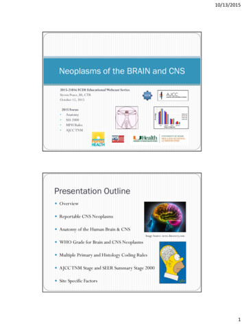
10/13/2015Neoplasms of the BRAIN and CNS2015-21016 FCDS Educational Webcast SeriesSteven Peace, BS, CTROctober 15, 20152015 Focus AnatomySSS 2000MPH RulesAJCC TNMPresentation Outline Overview Reportable CNS Neoplasms Anatomy of the Human Brain & CNSImage Source: news.discovery.com WHO Grade for Brain and CNS Neoplasms Multiple Primary and Histology Coding Rules AJCC TNM Stage and SEER Summary Stage 20002 Site Specific Factors1
10/13/2015OverviewBrain tumors are: Primary “brain” tumors - those that begin in thebrain or central nervous system (or its supporting tissues)and tend to stay in the brain - occur in people of allages, but they are statistically more frequent inchildren and older adults. Metastatic “brain” tumors – those that begin as acancer elsewhere in the body and spread to the brain– are more common in adults than in children.4Source: American Brain Tumor Association Facts and Statistics http://abta.org2
10/13/2015Brain tumors are: Usually described as intracranial neoplasms with varying behaviors (benign, borderline, malignant – ref. ICD-O-3)Include most identifiable structures within the craniumincluding the brain itself, small hormone-secreting ducts likethe pineal and pituitary glands, the cranial nerves (primarilythe optic nerve, olfactory nerve, acoustic nerve), the outerprotective lining of the brain (meninges), and the spinal cord.Metastatic neoplasms are excludedCertain benign “tumors” are excludedNo malignant neoplasms are excludedBenign bone tumors are excluded5Brain Tumor Characteristics Start in the brain and grow steadily there. Very rarely spread to other organs through the bloodstream. Are named for the anatomic location of tumor and/or the cellsfrom which they arise, each having a certain function essential tonormal physiological functioning of the brain. For example: 6Brain Stem Gliomas arise in the lowest part of the brain.Meningiomas arise in the meninges.Gliomas arise from glial cells that support the CNS.Astrocytomas arise from astrocytesEpendymomas arise from ependymal cells which line the ventriclesOligodendrogliomas arise from oligodentdrocyte cells which makeup the fatty substance called myelin that covers nerves like electricalinsulation.3
10/13/2015Characteristics of Brain Tumors7Source: medicalgeek.com/indian-post-graduate-examsRange of Tumors and Symptoms There are over 120 different types of brain/CNS tumors. CNS tumors are associated with a range of symptoms andcomplications such as edema, seizures, endocrinopathy,fatigue, psychiatric disorder, venous thromboembolism thatcan seriously impact quality of life. Symptoms depend very much on the size and location of thetumor. General symptoms include persistent headacheswhich tend to be worse with activity, at night or early in themorning, convulsions, vomiting, subtle changes inpersonality, memory, mental ability, drowsiness, lethargy.8SEER Training Modules4
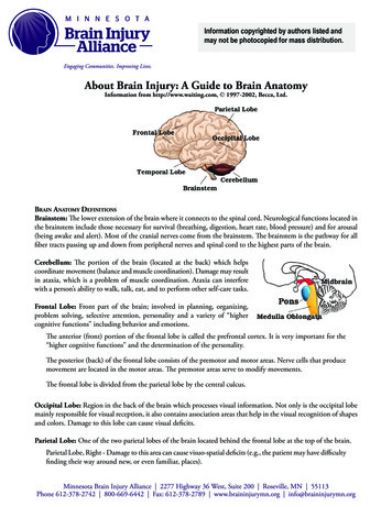
10/13/2015Brain Anatomy and Function9Tumor Location and Symptoms Symptoms are often tumor location specific or provide clues Symptoms on the right side of the body may occur if the tumor islocated on the left side of the brain and vice-versa. The speech center in most people is on the left side of thebrain. Symptoms of a tumor located here may include difficultysaying correct words while still capable of understanding whatis being said. If the tumor is located in the frontal lobe which controlsintellectual function, thought process, behavior and memory,those activities may be affected. Similarity to closed head injury victims (motorcycle crash).10SEER Training Modules5
10/13/2015ALL Brain Tumors are Reportable Public Law 107-260, the Benign Brain Tumor Cancer RegistriesAmendment Act, [PDF-185KB] requires programs participatingin the National Program for Cancer Registries (NPCR) to collectdata on benign and borderline tumors of the central nervoussystem in addition to the previously required data on malignanttumors. In addition to NPCR, the National Cancer Institute's (NCI)Surveillance, Epidemiology and End Results (SEER) program andthe American College of Surgeons' (ACoS) Commission on Cancerbegan requiring that these tumors be reported, starting with casesdiagnosed on January 1, 2004.11ALL Brain Tumors are Reportable BUT – Do Not Report Benign/Borderline Tumors that wereDiagnosed BEFORE 1/1/2004 – NOT EVEN HISTORICAL This creates an ERROR for FCDS at Call for Data FCDS then has to Delete the Case and Adjust Sequences126
10/13/2015ICD-O Topography Codes (Anatomic Site)13Brain Tumor Characteristics Benign – Borderline – Malignant Patient Age Tumor Location Tumor Histologic Type WHO Grade of Primary TumorAmerican Brain Tumor Association – http://www.abta.org147
10/13/2015Tumor Behavior Benign Tumors Slow growing Distinct borders Rarelyspread Malignant Tumors Usually rapid growing Invasive Lifethreatening Borderline Malignant Tumors Rare Likely to Recurfollowing Surgical Resection May become Life threatening15Common Histologic Types Astrocytoma, Grade I (Juvenile Pilocytic Astrocytoma) Astrocytoma, Grade II (Low Grade Pilocytic Astrocytoma) Astrocytoma, Grade III (Anaplastic Astrocytoma) Glioblastoma (Glioblastoma Multiforme/Astrocytoma, Grade IV) Ependymoma Medulloblastoma Meningioma Oligodendroglioma Oligoastrocytoma Pituitary Adenoma168
10/13/201517WHO Classification Groups Tumors of NeuroepithelialTissue Tumors of Cranial and Paraspinal Nerves Tumors of Meninges Tumors of Uncertain histogenesis Lymphomas and Hematopoietic Malignancies Germ Cell Tumors Cysts and Tumor-Like Lesions Tumors of the Sellar Region18 Metastatic Tumors9
10/13/2015192010
10/13/2015Distribution Primary Brain & CNS Tumors 2007-2011BehaviorMalignantNonmalignant21ALL ontent/16/suppl 4.tocDistribution Primary Brain & CNS Tumors 2007-2011Primary Site and BehaviorMalignant nt/16/suppl 4.toc11
10/13/2015ALL Tumors23Distribution Primary Brain & CNS Tumors 2007-2011Primary Site and BehaviorNon-Malignant nt/16/suppl 4.tocDistribution Primary Brain & CNS Tumors 2007-2011Histologic Type – All BehaviorALL ontent/16/suppl 4.toc12
10/13/2015Distribution Primary Brain & CNS Tumors 2007-2011Behavior and Histologic als.org/content/16/suppl 4.toc25Distribution Primary Brain & CNS Tumors 2007-2011Behavior and Histologic journals.org/content/16/suppl 4.toc13
10/13/2015Childhood Brain TumorsTentorium - extension of the dura mater separating the cerebellum from the occipital lobes 50% of childhood brain andCNS tumors are infratentorial,originating below the tentorium 20 % of childhood CNStumors are located in the sellaror suprasellar region around thesella turcica (the bone thatcontains the pituitary gland) Remainder of tumors occur inspinal cord, brain stem, cranialnerves, etc.27Childhood Brain TumorsSupratentorial - childhood Craniopharyngiomas. Diencephalic and hypothalamic gliomas. Germ cell tumors. Low-grade astrocytomas. Anaplastic astrocytomas. Glioblastoma multiforme. Mixed gliomas. Oligodendrogliomas. Primitive neuroectodermal tumors. Low-grade or anaplastic ependymomas.Infratentorial - childhood Cerebellar astrocytomas (usually high-grade). Medulloblastomas (primitive neuroectodermal tumors). Ependymomas (low-grade or anaplastic). Brain stem gliomas (high-grade or low-grade). Atypical teratoid tumors Meningiomas. Choroid plexus tumors.2814
10/13/2015Distribution Primary Brain & CNS Tumors 2007-2011Children and Adolescents by tent/16/suppl 4.tocDistribution Primary Brain & CNS Tumors 2007-2011Children and Adolescents by Histologic tent/16/suppl 10?etoc15
10/13/2015WHO Grade for Brain/CNS Tumors WHO Tumor Grades – Grade I, II, III, and IV Higher the grade – the more malignant the tumor A tumor can contain more than one grade of cell Always record and code the highest tumor grade noted Record WHO Grade in SSF1 NOT in tmlWHO Grade for Brain/CNS TumorsLOW GRADE Neoplasms Grade I: least malignant tumors associated with long-termsurvival. They grow slowly and have an almost normal appearancewhen viewed through a microscope. Surgery alone may be aneffective treatment for this grade tumor. Grade II: tumors are slow-growing and look slightly abnormalunder a microscope. Some can spread into nearby normal tissueand recur, sometimes as a higher grade n/diagnosis/grading-staging.html16
10/13/2015WHO Grade for Brain/CNS Tumors Grade III:These tumors are, by definition, malignant althoughthere is not always a big difference between grade II and grade IIItumors. The cells of a grade III tumor are actively reproducingabnormal cells, which grow into nearby normal brain tissue. Thesetumors tend to recur, often as a grade IV. Grade IV:The most malignant tumors. Tumors reproducerapidly, can have a bizarre appearance when viewed under themicroscope, and easily grow into nearby normal brain tissue.These tumors form new blood vessels so they can maintain theirrapid growth. They also have areas of dead cells in their ion/diagnosis/grading-staging.htmlWHO Grade for Brain/CNS n/diagnosis/grading-staging.html17
10/13/2015353618
ates19
10/13/2015ANATOMY OF THE HUMAN BRAIN3940Source: National Geographic, couretsy of Fred Hossler/Getty ImagesSource: University of Illinois20
10/13/2015Brain Lobes and Fissures414221
10/13/2015Ventricular System of the /brain-anatomy/structure.htmlVentricular System of the Brain44Source: solarnavigator.net/human brain22
10/13/2015Midline Shift and Mass Effect The bony cranium protects the brain fromoutside impacts to the head. When swellingoccurs in the brain, there isn’t much “give”. The swelling results in intracranial pressureand can cause a number of effects that beginto impact quality of life and comfort for thepatient. The easiest way to describe midline shift is45to bring to mind siting in a movie theater. Assoon as the person to one side of you putshis elbow onto the shared armrest betweenyou, you tend to shift away.Source: MedscsapeMidline Shift and Mass Effect Midline is a central boundary separatingthe left and right hemispheres. Midline Shift – Tumor crosses the brain toshift across the center line Mass Effect is – Edema or swelling causesthe brain to shift across center line Both create new symptoms at cross-over Depends on the size and location of hetumor and level of spread Edema caused by many things Either cause pushes midline out ofalignment46Source: Medscsape23
10/13/2015Meninges and Brain Stemtentorium47Cranial NervesOpticAcoustic4824
10/13/2015Cranial Nerve FunctionsCranial Nerve:Major Functions:I OlfactorysmellII OpticvisionIII Oculomotoreyelid and eyeball movementIV Trochlearturns eye downward and laterally, controls superior oblique musclesV Trigeminalchewing, face & mouth touch & painVI Abducensturns eye laterallyVII Facialfacial expressions, taste, tears, salivaVIII VestibulocochlearAlso referred to as Auditory Nerve: hearing, equilibrium sensationIX GlossopharyngealTaste, senses carotid blood pressureX Vagusaortic blood pressure, heart rate, stimulates digestive organs, tasteXI Spinal Accessorycontrols trapezius & sternocleidomastoid muscles, controlsswallowingXII Hypoglossalcontrols tongue movements49Sinus, Olfactory, Base of Skull Tumors Cancer Registries treat many of these as Head & Neck Neoplasms Some are intra-cranial but many are not intra-cranial or CNS Primary Site of Tumor Critical for Stage, Treatment, and Prognosis Histology is used to identify which are abstracted as Brain/CNS Highly specialized surgical procedures – location of tumor Some Prognostic Factors Overlap5025
10/13/2015Sinus, Olfactory, Base of Skull kull-base-ian-suk-sml.jpgHistologic Type - Glioma Most common category of primary brain tumors. They begin in glialcells (supporting cells of the CNS) – can be Grade I-IV not just III-IV. Often spread into surrounding brain tissue along nerve fibers invading thespaces between nearby normal brain cells. Some invade the surrounding brainmore than others. Difficulty obtaining complete surgical removal. MRI scans show the largestpart of the glioma, but cannot reliably show areas of the brain where tumorcells have invaded. Aggressive efforts to remove small numbers of tumor cellswithin the brain could cause loss of neurologic function. When it is not possible to remove the entire glioma, post-op radiation therapyand chemotherapy may be advised. Even with maximum safe resection followed by radiation and chemotherapy,gliomas can grow back.5226
10/13/2015Glioma – 3 Main Histologic SubTypes1. Astrocytoma: In adults most often arise in the cerebrum.In children they occur in the brain stem, cerebrum andcerebellum. Rarely in brain stem in adults. Felt to be mostaggressive of brain tumors. Grade I and II astrocytomas are low-grade astrocytomas. Grade III astrocytoma is an “anaplastic astrocytoma”. Grade IV astrocytoma is a “glioblastoma multiforme”.53Glioma – 3 Main Histologic SubTypes2. Oligodendroglioma: Rare tumor that usually occurs inthe cerebrum, grows slowly and usually does notspread into surrounding brain tissue like astrocytomadoes. Most common in middle-aged adults.3. Ependymoma: Most commonly arise in children andyoung adults. They are also seen with neurofibromatosisType II. (which we will discuss in a bit)5427
10/13/2015Glioma – Other SubtypesThere are other subtypes of gliomas, each with their ownspecific characteristics and modes of growth. Brain Stem Glioma Juvenile Pilocytic Astrocytoma Pleomorphic Xanthoastrocytoma Subependymoma Ganglioglioma55Glioma Tumor Markers5628
10/13/2015Glioma Chromosome Alterations57Non-Glial Tumors Medulloblastoma: Usually arises in the cerebrum, isthe most common brain tumor in children, and issometimes called a “primitive neuroectodermal tumor”or PNET. Meningioma: Arises from the meninges which are theoutside coverings of the brain between the skull andthe brain itself. It usually presses on the brain, butdoes not invade it and often grows slowly.5829
10/13/2015Meningioma Meningiomas are typically diagnosed by CT or MRI imaging Biopsy may be considered for confirmation Options stratified by presence/absence of symptoms and tumor size Most asymptomatic patients with small tumors ( 30mm) may justbe observed. If neurological impairment is imminent, surgery (ifaccessible) or radiotherapy (EBRT OR SRS) is feasible Asymptomatic tumors 30mm can be either resected or observed59Non-Glial Tumors Schwannoma: Arises from Schwann cells present incertain nerves, including those that control balance andhearing. May be called “neuroma”. A common site is the vestibular nerve which carriessignals from the inner ear to the brain stem. Tumors in this location are called “acoustic neuromas”(a.k.a. vestibular schwannoma), and occur most often inadults.6030
10/13/2015Non-Glial Tumors Craniopharyngioma: Grows at the base of the brain,arises from the tissue connecting the brain and thepituitary gland and occurs in both adults and children. Pituitary Adenoma: Arises from the pituitary gland andmay cause compression of the optic nerves causing visionproblems. Some produce excessive amounts ofhormones that can disrupt the body’s metabolism.61Roswell Park Cancer InsituteNeurofibromatosis The neurofibromatoses (NF) are a group of genetic disorderswhich cause tumors to grow along nerves and can also affect thedevelopment of non-nervous tissues such as bones and skin. Neurofibromatosis Type I (NF-I), also known as Peripheral NFand historically as von Recklinghausen Disease Occurs in 1:4,000 births Multiple cafe-au-lait spots (not reportable) Many, many neurofibromas on or under the skin (not reportable) Enlargement and deformation of bones and curvature of the spine Tumors may develop in brain, on cranial nerves, or the spinal cord62Neurofibromatosis Foundation31
10/13/2015NF Type I: First documented photo 187163Source Credit: Dr. Stanley B. Burnshttp://www.cbsnews.com/2300-204 162-10007019-6.html#ixzz1clEzAchIOther Manifestions of NF Type I64Lisch nodules on the eyeCafé-au-lait spots on skin Melanocytic hemartomas Discolored birth marksMedscape Source: Dermnet.com; Dermatologic Manifestations of NF Type I32
10/13/2015Neurofibromatosis Type II Neurofibromatosis Type II (NFII), also known as MultipleInherited Schwannomas, Meningiomas and Ependymomas(MISME) or Bilateral Acoustic Neurofibromatosis (BAN ). Is a genetically inherited diseasecaused by mutations of the"Merlin" gene, which appears to influences the form andmovement of cells Primary manifestation is a development of non-malignant braintumors in the region of the cranial nerves, frequently bilaterally.The eighth cranial nerve is the auditory-vestibular nerve whichtransmits sensory information from the inner ear to the brain andis commonly affected.65Source: California Ear InstituteAcoustic Neuroma/Schwannoma66Source: 3
10/13/2015Multiple Primary RulesHistology Coding Rules67Different Rules for Benign and Malignant6834
10/13/2015Sequence Numbering for Brain Tumors Malignant primary brain and CNS tumors are assignedSequence Codes in the range 00-35 Sequence Chronologically 00-35Only count malignant tumors in the sequenceIf only one malignant tumor occurs, it is coded 00If subsequent (multiple) primary malignant and/or in situ neoplasms,the sequence number for the first tumor begins at 01, the sequencenumber for the second primary tumor is 02, and so forth. Non-malignant primary brain and CNS tumors are assignedSequence Codes in the range 60-87. Sequence Chronologically 60-87 Only count benign/borderline or reportable by agreement neoplasms69in the sequence If only one non-malignant tumor occurs, it is coded 60. If subsequent (multiple) non-malignant neoplasms are diagnosed, thefirst tumor should be sequenced as 61, the second 62 and so forth.Benign and Borderline Tumor Rules7035
10/13/2015Benign and Borderline Tumor Rules When multiple tumors are present registrars should identifyand document specific characteristics for MPH Rules Text Date of Diagnosis (Timing is not used to determine number ofabstracts or primary neoplasms to abstract) Method and Details of Diagnosis (some are never resected) Location of Tumor Laterality Histologic Type – refer to Chart 1 Tumor Behavior Multiple Meningioma’s (meningiomatosis) Neurofibromatosis Characteristics (when applicable)71Malignant Tumor Rules7236
10/13/2015Malignant Tumor Rules73Malignant Tumor Rules When multiple tumors are present registrars should identify anddocument specific characteristics for MPH Rules Text Date of Diagnosis (Timing is not used to determine number of abstracts or primary neoplasms to abstract)Method and Details of Diagnosis (most attempt resection)Location of Tumor (not spread or invasion – but bulk of tumor)Histologic Type – refer to Chart 1 and/or Chart 2Tumor BehaviorVariations or Combinations of One or More Glial Tumors Over Lifetime –astrocytoma, glioblastoma, ependymoma, or oligodendroglioma Special rules for determining # abstracts Special rules for determining whether or not is mixed glioma Note: Recurrence, progression, or any reappearance of histologies on thesame branch in Chart 1 or Chart 2 is always the same disease process.7437
10/13/2015Do NOT Report Benign Tumors Dx’d 2004REMINDER: Sequence numbers for malignant neoplasms and for benign, borderline, andother reportable-by-agreement cases are usually assigned over a lifetime. HOWEVER,WHEN A PATIENT WAS DIAGNOSED WITH A NON-MALIGNANT CNSNEOPLASM BEFORE REPORTING WAS REQUIRED (January 1, 2004),THE FIRST NEOPLASM IS NOT REPORTABLE TO FCDS EVENAS A HISTORICAL CASETHE NEW (SECOND) NEOPLASM SHOULD BE ASSIGNED SEQUENCE NUMBER 60DO NOT REPORT THE BENIGN NEOPLASM IF DIAGNOSED BEFORE 1/1/2004THIS HAS BEEN A SOURCE OF CONFUSION – PLEASE REVIEW AND SAVE75FCDS Data Acquisition Manual and CDC Data Collection of Primary Central Nervous System TumorsStaging Brain and CNS Neoplasms7638
10/13/2015AJCC TNM, 7th edition“Attempts at developing a TNM-based classification and staging system for tumors of thecentral nervous system have not been successful.” Basic TNM Concepts are not applicableto brain and CNS sites. “It continues to be the recommendation of the ASJCC CNSTumor Task Force that a formal classification and staging system not be attempted.”AJCC Stage 88 (Not Applicable)Includes: ANY Benignor ANY Malignant Neoplasm77AJCC TNM, 7th editionFactors Felt to be of Prognostic and/or Clinical Significant Include: Tumor HistologyLocation of TumorUnifocal or MultifocalWHO Grade of TumorPatient Age at DiagnosisFunctional Neurologic Status *KPS)Primary or Recurrent TumorExtent of ResectionMetastatic Spread Proliferative Fraction (Ki-67, M1 B-1)Gene Deletions (1p, 19q)MGMT Methylation7839
10/13/2015SEER Summary Stage 2000To obtain a FREE electronic copy of the SS2000 22012.pdf79SS20008040
10/13/2015Steps to Assign SS2000Three summary stage groups can be ruled out quickly:in situ, distant, and localizedFIRSTIn Situ Stage is Not Applicable for Brain/CNS Tumors81In Situ Stage is Not the Same as Benign or Borderline BehaviorSS2000 for Benign/Borderline Tumors 88241
10/13/2015Steps to Assign SS2000Three summary stage groups can be ruled out quickly:in situ, distant, and localizedSECOND83Brain and CNS these are usually CSF Involvement – cells in fluidOn rare occasion you may see “drop metastasis” – code as Distant StageSteps to Assign SS2000Three summary stage groups can be ruled out quickly:in situ, distant, and localizedTHIRD84Most common Summary Stage – unless tumor crosses midline42
10/13/2015Steps to Assign SS2000Three summary stage groups can be ruled out quickly:in situ, distant, and localizedFINAL (if needed)85Additional Resources The 2007 WHO Classification of Tumours of the Central Nervous System, David N.Louis, Hiroko Ohgaki, Otmar D. Wiestler, Webster K. Cavenee, Peter C. Burger, Anne Jouvet,Bernd W. Scheithauer and Paul Kleihues, World Health Organization, Lyon, France, 2007 Central Brain Tumor Registry of the United States (CBTRUS), 2015 NCCN Evidence Based Treatment Guidelines, nccn.org, 2015 Data collection of primary central nervous system tumors. National Program ofCancer Registries Training Materials. Department of Health and Human Services,Centers for Disease Control and Prevention. Atlanta, Georgia, 2004. Multiple Primary and Histology Coding Rules, SEER 2007 AJCC Cancer Staging Manual, 7th ed., AJCC, 2010 SEER Summary Staging Manual 20008643
10/13/20158744
10/13/2015 5 9 Brain Anatomy and Function Tumor Location and Symptoms 10 Symptoms are often tumor location specific or provide clues Symptoms on the right side of the body may occur if the tumor is located on the left side of the brain and vice-versa. The speech center in most people is on the left side of the brain