Transcription
CLINICAL ARTICLEJ Neurosurg 130:2055–2062, 2019Laser ablation for mesial temporal epilepsy: a multi-site,single institutional seriesSanjeet S. Grewal, MD,1 Richard S. Zimmerman, MD,2 Gregory Worrell, MD, PhD,3,4Benjamin H. Brinkmann, PhD,3,4 William O. Tatum DO,5 Amy Z. Crepeau, MD,6David A. Woodrum, MD, PhD,7 Krzysztof R. Gorny, PhD,7 Joel P. Felmlee, PhD,7Robert E. Watson, MD, PhD,7 Joseph M. Hoxworth, MD,8 Vivek Gupta, MD,9Prasanna Vibhute, MD,9 Max R. Trenerry, PhD,10 Timothy J. Kaufmann, MD,7W. Richard Marsh, MD,11 Robert E. Wharen Jr., MD,1 and Jamie J. Van Gompel, MD11Departments of 1Neurosurgery, 5Neurology, and 9Radiology, Mayo Clinic, Jacksonville, Florida; Departments of 2NeurologicalSurgery, 6Neurology, and 8Radiology, Mayo Clinic, Phoenix, Arizona; and Departments of 3Neurology, 4Biomedical Engineering,7Radiology, 10Psychology, and 11Neurosurgery, Mayo Clinic, Rochester, MinnesotaOBJECTIVE Although it is still early in its application, laser interstitial thermal therapy (LiTT) has increasingly been employed as a surgical option for patients with mesial temporal lobe epilepsy. This study aimed to describe mesial temporallobe ablation volumes and seizure outcomes following LiTT across the Mayo Clinic’s 3 epilepsy surgery centers.METHODS This was a multi-site, single-institution, retrospective review of seizure outcomes and ablation volumesfollowing LiTT for medically intractable mesial temporal lobe epilepsy between October 2011 and October 2015. Preablation and post-ablation follow-up volumes of the hippocampus were measured using FreeSurfer, and the volume ofablated tissue was also measured on intraoperative MRI using a supervised spline-based edge detection algorithm. Todetermine seizure outcomes, results were compared between those patients who were seizure free and those who continued to experience seizures.RESULTS There were 23 patients who underwent mesial temporal LiTT within the study period. Fifteen patients (65%)had left-sided procedures. The median follow-up was 34 months (range 12–70 months). The mean ablation volumewas 6888 mm3. Median hippocampal ablation was 65%, with a median amygdala ablation of 43%. At last follow-up, 11(48%) of these patients were seizure free. There was no correlation between ablation volume and seizure freedom (p 0.69). There was also no correlation between percent ablation of the amygdala (p 0.28) or hippocampus (p 0.82) andseizure outcomes. Twelve patients underwent formal testing with computational visual fields. Visual field changes wereseen in 67% of patients who underwent testing. Comparing the 5 patients with clinically noticeable visual field deficits tothe rest of the cohort showed no significant difference in ablation volume between those patients with visual field deficitsand those without (p 0.94). There were 11 patients with follow-up neuropsychological testing. Within this group, verballearning retention was 76% in the patients with left-sided procedures and 89% in those with right-sided procedures.CONCLUSIONS In this study, there was no significant correlation between the ablation volume after LiTT and seizureoutcomes. Visual field deficits were common in formally tested patients, much as in patients treated with open temporallobectomy. Further studies are required to determine the role of amygdalohippocampal .JNS171873TKEYWORDS intractable epilepsy; laser interstitial thermal therapy; laser ablation; LiTT; neuropsychologyraditionally,temporal lobectomies, or variantssuch as selective amygdalohippocampectomy, havebeen employed as successful treatment options forpatients with medically refractory temporal lobe epilepsy.18 While laser interstitial thermal therapy (LiTT) has be-come increasingly common as a minimally invasive surgical option for these patients, unlike with temporal lobectomies, there are very limited data in the literature regardingfactors associated with seizure outcomes after LiTT.9,17,19Resective surgery has become relatively standard in theABBREVIATIONS AVLT Auditory Verbal Learning Test; BNT Boston Naming Test; CVLT California Verbal Learning Test; LiTT laser interstitial thermal therapy;MPRAGE magnetization-prepared rapid acquisition gradient echo; MTS mesial temporal sclerosis.SUBMITTED August 17, 2017. ACCEPTED February 16, 2018.INCLUDE WHEN CITING Published online July 6, 2018; DOI: 10.3171/2018.2.JNS171873. AANS 2019, except where prohibited by US copyright lawJ Neurosurg Volume 130 June 20192055Unauthenticated Downloaded 09/07/22 05:01 PM UTC
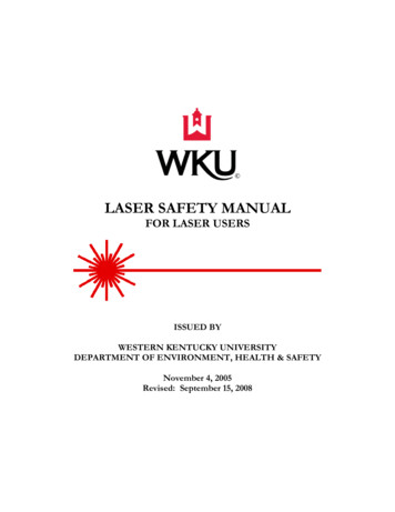
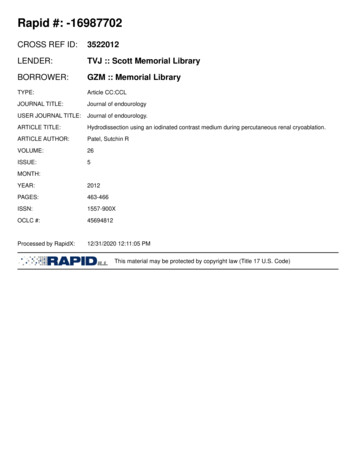
Grewal et al.FIG. 1. A: Axial T1-weighted MPRAGE Gd-enhanced image showing a laser cannula in the hippocampus with a zone of nonenhancing ablated tissue surrounded by a thin rim of contrast enhancement. B: Axial T2-weighted FLAIR image showing theexpected peripheral zone of T2 hyperintense signal around the area of ablation. C: Coronal T1-weighted MPRAGE Gd-enhancedimage showing a peripheral zone of enhancing tissue with the catheter in the center of the zone of enhancement.amount of amygdala and hippocampus that are removedby the procedure, and LiTT is capable of ablating a cylindrically shaped volume of the amygdala and hippocampus. As ablation volumes are not as large as resectionvolumes, there is speculation that the volume of ablationcorrelates with improved seizure outcome, with the assumption that larger ablations would have better seizureoutcomes. While there is an emphasis on volume, it is important to note that accuracy may be equally important.Large ablations or resections that eliminate neocorticalstructures but not the hippocampus are unlikely to be assuccessful as targeted resections or ablations of the mesialstructures. As of now, the optimum volume of hippocampal ablation with LiTT has not been characterized. Theprimary objective of this study was to test the hypothesisthat larger volumes of hippocampal ablation would correlate with better seizure outcomes after LiTT. We furtherevaluated comorbidities in this cohort, such as visual fieldand neuropsychological changes following LiTT.MethodsThis was a multi-site, single-institution, retrospectivereview of seizure outcomes and ablation volumes following LiTT for medically intractable mesial temporal lobeepilepsy performed between October 2011 and October2015. This study was approved by the Mayo Clinic institutional review board. The LiTT procedure was performedby 4 different surgeons at the 3 Mayo Clinic epilepsy centers (in Arizona, Florida, and Minnesota). At all sites thesame general process was used for targeting the medialtemporal lobe. The trajectory started with an occipital approach, avoiding cortical vessels and the ventricle, and targeting the body and head of the hippocampus and as muchof the amygdala as possible. Stereotactic guidance included 3 technologies: conventional frame-based (Compass orLeksell), frameless (Stealth) with in-bone fiducial markers, or intraoperative MR-guided placement (Clearpoint).In all cases, multiple overlapping ablations were used.Pre-ablation volumetric (3D) T1-weighted MR images were acquired in all cases for treatment planning.2056The ablations were monitored in real time with MR thermometry. The individual ablations were repeated as theapplicator was systematically pulled back until the thermal dose computed in the workstation sufficiently covered the entire targeted volume. Immediately after theablations, (3D) gadolinium-enhanced T1-weighted imageswere acquired to delineate tissue damage by gadoliniummediated signal enhancement around the ablation margin(Fig. 1). Pre-ablation and post-ablation volumes of mesialtemporal structures (hippocampus amygdala) in the T1weighted magnetization-prepared rapid acquisition gradient echo (MPRAGE) images were calculated using an automated atlas-based segmentation algorithm (FreeSurfer4.2) (Fig. 2). The volume of tissue ablated during LiTTwas measured in gadolinium-enhanced T1-weighted MRimages acquired perioperatively after tissue ablation usinga manually supervised spline-based edge detection algorithm (Analyze 12.0, Biomedical Imaging Resource). Inall volumetric measurements, hippocampal and amygdalavolumes were aggregated, as post-ablation tissue damagemade differentiation between amygdala and hippocampusdifficult and potentially inaccurate in follow-up imaging.Post-ablation automated segmentations of mesial temporal structures were visually reviewed for accuracy, and ifinaccurate, the segmentation was repeated manually withthe spline-based edge detection algorithm.To determine seizure outcomes, results were comparedbetween those patients who were seizure free and thosewho continued to experience seizures. Engel classificationwas also used to evaluate seizure outcomes, which wereverified by an epileptologist. Visual field assessments wereperformed postoperatively using standard computationalperimetry, with a dedicated ophthalmological consultation.Neuropsychological testing was performed preoperatively in all cases, and the intent was for all patients toreceive postoperative testing. However, only half of thecohort completed postoperative testing. Neuropsychological testing was specific for verbal memory and includedthe Boston Naming Test (BNT), Auditory Verbal Learning Test (AVLT), and the California Verbal Learning TestJ Neurosurg Volume 130 June 2019Unauthenticated Downloaded 09/07/22 05:01 PM UTC
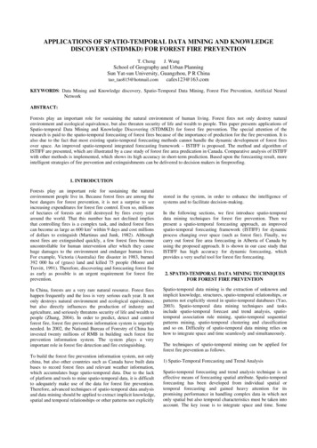
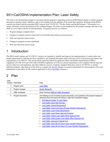
Grewal et al.FIG. 2. Coronal and axial T1-weighted pre- and post-LiTT MR images showing segmentation of mesial temporal structures withthe FreeSurfer program. These images reveal expected postoperative changes after right mesial temporal lobe ablation. Figure isavailable in color online only.(CVLT). All statistical analysis was performed using JMPsoftware (SAS International Inc.) with significance set atp 0.05.ResultsTwenty-five patients underwent LiTT treatment for mesial temporal lobe epilepsy. In 2 cases, adequate immediatepost-ablation imaging was not available, and these patientswere excluded from the study. Twelve (52%) patients weretreated at Mayo Clinic Arizona, 6 (26%) at Mayo ClinicRochester, and 5 (22%) at Mayo Clinic Florida. There wasno correlation between site and seizure outcomes (p 0.66). Ten patients (43%) were male. The patients’ averageage was 43.9 years (Table 1). A majority of the patients hadablation for presumed mesial temporal sclerosis (MTS) asdetermined by MRI (increased T2 signal and hippocampal volume loss); however, 5 patients (22%) had normalMRI findings, without imaging consistent with MTS.These patients had presumed mesial temporal lobe epilepsy based on clinical seizure semiology and ictal EEG.Pre-ablation mesial temporal volumes are shown in Fig. 3.Most patients had multiple seizure types, but the predominant seizure type for each patient was focal seizures withimpaired awareness (formally called complex partial seizures) (74% of patients) (Table 1). Fifteen patients (65%)had left-sided procedures. The median follow-up was 34months (range 12–70 months).Ablation VolumeThe mean volume of ablated tissue was 6888 mm3(range 3045.41–11,560.64 mm3) (Fig. 4). Median hippocampal ablation was 65% (range 28%–85%) with a medi-an amygdala ablation of 43% (range 1%–80%). At lastfollow-up, 11 (48%) of these patients were seizure free(Table 2). An additional 4 patients (17%) had Engel Ib outcomes, with no disabling seizures after surgery. Presenceof MTS on preoperative imaging was not a predictor ofseizure freedom, as 9 of 18 (50%) patients with MTS wereseizure free compared to 2 of 5 (40%) patients whose pre-FIG. 3. Mesial temporal volumes measured by FreeSurfer in pre-ablation T1-weighted MPRAGE imaging show lower overall volume on theside undergoing ablation. The majority of patients studied (88%) had adiagnosis of MTS with volume loss and/or hyperintense signal on FLAIRimaging. Box-and-whisker plots show the 25th and 75th quartiles asdemarcated by the box, with the range demarcated by the lines aboveand below. Figure is available in color online only.J Neurosurg Volume 130 June 20192057Unauthenticated Downloaded 09/07/22 05:01 PM UTC
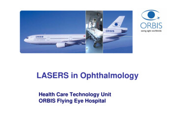
Grewal et al.TABLE 1. Summary of patient demographic and clinicalcharacteristicsAgeCase 8, M57, F25, F33, F42, F44, F57, F46, M48, M52, M58, M58, F43, F32, F44, M38, M42, M48, F22, M46, M20, F60, F37, FSeizuresPathologyFocal seizures w/o impaired awarenessFocal seizures w/o impaired awarenessFocal seizures w/ impaired awarenessFocal seizures w/ impaired awarenessFocal seizures w/ impaired awarenessFocal seizures w/o impaired awarenessFocal seizures w/ impaired awarenessFocal seizures w/ impaired awarenessFocal seizures w/ impaired awarenessFocal seizures w/o impaired awarenessFocal seizures w/ impaired awarenessFocal seizures w/ impaired awarenessFocal seizures w/o impaired awarenessFocal seizures w/ impaired awarenessFocal seizures w/o impaired awarenessFocal seizures w/ impaired awarenessFocal seizures w/ impaired awarenessFocal seizures w/ impaired awarenessFocal seizures w/ impaired awarenessFocal seizures w/ impaired awarenessFocal seizures w/ impaired awarenessFocal seizures w/ impaired awarenessFocal seizures w/ impaired TSNon-lesionalMTSMTSMTSFIG. 4. Left: Bar graph describing the variance in ablation volumeacross patients. Right: Quartile box plot describing the median ablationvolume as well as the 25th and 75th quartiles, with the lines in the whisker plot showing the 10th and 90th quartiles and the whiskers indicatingthe full range.operative imaging showed no lesion (p 0.69). There wasno correlation between ablation volume and seizure freedom (p 0.69). There was no correlation between Engelclass and ablation volume (p 0.93). There was no correlation between percent ablation of the amygdala (p 0.28)or hippocampus (p 0.82) and seizure outcomes. Whenanalysis was restricted to data from the 18 patients withimaging consistent with MTS, ablation volume also didnot correlate with seizure outcomes (p 0.39). Thirteenpatients had high-resolution follow-up MRI scans afterablation. These scans were performed an average of 70days after ablation (range 10–120 days). In this subset ofpatients, comparison of follow-up MRI studies to the immediate pre-procedure MRI studies revealed an averagedecline in the volume of the mesial temporal structures of1095 mm3. Four patients in the cohort had multiple followup MRI examinations, and the longest follow-up obtainedwas 788 days (Fig. 5). A regression analysis performedon the ablated and contralateral mesial temporal volumesshowed an average rate of volume decrease of 3.03 mm3per day for the ablated mesial temporal structures, whilethe contralateral mesial temporal volumes remained essentially unchanged (slope 0.134 mm3/day).ing with computational visual fields. Of these patients,two had near-complete hemianopsias, and 3 patients hadquadrantanopsias that they noticed but did not find to bebothersome. Additionally, there were 3 “silent” quadrantanopsias. The remaining 4 patients had normal visualfields. We compared ablation volumes in the 5 patientswith clinically noticeable deficits to ablation volumes inthe group without clinically noticeable visual field deficits, and there were no statistically significant differencesbetween the 2 groups (6972.27 mm3 vs 6864.57 mm3, p 0.94). We repeated this analysis to compare patients withhemianopsias, clinically noticeable quadrantanopsias, silent quadrantanopsias, and no visual field deficits. Therewas no correlation between ablation volumes and thesevisual field deficits (p 0.54), or between percentage ofhippocampus ablated (p 0.96) or percentage of amygdala ablated (p 0.35) and visual field deficits. In the 2patients who developed hemianopsias, further analysis ofthe lesion was performed. In the first patient, it appears thelesion was too superior and there was thermal damage tothe lateral geniculate nucleus (Fig. 6A), while in the second patient, the optic radiations were damaged at their exitfrom the lateral geniculate nucleus (Fig. 6B).Visual OutcomeTwelve patients underwent formal postoperative test-Neuropsychometric OutcomeEleven patients completed preoperative and postop-2058J Neurosurg Volume 130 June 2019Unauthenticated Downloaded 09/07/22 05:01 PM UTC
Grewal et al.TABLE 2. Ablation side and seizure outcomesCase oYesNoYesYesNoYesNoYesNoNoYesYesNoNoYesYeserative neuropsychological testing. The average timeof follow-up for the neuropsychological testing was 7.8months after LiTT. Of the 11 patients, 7 patients had leftsided procedures. In the group of patients with left-sidedprocedures, the average verbal learning score (AVLT orCVLT) at follow-up assessment was 76% of the preoperative score. A smaller decline was noted in the 4 patientswith right-sided procedures, with an average verbal learning score of 89% of the preoperative score. There were 4patients with declines in their BNT: 2 patients had a decline of 1 point and 2 patients had a decline of 2 points.We consider a decline of 4 points or greater in the BNT tobe clinically significant. There was no difference betweenthe sides for those patients with a decline in their BNT aseach group had 1 patient with a decline of 2 points and 1patient with a decline of 1 point.DiscussionPenfield reported anterior temporal lobectomy as amethod to treat medically refractory temporal lobe epilepsy as early as 1950.11 A study by Wiebe et al. in 2001provided Class I evidence of the superiority of anteriortemporal lobectomy when compared to medical management in a randomized, prospective controlled trial.18 Thepresent study revealed a 58% seizure freedom rate at 1year after the procedure. Other studies have corroboratedthese results, with a meta-analysis showing a 75% seizurefreedom rate at 1 year.8However, concerns regarding neurocognitive deficitsafter anterior temporal lobectomy have prompted the desire for more precise procedures, such as selective amygdalohippocampectomy. These selective procedures haveled to similar rates of seizure freedom (67%).8 There isrelatively little data regarding seizure outcomes after LiTTfor mesial temporal lobe epilepsy. In patients with at least 6months of follow-up, the reported rates of seizure freedomare around 54%.9,19 Our results show a slightly lower rate ofseizure freedom when compared to traditional approachessuch as anterior temporal lobectomy but are in concordance with the literature on LiTT. However, our study involved a mixed cohort of patients, as not all included patients had mesial temporal lobe sclerosis, and this couldpartially explain the lower seizure freedom rate as patientswithout identifiable temporal lobe lesions have lower ratesof seizure freedom overall.3,15Visual field deficits are among the most commonlyreported adverse outcomes after anterior temporal lobectomy, occurring in between 28% and 52% of patients.5,14 Infact, in cases in which detailed visual field analysis is performed, nearly all hippocampal resections result in somevisual field deficit.16 However, many of these deficits areclinically “silent.”10 LiTT theoretically has the potentialto decrease the rate of visual field deficits, as it is allowssurgeons to traverse the optic radiations less in order toperform an ablation. However, there has not been a studyto formally assess for visual field cuts in this cohort.19 Inour study, formal visual field testing was performed in 12patients, two of whom had clinically significant visual fielddeficits. Only 4 patients had normal visual fields, and ablation volumes were not correlative with postoperative visual field deficits. In the 2 patients with hemianopsia, it wascaused in one case by damage to the laternal geniculatenucleus, while in the other it was caused by damage to theoptic radiations as they exit the lateral geniculate nucleus.We believe that the first case was due to a trajectory thatwas too high in the hippocampus, leading to damage to theinferolateral aspect of the lateral geniculate nucleus, whilethe second ablation was carried too far posteriorly, leadingto damage of the optic radiations. This warrants furtherstudy, with more detailed analysis of volume of ablationwith respect to the location of the optic apparatus; however, due to these visual field deficits, we currently aim fora trajectory that leads to the middle or inferior aspect ofthe hippocampus and complete our ablation once we havereached the level of the lateral mesencephalic sulcus.There is a relative paucity of data regarding neuropsychological outcomes and LiTT, and there is unfortunatelynot an agreed-upon standardized battery of tests performedat each center. Similar to results reported in previouslypublished literature,4,6,9 in our study, LiTT was associatedwith a slight decrease in verbal learning with preservationof neocortical functions such as tested by the BNT. Consistent with prior studies, this study showed that decline inverbal memory was more prominent in patients undergoing procedures on their dominant side.6 However, a smalldecline was also noted in verbal memory for patients whounderwent procedures on their nondominant hemisphere.J Neurosurg Volume 130 June 20192059Unauthenticated Downloaded 09/07/22 05:01 PM UTC
Grewal et al.FIG. 5. Mesial temporal volume changes following ablation on the ablated side (red), while the non-ablated side volumes remainconstant. A: Volume changes pre- (time 0) and post-ablation as raw (mm3) volumes. A regression line fit to the volume measurements has a slope of -3.03 mm3/day and a y-intercept of 4875 mm3 for the ablated side, while the non-ablated side’s regressionhas a slope of 0.134 mm3/day and a y-intercept of 5783 mm3. B: Post-ablation measurements normalized to each patient’s preablation volume are plotted. A regression line fit to the normalized measurements has a slope of -4.9 10-4 units/day and a yintercept of 0.948 for the ablated side, while the non-ablated side’s regression has a slope of 5.5 10-5 units/day and a y-interceptof 1.01. Figure is available in color online only.In the surgical literature, there are conflicting data regarding the extent of resection and seizure outcomes intemporal lobe epilepsy. Certain studies have demonstratedthat a greater extent of resection leads to improved sei2060zure outcomes,2,13 while other studies show no correlationbetween extent of resection and seizure outcomes.1 Whenthe specific aspects of resection are analyzed in detail, results suggest that the amount of hippocampal resection isJ Neurosurg Volume 130 June 2019Unauthenticated Downloaded 09/07/22 05:01 PM UTC
Grewal et al.FIG. 6. A: Coronal T1-weighted MR image revealing the location of thelateral geniculate nucleus contralateral to the lesion (yellow arrow); postablation changes are noted in the area of the ipsilateral lateral genticulate nucleus (red arrow). B: Axial Gd-enhanced T1-weighted MR imagerevealing thermal damage to the optic radiations as they exit the lateralgeniculate nucleus (red arrow). Figure is available in color online only.potentially a better correlate of seizure outcomes.7 Whenanalyzing other minimally invasive methods such as radiosurgery, preliminary studies revealed that there was avery tight window of 5.5 to 7.5 ml, below which efficacydecreased and above which resulted in excessive toxicity.12In our study, there was no correlation between seizure outcomes and ablation volumes.One notable point from this retrospective review isthe variance in percent ablation of the amygdala (range1%–80%). Based upon prior temporal lobectomy results,the surgical goal with LiTT was to achieve as large a hippocampal ablation as possible without causing increasedrisk to the visual pathways. Due to the curvature of certainhippocampi, a greater degree of ablation of the amygdalawas impossible without the addition of a second trajectory.The risk of a secondary trajectory was deemed excessivein these cases, as there were no data to support increasedseizure freedom with a greater percentage ablation of theamygdala.This initial multi-site study, while valuable, is limiteddue to its retrospective nature as well as its sample size,leading to a low statistical power. This study could alsobe limited by a narrow range in the variation of ablationvolumes, considering that they were all performed by 3surgeons. This could be improved in a larger multicentermultiple-surgeon trial. Due to the retrospective design ofthis study, we were unable to assess postoperative visual fields and neuropsychometric testing in the entire cohort, introducing a likely selection bias, as patients whoreceived postoperative testing and had longer follow-upperiods could potentially have had these due to pooreroutcomes requiring additional care. In addition, while thegeneral techniques were similar across the 3 sites, therewas no standardization regarding operative technique ortarget volumes for ablation.ConclusionsThis retrospective multi-site study did not show a correlation between seizure outcomes and ablation volumesafter LiTT. Two patients had clinically significant visualfield deficits, and there was a slight decrease in verballearning associated with LiTT of the mesial temporallobe. The results of our study suggest that larger ablationsor increased ablation of either the hippocampus or amygdala does not necessarily correlate with seizure outcomes.However, larger ablations were also not correlated withincreased complications such as visual field deficits. Future prospective multicenter studies are needed to validatethese results in a larger cohort, with further breakdown ofthe mesial structures, assessing ablation of the subiculum,entorhinal cortex, and uncus as well as assessing postoperative computational visual fields and neuropsychometrictesting.References1. Alsaadi TM, Ulmer JL, Mitchell MJ, Morris GL, SwansonSJ, Mueller WM: Magnetic resonance analysis of postsurgical temporal lobectomy. J Neuroimaging 11:243–247, 20012. Awad IA, Katz A, Hahn JF, Kong AK, Ahl J, Lüders H:Extent of resection in temporal lobectomy for epilepsy. I.Interobserver analysis and correlation with seizure outcome.Epilepsia 30:756–762, 19893. Bell ML, Rao S, So EL, Trenerry M, Kazemi N, Stead SM, etal: Epilepsy surgery outcomes in temporal lobe epilepsy witha normal MRI. Epilepsia 50:2053–2060, 20094. Drane DL, Loring DW, Voets NL, Price M, Ojemann JG,Willie JT, et al: Better object recognition and namingoutcome with MRI-guided stereotactic laser amygdalohippocampotomy for temporal lobe epilepsy. Epilepsia 56:101–113, 20155. Egan RA, Shults WT, So N, Burchiel K, Kellogg JX, Salinsky M: Visual field deficits in conventional anterior temporallobectomy versus amygdalohippocampectomy. Neurology55:1818–1822, 20006. Jermakowicz WJ, Kanner AM, Sur S, Bermudez C, D’HaesePF, Kolcun JPG, et al: Laser thermal ablation for mesiotemporal epilepsy: analysis of ablation volumes and trajectories.Epilepsia 58:801–810, 20177. Joo EY, Han HJ, Lee EK, Choi S, Jin JH, Kim JH, et al: Resection extent versus postoperative outcomes of seizure andmemory in mesial temporal lobe epilepsy. Seizure 14:541–551, 20058. Josephson CB, Dykeman J, Fiest KM, Liu X, Sadler RM,Jette N, et al: Systematic review and meta-analysis of standard vs selective temporal lobe epilepsy surgery. Neurology80:1669–1676, 20139. Kang JY, Wu C, Tracy J, Lorenzo M, Evans J, Nei M, et al:Laser interstitial thermal therapy for medically intractablemesial temporal lobe epilepsy. Epilepsia 57:325–334, 201610. Katz A, Awad IA, Kong AK, Chelune GJ, Naugle RI, Wyllie E, et al: Extent of resection in temporal lobectomy forepilepsy. II. Memory changes and neurologic complications.Epilepsia 30:763–771, 198911. Penfield W, Flanigin H: The surgical therapy of temporallobe seizures. Trans Am Neurol Assoc 51:146–149, 195012. Quigg M, Rolston J, Barbaro NM: Radiosurgery for epilepsy:clinical experience and potential antiepileptic mechanisms.Epilepsia 53:7–15, 201213. Shamim S, Wiggs E, Heiss J, Sato S, Liew C, Solomon J, etal: Temporal lobectomy: resection volume, neuropsychological effects, and seizure outcome. Epilepsy Behav 16:311–314, 200914. Tecoma ES, Laxer KD, Barbaro NM, Plant GT: Frequencyand characteristics of visual field deficits after surgery formesial temporal sclerosis. Neurology 43:1235–1238, 199315. Téllez-Zenteno JF, Hernández Ronquillo L, Moien-Afshari F,Wiebe S: Surgical outcomes in lesional and non-lesional epiJ Neurosurg Volume 130 June 20192061Unauthenticated Downloaded 09/07/22 05:01 PM UTC
Grewal et al.16.17.18.19.lepsy: a systematic review and meta-analysis. Epilepsy Res89:310–318, 2010Van Gompel JJ, Welker KM: Visual field mapping to preventvisual field deficits in epilepsy surgery: Seeing the problem.Neurology 83:578–579, 2014Waseem H, Osborn KE, Schoenberg MR, Kelley V, BozorgA, Cabello D, et al: Laser ablation therapy: an alternativetreatment for medically resistant mesial temporal lobe epilepsy after age 50. Epilepsy Behav 51:152–157, 2015Wiebe S, Blume WT, Girvin JP, Eliasziw M: A randomized,controlled trial of surgery for temporal-lobe epilepsy. N EnglJ Med 345:311–318, 2001Willie JT, Laxpati NG, Drane DL, Gowda A, Appin C, HaoC, et al: Real-time magnetic resonance-guided stereotacticlaser amygdalohippocampotomy for mesial temporal lobeepilepsy. Neurosurgery 74:569–585, 2014Disclosuresence and NeuroOne Inc. Dr. Kaufmann reports a consultant relationship with SpineThera.Author ContributionsConception and design: Van Gompel, Grewal, Zimmerman, Worrell, Brinkmann, Tatum,
review of seizure outcomes and ablation volumes follow-ing LiTT for medically intractable mesial temporal lobe epilepsy performed between October 2011 and October 2015. This study was approved by the Mayo Clinic institu - tional review board. The LiTT procedure was performed by 4 different surgeons at the 3 Mayo Clinic epilepsy cen -