Transcription
Rapid #: -16987702CROSS REF ID:3522012LENDER:TVJ :: Scott Memorial LibraryBORROWER:GZM :: Memorial LibraryTYPE:Article CC:CCLJOURNAL TITLE:Journal of endourologyUSER JOURNAL TITLE:Journal of endourology.ARTICLE TITLE:Hydrodissection using an iodinated contrast medium during percutaneous renal cryoablation.ARTICLE AUTHOR:Patel, Sutchin 1557-900XOCLC #:45694812Processed by RapidX:12/31/2020 12:11:05 PMThis material may be protected by copyright law (Title 17 U.S. Code)
Tricks of the TradeJOURNAL OF ENDOUROLOGYVolume 26, Number 5, May 2012ª Mary Ann Liebert, Inc.Pp. 463–466DOI: 10.1089/end.2011.0516Downloaded by Jefferson (Philadelphia University Thomas Jefferson University) from www.liebertpub.com at 12/31/20. For personal use only.Hydrodissection Using an Iodinated Contrast MediumDuring Percutaneous Renal CryoablationSutchin R. Patel, M.D.,1 J. Louis Hinshaw, M.D.,2 Meghan G. Lubner, M.D.,2 Fred T. Lee Jr., M.D.,2Stephen Y. Nakada, M.D.,1 and Sean P. Hedican, M.D.1AbstractWe describe our experience using an iodinated contrast solution to hydrodissect adjacent structures beforepercutaneous renal cryoablation. Hydrodissection was performed before cryoablation with placement of a20-gauge, 15-cm introducer needle into the retroperitoneum under CT or ultrasonographic guidance followed byinfusion of 5% dextrose in water and 2% iodinated contrast between the kidney and the adjacent organ. Tenpatients underwent hydrodissection with an iodinated contrast solution at our institution. The mean tumor sizewas 3.1 – 1.2 cm. The organs displaced included colon (n 7), small bowel (n 1), pancreas (n 1), and in onecase, both the colon and ureter were displaced. The average displacement of all organs from the kidney was2.8 cm (range 2.2–3.5 cm). There were no complications and no injuries to any adjacent structures. The injectionof iodinated contrast allows for safe mobilization and differentiation of adjacent structures from the renal tumorand parenchyma leading to potentially safer cryoablation.IntroductionTechniqueTThis study was approved by our Institutional ReviewBoard, and the need for informed consent was waived. Weperformed a retrospective review of all percutaneous renalcryoablation cases performed at our institution from 2003 to2010 for patients undergoing hydrodissection as a protectivetechnique. Patient medical records and imaging studies werereviewed, and demographic data, tumor size, and detailsregarding hydrodissection of adjacent organs on CT wereextracted.Percutaneous cryoablation procedures were performed bythe same team of radiologists and urologists working together.12 Patients were placed in either the prone or decubitusposition in the CT gantry after intubation and induction ofgeneral anesthesia. Hydrodissection was performed beforecryoablation by placement of a 20-gauge, 15-cm introducerneedle into the retroperitoneum and the infusion of D5W and2% iodinated contrast between the kidney and the adjacentorgan. The introducer needle was placed under CT guidance,and displacement of the adjacent organ was verified via CTbefore cryoablation (Figs. 1 and 2). Because of migration offluid in the retroperitoneum or peritoneal cavity, differentvolumes of fluid were infused to maintain adequate displacement of adjacent structures away from the renal tumor.All patients were treated with 1.7-mm cryoprobes(CryoCare, Endocare). Real-time ultrasonography was alsoperformed for targeting and placement of cryoprobes. Acombination of ultrasonography and unenhanced CT washe first reported percutaneous renal cryoablation wasperformed by Uchida and associates1 in 1995. Since thatinitial case, percutaneous renal cryoablation has become awidespread nephron-sparing alternative to partial nephrectomy in selected patients for small renal masses because of thelack of invasiveness, low morbidity, and rapid patient recovery.2,3 The increasing usage of cryoablation has led tocomplications that are novel to the procedure, includingthermal injury to adjacent structures.4,5The anatomy of the retroperitoneum dictates that otherstructures, such as the colon, duodenum, ureter, psoas muscle, and pancreas, may be immediately adjacent to the renaltumor. Given that the ice ball must exceed the tumor marginby ‡ 3 mm to cause complete cell death, adjacent organs mayneed to be displaced to decrease the risk of injury to adjacentstructures.6–9Hydrodissection using 5% dextrose in water (D5W), saline,or sterile water has been described as a method to displacestructures at risk of injury during thermal ablation.10,11 Whenused in combination with CT as a guidance and monitoringtechnique, however, these solutions may be difficult to differentiate from surrounding organs and muscles. To increase the visibility of injected fluids at CT, we combine D5Wwith iodinated contrast material. We describe our experienceusing D5W doped with iodinated contrast to hydrodissectcolon, pancreas, and ureter during percutaneous renalcryoablation.Departments of 1Urology and 2Radiology, University of Wisconsin School of Medicine and Public Health, Madison, Wisconsin.463
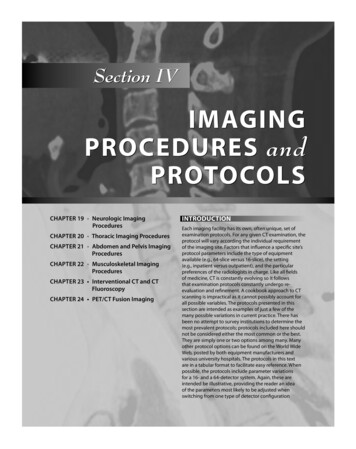
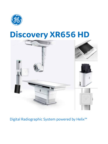
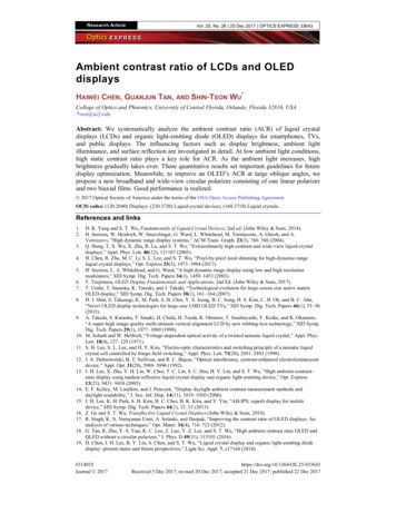
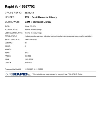
Downloaded by Jefferson (Philadelphia University Thomas Jefferson University) from www.liebertpub.com at 12/31/20. For personal use only.464PATEL ET AL.FIG. 1. Hydrodissection of the colon from the right kidney (patient in prone position). (A) Colon (*) and psoas muscleadjacent to a lower pole renal tumor; (B) placement of a 20-gauge needle; (C) hydrodissection using iodinated contrastsolution to displace both structures; (D) safe placement of cryoprobe. (E) contrast-enhanced CT after completion of ablationand removal of cryoprobe.used to monitor ice ball formation. Our standard treatmentregimen consists of two 10-minute freeze cycles with anintervening 5-minute passive thaw. The ice ball size wasclosely monitored on ultrasonography and CT, and thepercentage of argon flow was modified to obtain a 5- to10-mm margin. After probe removal, a contrast-enhancedCT with delayed images was performed to evaluate forhematoma or acute bleed and assess the renal collectingsystem in cases of endophytic tumors.Measurement of organ displacement before and after instillation of the hydrodissection fluid was performed on CTfluoroscopic images. Region of interest measurements wereperformed of the renal tumor and adjacent hydrodissectionfluid using a picture archiving and communication systemsworkstation (McKesson, Inc, San Francisco, CA). Follow-upimaging consisted of a contrast-enhanced MRI within6 months of the ablation, and ablation success was defined asadequate coverage of the entire tumor by the ablation zone,and a lack of tumor enhancement. Statistical analysis wasperformed using paired Student t test; P value 0.05 wasconsidered significant.Of the 101 percutaneous renal cryoablations performed atour institution in the study period, 10 patients underwent hydrodissection using D5W and iodinated contrast. The meanpatient age was 64 years. The mean tumor size was 3.1 – 1.2 cm.Fluid was injected into the retroperitoneum to displaceseveral different organs, including colon (n 7), small bowel(n 1), pancreas (n 1), and in one case, both the colon andureter were displaced. The mean distance between the renaltumor and the adjacent organ before hydrodissection was0.15 cm (range 0.01–0.80 cm). A mean of 389 mL of fluid (range100–1000 mL) was injected for displacement resulting in amean postinjection displacement of 2.7 cm (range 2.2–3.5 cm)away from the target tumor. The mean attenuation of thehydrodissection fluid was 305 Hounsfield units (HU) (range73–883 HU) and statistically different compared with 48 HU(range 16–75 HU) for the kidney (P 0.01). There wereno intraprocedure or postprocedure complications, and noknown injuries to any adjacent structure. No recurrenceswere noted on follow-up imaging with a mean follow-up of5.4 months.DiscussionCryoinjury to adjacent structures during percutaneouscryoablation has been described and can have devastatingconsequences.4 The anatomy of the retroperitoneum in reference to the renal tumor and the need for the ice ball toextend at least 3 mm beyond the outer tumor margin to resultin complete cell death are important factors that may lead toinjury of adjacent structures.6,7 Damage to hollow thin-walledstructures (bowel, ureter, gallbladder) is more severe thanfreezing adjacent solid organs (liver, spleen) because of therisk of mural necrosis and secondary abscess or stricture.13,14Large blood vessels such as the inferior vena cava tend to be atlower risk for cryoinjury because of the heat sink effect ofblood flow.15,16The location of the renal tumor frequently dictates the needfor hydrodissection. The colon is the most commonly displaced structure and is generally located anterior and inferiorto the kidneys. Lower pole or anterior renal tumors may thusbe more likely to need hydrodissection. Lower pole tumorsmay also need hydrodissection and/or stent placement because of the close proximity of the ureter.11 In the past, weFIG. 2. Hydrodissection of the pancreas from the right kidney (patient in decubitus position). (A) Pancreas (p) adjacent toan anterior renal tumor; (B) placement of a 20-gauge needle; (C) hydrodissection using iodinated contrast solution to displacethe pancreas from the anterior renal tumor; (D) safe placement of cryoprobe; (E) contrast-enhanced CT after completion ofablation and removal of cryoprobe.
Downloaded by Jefferson (Philadelphia University Thomas Jefferson University) from www.liebertpub.com at 12/31/20. For personal use only.HYDRODISSECTION DURING CRYOABLATIONapproached many of these lesions under laparoscopic assistance because of concerns regarding adjacent organ injurywithout previous mobilization. Whenever feasible, however,we favor a percutaneous approach because of the decreasedmorbidity and costs when compared with laparoscopiccryoablation, and the use of hydrodissection techniques hasincreased the number of cases that can be performed percutaneously.12Hydrodissection with the use of sterile water or D5W hasbeen described.10 Both of these fluids, however, are similar inattenuation to the renal tumor, normal renal parenchyma, andadjacent structures (including bowel, which is often fluid filled) making accurate targeting and monitoring difficult. Arecent retrospective study by Bodily and associates11 described hydrodissection in 50 cases of percutaneous renalcryoablation. The structures displaced included the colon(n 41), body wall (n 3), duodenum (n 2), jejunum and ileum (n 2), ureter (n 1), and psoas muscle (n 1). Hydrodissection was successful in 50 of 52 cases, displacingstructures a mean distance of 1.6 cm. The two failed casesoccurred early in the series and were because of insufficientdisplacement of the colon from the index tumor. There wasone complication of hemorrhage resulting from injury to anintercostal artery branch that necessitated termination of theprocedure before fluid infusion and endovascular intervention to embolize the injured vessel.DeBenedectis and colleagues17 described using a mixture ofD5W and iodinated contrast medium to aid hydrodissectionfor radiofrequency ablation. They performed hydrodissectionusing an iodinated contrast medium in 21 patients who wereundergoing percutaneous radiofrequency ablation. Organsdisplaced included colon (n 19), small bowel (n 4), pancreas(n 3), and spleen (n 1), with a mean displacement distanceof 2.6 cm. Ablation was performed successfully in all 21 caseswith no injury to adjacent structures. All patients also underwent a follow-up CT scan an average of 3 weeks after theprocedure, and no residual hydrodissection fluid was noted.The choice of hydrodissection fluid varies with ablationmodality. For radiofrequency ablation, tissue heating is created by ionic agitation as current is conducted between electrodes and ground pads. Thus, maximum protection ofvulnerable structures is gained by the choice of nonionic,poorly conducting fluids, such as sterile water or dextrose inwater. Saline is an ionic fluid that conducts electricity and isthus poorly suited for this purpose when used during radiofrequency ablation.10,18For cryoablation, the choice of fluid type is less importantbecause the object of hydrodissection is solely to increasedisplacement between the ice ball and vulnerable structure.We used a 2% iodinated contrast solution based on the resultsof Roen and coworkers,19 which demonstrated that the idealconcentration of 300 mg/mL of iohexol in physiologic salineand D5W is between 0.5% and 2%. At these concentrations,there is sufficient contrast between the infused fluid and abdominal organs without producing beam hardening artifacts.19 We found that as the hydrodissection fluid dissipatesin the abdomen, there will be some variability in the HU attenuation of the fluid.In our study, the amount of displacement of adjacentstructures (2.8 cm) was similar to that reported by otherstudies (range 1.6–2.6 cm).10,11,18 We show that the use ofa D5W with 2% iodinated contrast allows for a clear differ-465entiation between the renal tumor (mean attenuation of48 HU) and the hydrodissection fluid (mean attenuation of305 HU). The ability to differentiate between the hydrodissection fluid and the renal tumor and adjacent structuresis helpful to prevent inadvertent freezing of nontargetedstructures and assure adequate coverage of the tumor by theablation zone.Limitations of our study include a small number of patientsand the retrospective nature of this study. Another potentiallimitation is the subjectivity of the improved visualizationwith an iodinated contrast solution vs D5W, although a significant difference in HU attenuation was noted between thetumor and hydrodissection fluid.ConclusionThe injection of D5W 5 mixed with 2% iodinated contrastallows for safe mobilization and differentiation of adjacentorgans from the renal tumor and parenchyma during percutaneous cryoablation.Disclosure StatementDr. Henshaw is a stockholder and on the Board of MedicalAdvisors for Neuwave Medical. For all other authors, nocompeting financial interests exist.References1. Uchida M, Imaide Y, Sugimoto K, et al. Percutaneouscryosurgery for renal tumours. Br J Urol 1995;75:132–137.2. Kunkle DA, Egleston BL, Uzzo RG. Excise, ablate or observe.The small renal mass dilemma—a meta-analysis and review.J Urol 2008;179:1227–1234.3. Campbell SC, Novick AC, Belldegrun A, et al. Guideline formanagement of the clinical T1 renal mass. J Urol 2009;182:1271–1279.4. Tsivian M, Chen VH, Kim CY, et al. Complications of laparoscopic and percutaneous renal cryoablation in a singletertiary referral center. Eur Urol 2010;58:142–147.5. Igor Pinkhasov G, Raman JD. Management and preventionof renal ablative therapy complications. World J Urol 2010;28:559–564.6. Campbell SC, Krihnamurthi V, Chow G, et al. Renal cryosurgery: Experimental evaluation of treatment parameters.Urology 1998;52:29–34.7. Chosy SG, Nakada SY, Lee FT Jr, Warner TF. Monitoringrenal cryosurgery: Predictors of tissue necrosis in swine.J Urol 1998;159:1370–1374.8. Littrup PJ, Jallad B, Vorugu V, et al. Lethal isotherms ofcryoablation in a phantom study: Effects of heat load, probesize, and number. J Vasc Interv Radiol 2009;20:1343–1351.9. Young JL, Kolla SB, Pick DL, et al. In vitro, ex vivo and in vivoisotherms for renal cryotherapy. J Urol 2010;183:752–758.10. Farrell MA, Charboneau JW, Callstrom MR, et al. Paranephric water instillation: A technique to prevent bowelinjury during percutaneous renal radiofrequency ablation.AJR Am J Roentgenol 2003;181:1315–1317.11. Bodily KD, Atwell TD, Mandrekar JN, et al. Hydrodisplacement in the percutaneous cryoablation of 50 renaltumors. AJR Am J Roentgenol 2010;194:779–783.12. Hinshaw JL, Shadid AM, Nakada SY, et al. Comparison ofpercutaneous and laparoscopic cryoablation for the treatment of solid renal masses. AJR Am J Roentgenol 2008;191:1159–1168.
Downloaded by Jefferson (Philadelphia University Thomas Jefferson University) from www.liebertpub.com at 12/31/20. For personal use only.46613. Littrup PJ, Ahmed A, Aoun HE, et al. CT-guided percutaneous cryotherapy of renal masses. J Vasc Interv Radiol2007;18:383–392.14. Silverman SG, Tuncali K, van Sonnenberg E, et al. Renaltumors: MR imaging-guided percutaneous cryotherapy—initial experience in 23 patients. Radiology 2005;236:716–724.15. Gage AA, Fazekas G, Riley EE Jr. Freezing injury to largeblood vessels in dogs. With comments on the effect ofexperimental freezing of bile ducts. Surgery 1967;61:748–754.16. Lagerveld BW, van Horssen P, Pes MP, et al. Immediateeffect of kidney cryoablation on renal arterial structure in aporcine model studied by imaging cryomicrotome. J Urol2010;183:1221–1226.17. DeBenedectis CM, Beland MD, Dupuy DE, Mayo-SmithWW. Utility of iodinated contrast medium in hydrodissection fluid when performing renal tumor ablation. J VascInterv Radiol 2010;21:745–747.18. Brace CL, Laeseke PF, Prasad V, Lee FT. Electrical isolationduring radiofrequency ablation: 5% dextrose in water provides better protection than saline. Conf Proc IEEE Eng MedBiol Soc 2006;1:5021–5024.PATEL ET AL.19. Roen C, Hinshaw JL, Lubner MG, et al. Contrast-dopedhydrodissection fluids for thermal ablation: Optimization ofiohexol concentration. World Conference on InterventionalOncology, June 9-12, 2010, Philadelphia, PA. Abstract 35.Address correspondence to:Sean P. Hedican, M.D.Department of UrologyUniversity of WisconsinSchool of Medicine and Public Health1685 Highland AvenueMadison WI, 53705E-mail: hedican@urology.wisc.eduAbbreviations UsedCT ¼ computed tomographyD5W ¼ 5% dextrose in waterHU ¼ Hounsfield unitMRI ¼ magnetic resonance imaging
Downloaded by Jefferson (Philadelphia University Thomas Jefferson University) from www.liebertpub.com at 12/31/20. For personal use only.This article has been cited by:1. Takafumi Yanagisawa, Jun Miki, Kanichiro Shimizu, Wataru Fukuokaya, Fumihiko Urabe, Keiichiro Mori, Hiroshi Sasaki,Takahiro Kimura, Kenta Miki, Shin Egawa. 2020. Functional and oncological outcome of percutaneous cryoablation versuslaparoscopic partial nephrectomy for clinical T1 renal tumors: A propensity score-matched analysis. Urologic Oncology: Seminarsand Original Investigations 38:12, 938.e1-938.e7. [Crossref]2. Takuji Yamagami, Rika Yoshimatsu, Kenji Kajiwara, Tomoaki Yamanishi, Hiroki Minamiguchi, Takashi Karashima, Keiji Inoue.2019. Protection from injury of organs adjacent to a renal tumor during percutaneous cryoablation. International Journal of Urology26:8, 785-790. [Crossref]3. Ali Devrim Karaosmanoglu, Mehmet Ruhi Onur, Okan Akhan. Role of Interventional Radiology in Management ofGastrointestinal Cancers and Neuroendocrine Tumors 551-572. [Crossref]4. Mustafa M. Haddad, Grant D. Schmit, A. Nicholas Kurup, John J. Schmitz, Stephen A. Boorjian, Jennifer Geske, R. HoustonThompson, Matthew R. Callstrom, Thomas D. Atwell. 2018. Percutaneous Cryoablation of Solitary, Sporadic Renal CellCarcinoma: Outcome Analysis Based on Clear-Cell versus Papillary Subtypes. Journal of Vascular and Interventional Radiology29:8, 1122-1126. [Crossref]5. Ronald S. Arellano. 2018. Ablation of Renal Cell Carcinoma: An Assessment of Currently Available Techniques. Journal ofRadiology Nursing 37:1, 30-35. [Crossref]6. Sutchin R. Patel, Stephen Y. Nakada. The History of Percutaneous Renal Cryoablation 163-175. [Crossref]7. Bing Li, Chuan Liu, Xiao-Xue Xu, Yang Li, Yong Du, Chuan Zhang, Hou-Jun Zheng, Han-Feng Yang. 2017. Clinical Applicationof Artificial Ascites in assisting CT-guided Percutaneous Cryoablation of Hepatic Tumors Adjacent to the Gastrointestinal Tract.Scientific Reports 7:1. . [Crossref]8. J. Garnon, T. Tricard, R.L. Cazzato, X. Cathelineau, A. Gangi, H. Lang. 2017. Traitement ablatif pour cancer du rein : modalitésd’évaluation pré-, per-, post-intervention et prise en charge adaptée. Progrès en Urologie 27:15, 971-993. [Crossref]9. Julien Garnon, Guillaume Koch, Jean Caudrelier, Nitin Ramamurthy, Pramod Rao, Georgia Tsoumakidou, Roberto LuigiCazzato, Afshin Gangi. 2016. Percutaneous Image-Guided Cryoablation of Challenging Mediastinal Lesions Using LargeVolume Hydrodissection: Technical Considerations and Outcomes. CardioVascular and Interventional Radiology 39:11, 1636-1643.[Crossref]10. Miyuki Sone, Yasuaki Arai, Shunsuke Sugawara, Koji Tomita, Keishi Fujiwara, Hiroaki Ishii, Shinichi Morita. 2016. Angio-CT–Assisted Balloon Dissection: Protection of the Adjacent Intestine during Cryoablation for Patients with Renal Cancer. Journal ofVascular and Interventional Radiology 27:9, 1414-1419. [Crossref]11. Stephen R. Lee, Aoife Kilcoyne, Avinash Kambadakone, Ronald Arellano. 2016. Interventional oncology: pictorial review of postablation imaging of liver and renal tumors. Abdominal Radiology 41:4, 677-705. [Crossref]12. Timothy J. Ziemlewicz, Shane A. Wells, Meghan G. Lubner, Christopher L. Brace, Fred T. Lee, J. Louis Hinshaw. 2016. HepaticTumor Ablation. Surgical Clinics of North America 96:2, 315-339. [Crossref]13. J. Louis Hinshaw, Meghan G. Lubner, Timothy J. Ziemlewicz, Fred T. Lee, Christopher L. Brace. 2014. Percutaneous TumorAblation Tools: Microwave, Radiofrequency, or Cryoablation—What Should You Use and Why?. RadioGraphics 34:5, 1344-1362.[Crossref]14. Anil Kapoor, Yuding Wang, Brad Dishan, Stephen E. Pautler. 2014. Update on Cryoablation for Treatment of Small Renal Mass:Oncologic Control, Renal Function Preservation, and Rate of Complications. Current Urology Reports 15:4. . [Crossref]15. F. Khan, S. Sriprasad, F.X. Keeley. 2012. Cryosurgical ablation for small renal masses, current status and future prospects. BritishJournal of Medical and Surgical Urology 5, S28-S34. [Crossref]
workstation (McKesson, Inc, San Francisco, CA). Follow-up imaging consisted of a contrast-enhanced MRI within 6 months of the ablation, and ablation success was defined as adequate coverage of the entire tumor by the ablation zone, and a lack of tumor enhancement. Statistical analysis was performed using paired Student t test; P value 0.05 was