Transcription
ArcturusXT Laser Capture Microdissection(LCM) SystemUSER GUIDE Nikon Eclipse Ti-E Microscope Basefor use with:CapSure LCM MicroCapsCapSure HS LCM CapsArcturusXT Software v3.4Publication Number MAN0016091Revision A.0For Research Use Only. Not for use in diagnostic procedures.
The information in this guide is subject to change without notice.DISCLAIMER: TO THE EXTENT ALLOWED BY LAW, LIFE TECHNOLOGIES AND/OR ITS AFFILIATE(S) WILL NOT BE LIABLE FOR SPECIAL, INCIDENTAL,INDIRECT, PUNITIVE, MULTIPLE, OR CONSEQUENTIAL DAMAGES IN CONNECTION WITH OR ARISING FROM THIS DOCUMENT, INCLUDING YOURUSE OF IT.Revision history: Pub. no. MAN0016091RevisionDateA.022 September 2016DescriptionNew document. Supports CapSure LCM MicroCaps and ArcturusXTSoftware v3.4. See Pub. No. 0112-0153 for instructions for CapSure Macro LCM Caps XTand ArcturusSoftware v3.3. Limited Use Label License No. 358: Research Use Only: Notice to Purchaser: The purchase of this product conveys to the purchaser the limited, nontransferable right to use the product only to perform internal research for the sole benefit of the purchaser. No right to resell this product or any of itscomponents is conveyed expressly, by implication, or by estoppel. This product is for internal research purposes only and is not for use in commercialapplications of any kind, including, without limitation, quality control and commercial services such as reporting the results of purchaser's activitiesfor a fee or other form of consideration. For information on obtaining additional rights, please contact outlicensing@thermofisher.com or OutLicensing, Life Technologies Corporation (part of Thermo Fisher Scientific), 5823 Newton Drive, Carlsbad, California 92008.Corporate entity: Life Technologies Corporation Carlsbad, CA 92008 USA Toll Free in USA 1 800 955 6288Trademarks: All trademarks are the property of Thermo Fisher Scientific and its subsidiaries unless otherwise specified. Nikon and Eclipse aretrademarks of Nikon Corporation. Pentium is a trademark of Intel Corporation. Microsoft, Windows, and Internet Explorer are trademarks of MicrosoftCorporation. Cy is a trademark of GE Healthcare. 2016 Thermo Fisher Scientific Inc. All rights reserved.
ContentsAbout this guide . . . . . . . . . . . . . . . . . . . . . . . . . . . . . . . . . . . . . . . . . . . . 9Purpose of this guide . . . . . . . . . . . . . . . . . . . . . . . . . . . . . . . . . . . . . . . . . . . . . . . . . . . . . .9Prerequisites . . . . . . . . . . . . . . . . . . . . . . . . . . . . . . . . . . . . . . . . . . . . . . . . . . . . . . . . . . . .9CHAPTER 1System overview . . . . . . . . . . . . . . . . . . . . . . . . . . . . . . . . . . . . . . . . . . 11About the ArcturusXT Instrument . . . . . . . . . . . . . . . . . . . . . . . . . . . . . . . . . . . . . . . . . . .11About laser capture microdissection . . . . . . . . . . . . . . . . . . . . . . . . . . . . . . . . . . . . . . . .12Types of cut and capture . . . . . . . . . . . . . . . . . . . . . . . . . . . . . . . . . . . . . . . . . . . . . . . . . . . . 12Outline of the microdissection process . . . . . . . . . . . . . . . . . . . . . . . . . . . . . . . . . . . . . . . . 12Using the ArcturusXT operating software . . . . . . . . . . . . . . . . . . . . . . . . . . . . . . . . . . . .13The primary screen . . . . . . . . . . . . . . . . . . . . . . . . . . . . . . . . . . . . . . . . . . . . . . . . . . . . . . . . 13Viewing tool tips . . . . . . . . . . . . . . . . . . . . . . . . . . . . . . . . . . . . . . . . . . . . . . . . . . . . . . . . . . . 14Making selections from pop-up menus . . . . . . . . . . . . . . . . . . . . . . . . . . . . . . . . . . . . . . . 14Using the options dialog boxes . . . . . . . . . . . . . . . . . . . . . . . . . . . . . . . . . . . . . . . . . . . . . . . 15Finding the version number . . . . . . . . . . . . . . . . . . . . . . . . . . . . . . . . . . . . . . . . . . . . . . . . . 17Using menus and commands . . . . . . . . . . . . . . . . . . . . . . . . . . . . . . . . . . . . . . . . . . . . . . . . 17Key commands . . . . . . . . . . . . . . . . . . . . . . . . . . . . . . . . . . . . . . . . . . . . . . . . . . . . . . . . . . . . 18Using the ArcturusXT Instrument as a stand-alone microscope . . . . . . . . . . . . . . . . . . . . . . . 19Getting into and out of manual mode . . . . . . . . . . . . . . . . . . . . . . . . . . . . . . . . . . . . . . . . . 19Using manual mode . . . . . . . . . . . . . . . . . . . . . . . . . . . . . . . . . . . . . . . . . . . . . . . . . . . . . . . . 19CHAPTER 2Preparing samples . . . . . . . . . . . . . . . . . . . . . . . . . . . . . . . . . . . . . . . . 21Summary of chapter topics . . . . . . . . . . . . . . . . . . . . . . . . . . . . . . . . . . . . . . . . . . . . . . . .21Choosing slides and Petri dishes . . . . . . . . . . . . . . . . . . . . . . . . . . . . . . . . . . . . . . . . . . .21Acquiring slides and Petri dishes . . . . . . . . . . . . . . . . . . . . . . . . . . . . . . . . . . . . . . . . . . . . 21For information about using these products . . . . . . . . . . . . . . . . . . . . . . . . . . . . . . . . . . . 22Preparing tissue samples . . . . . . . . . . . . . . . . . . . . . . . . . . . . . . . . . . . . . . . . . . . . . . . . .22Using frozen tissue samples . . . . . . . . . . . . . . . . . . . . . . . . . . . . . . . . . . . . . . . . . . . . . . . . . 22Using formalin-fixed, paraffin-embedded tissue samples . . . . . . . . . . . . . . . . . .23Using other types of samples . . . . . . . . . . . . . . . . . . . . . . . . . . . . . . . . . . . . . . . . . . . . . . . . 23CHAPTER 3Starting the system and loading samples . . . . . . . . . . . . . . . . . . . . 25Summary of chapter topics . . . . . . . . . . . . . . . . . . . . . . . . . . . . . . . . . . . . . . . . . . . . . . . .25Introduction to the Nikon Eclipse Ti-E microscope controls . . . . . . . . . . . . . . . . . . .25Using the front operation panel . . . . . . . . . . . . . . . . . . . . . . . . . . . . . . . . . . . . . . . . . . . . . . 25Using the left operation panel . . . . . . . . . . . . . . . . . . . . . . . . . . . . . . . . . . . . . . . . . .28Using the right operation panel . . . . . . . . . . . . . . . . . . . . . . . . . . . . . . . . . . . . . . . . . . . . . . 29ArcturusXT Laser Capture Microdissection System User Guide (CapSure LCM MicroCaps)3
ContentsStarting the ArcturusXT operating software . . . . . . . . . . . . . . . . . . . . . . . . . . . . . . . . . . 30Loading materials onto the ArcturusXT Instrument . . . . . . . . . . . . . . . . . . . . . . . . . . . 31Prepare the work surface . . . . . . . . . . . . . . . . . . . . . . . . . . . . . . . . . . . . . . . . . . . . . . . . . . . 31(Optional) selecting load options for slides . . . . . . . . . . . . . . . . . . . . . . . . . . . . . . . . . . . . 31Selecting load options for CapSure Caps . . . . . . . . . . . . . . . . . . . . . . . . . . . . . . . . . . . . 32Selecting file path options . . . . . . . . . . . . . . . . . . . . . . . . . . . . . . . . . . . . . . . . . . . . . . . . . . . 33Selecting static image options . . . . . . . . . . . . . . . . . . . . . . . . . . . . . . . . . . . . . . . . . . . . . . . 33Implementing your selections . . . . . . . . . . . . . . . . . . . . . . . . . . . . . . . . . . . . . . . . . . . . . . . 34Saving images automatically . . . . . . . . . . . . . . . . . . . . . . . . . . . . . . . . . . . . . . . . . . . . . . 34CHAPTER 4Inspecting slides . . . . . . . . . . . . . . . . . . . . . . . . . . . . . . . . . . . . . . . . . . 37Summary of chapter topics . . . . . . . . . . . . . . . . . . . . . . . . . . . . . . . . . . . . . . . . . . . . . . . 37Using the Inspect tools . . . . . . . . . . . . . . . . . . . . . . . . . . . . . . . . . . . . . . . . . . . . . . . . . . . 37Viewing the slides . . . . . . . . . . . . . . . . . . . . . . . . . . . . . . . . . . . . . . . . . . . . . . . . . . . 38Adjusting the brightness . . . . . . . . . . . . . . . . . . . . . . . . . . . . . . . . . . . . . . . . . . . . . . . . . . . . 38Focusing the image . . . . . . . . . . . . . . . . . . . . . . . . . . . . . . . . . . . . . . . . . . . . . . . . . . 39Magnifying the image . . . . . . . . . . . . . . . . . . . . . . . . . . . . . . . . . . . . . . . . . . . . . . . . . . . . . . 39Working with the Bright Field lamp . . . . . . . . . . . . . . . . . . . . . . . . . . . . . . . . . . . . . . . . . . . . . . . 40Adjusting the Bright Field lamp . . . . . . . . . . . . . . . . . . . . . . . . . . . . . . . . . . . . . . . . . . . . . . 40Adjusting the video camera properties . . . . . . . . . . . . . . . . . . . . . . . . . . . . . . . . . . 41Changing the autobrightness settings . . . . . . . . . . . . . . . . . . . . . . . . . . . . . . . . . . 42Performing phase contrast and DIC imaging . . . . . . . . . . . . . . . . . . . . . . . . . . . . . . . . . . . . . . . 43Using phase contrast imaging . . . . . . . . . . . . . . . . . . . . . . . . . . . . . . . . . . . . . . . . . . . . . . . 43Using differential interference contrast (DIC) imaging . . . . . . . . . . . . . . . . . . . . . . . . . . 45Working with fluorescence . . . . . . . . . . . . . . . . . . . . . . . . . . . . . . . . . . . . . . . . . . . . . . . . . . . . . . 47Before you begin . . . . . . . . . . . . . . . . . . . . . . . . . . . . . . . . . . . . . . . . . . . . . . . . . . . . . . . . . . . 47Getting set up for fluorescence . . . . . . . . . . . . . . . . . . . . . . . . . . . . . . . . . . . . . . . . . . . . . . 47Working with fluorescently labeled samples . . . . . . . . . . . . . . . . . . . . . . . . . . . . . 48Working with Fluorescence timed exposure . . . . . . . . . . . . . . . . . . . . . . . . . . . . . . . . . . . 49Working with slides . . . . . . . . . . . . . . . . . . . . . . . . . . . . . . . . . . . . . . . . . . . . . . . . . . . . . . 50Displaying a different slide . . . . . . . . . . . . . . . . . . . . . . . . . . . . . . . . . . . . . . . . . . . . . . . . . . 50Viewing slide properties . . . . . . . . . . . . . . . . . . . . . . . . . . . . . . . . . . . . . . . . . . . . . . . . . . . . 50Working with images and videos . . . . . . . . . . . . . . . . . . . . . . . . . . . . . . . . . . . . . . . . . . . . . . . . . 51Capturing and saving images . . . . . . . . . . . . . . . . . . . . . . . . . . . . . . . . . . . . . . . . . . . . . . . . 51Capturing, saving, and viewing videos . . . . . . . . . . . . . . . . . . . . . . . . . . . . . . . . . . . . . . . . 52CHAPTER 5Selecting cells for microdissection . . . . . . . . . . . . . . . . . . . . . . . . . . 53Summary of chapter topics . . . . . . . . . . . . . . . . . . . . . . . . . . . . . . . . . . . . . . . . . . . . . . . 53Marking cells for microdissection . . . . . . . . . . . . . . . . . . . . . . . . . . . . . . . . . . . . . . . . . . 54Working with drawing items . . . . . . . . . . . . . . . . . . . . . . . . . . . . . . . . . . . . . . . . . . . . . . . . . . . . . 56Moving drawing items . . . . . . . . . . . . . . . . . . . . . . . . . . . . . . . . . . . . . . . . . . . . . . . . . . . . . . 56Moving drawing items to a different Capture Group . . . . . . . . . . . . . . . . . . . . . . . . . . . . . 56Deleting drawing items . . . . . . . . . . . . . . . . . . . . . . . . . . . . . . . . . . . . . . . . . . . . . . . . . . . . . 56Deleting IR Capture Spots from a drawing item . . . . . . . . . . . . . . . . . . . . . . . . . . . . . . . . 574ArcturusXT Laser Capture Microdissection System User Guide (CapSure LCM MicroCaps)
ContentsChanging the microdissection properties of drawing items . . . . . . . . . . . . . . . . . . . . . . 57Viewing information about a drawing item . . . . . . . . . . . . . . . . . . . . . . . . . . . . . . .58Setting drawing tools options . . . . . . . . . . . . . . . . . . . . . . . . . . . . . . . . . . . . . . . . . . . . . . . . 58Measuring distances and objects . . . . . . . . . . . . . . . . . . . . . . . . . . . . . . . . . . . . . . . . . . .59Setting the IR Capture Spot size . . . . . . . . . . . . . . . . . . . . . . . . . . . . . . . . . . . . . . . . . . . .59Setting up the Test Fire . . . . . . . . . . . . . . . . . . . . . . . . . . . . . . . . . . . . . . . . . . . . . . . . . . . . . 59Checking the spot diameter . . . . . . . . . . . . . . . . . . . . . . . . . . . . . . . . . . . . . . . . . . . . . . . . . 60Working with overlays . . . . . . . . . . . . . . . . . . . . . . . . . . . . . . . . . . . . . . . . . . . . . . . . . . . .61Saving an overlay . . . . . . . . . . . . . . . . . . . . . . . . . . . . . . . . . . . . . . . . . . . . . . . . . . . . . . . . . . 61Using a saved overlay . . . . . . . . . . . . . . . . . . . . . . . . . . . . . . . . . . . . . . . . . . . . . . . . . . . . . . 61Working with Capture Groups . . . . . . . . . . . . . . . . . . . . . . . . . . . . . . . . . . . . . . . . . . . . . . . . . . . . 62Viewing a capture group . . . . . . . . . . . . . . . . . . . . . . . . . . . . . . . . . . . . . . . . . . . . . . . . . . . . 62Setting formatting properties for a Capture Group . . . . . . . . . . . . . . . . . . . . . . . . . . . . . . 62Working with stored positions . . . . . . . . . . . . . . . . . . . . . . . . . . . . . . . . . . . . . . . . . . . . . .63CHAPTER 6Microdissecting cells and tissue . . . . . . . . . . . . . . . . . . . . . . . . . . . . 65Summary of chapter topics . . . . . . . . . . . . . . . . . . . . . . . . . . . . . . . . . . . . . . . . . . . . . . . .65Capturing cells by microdissection . . . . . . . . . . . . . . . . . . . . . . . . . . . . . . . . . . . . . . . . . . . . . . . . 66Capturing cells in one step . . . . . . . . . . . . . . . . . . . . . . . . . . . . . . . . . . . . . . . . . . . . . . . . . . 66Capturing cells using the capture and cutting tools separately . . . . . . . . . . . . . . . . . . . 67Repeating microdissection . . . . . . . . . . . . . . . . . . . . . . . . . . . . . . . . . . . . . . . . . . . . . . . . . . 68Inspecting microdissected material . . . . . . . . . . . . . . . . . . . . . . . . . . . . . . . . . . . . . . . . . . . . . . . 69Unloading materials . . . . . . . . . . . . . . . . . . . . . . . . . . . . . . . . . . . . . . . . . . . . . . . . . . . . . . . . . . . . 70Locating the lasers . . . . . . . . . . . . . . . . . . . . . . . . . . . . . . . . . . . . . . . . . . . . . . . . . . . . . . . . . . . . . 71Locating the UV cutting laser . . . . . . . . . . . . . . . . . . . . . . . . . . . . . . . . . . . . . . . . . . . . . . . . 71Locating the IR capture laser . . . . . . . . . . . . . . . . . . . . . . . . . . . . . . . . . . . . . . . . . .72Selecting preferences for cut and capture . . . . . . . . . . . . . . . . . . . . . . . . . . . . . . . . . . .73Changing the cut and capture order . . . . . . . . . . . . . . . . . . . . . . . . . . . . . . . . . . . . . . . . . . 73Setting properties for cut and capture . . . . . . . . . . . . . . . . . . . . . . . . . . . . . . . . . . . . . . . . 74Working with CapSure Caps . . . . . . . . . . . . . . . . . . . . . . . . . . . . . . . . . . . . . . . . . . . . . .75Viewing a CapSure Cap In the QC area . . . . . . . . . . . . . . . . . . . . . . . . . . . . . . . . . . . . . . 75Viewing and updating CapSure Cap properties . . . . . . . . . . . . . . . . . . . . . . . . . . . . . . . 76Viewing the cap interaction history . . . . . . . . . . . . . . . . . . . . . . . . . . . . . . . . . . . . . . . . . . . 76Using the Laser Bypass feature . . . . . . . . . . . . . . . . . . . . . . . . . . . . . . . . . . . . . . . . . . . .76CHAPTER 7Extracting cells and tissue . . . . . . . . . . . . . . . . . . . . . . . . . . . . . . . . . 79Summary of chapter topics . . . . . . . . . . . . . . . . . . . . . . . . . . . . . . . . . . . . . . . . . . . . . . . .79Choosing an extraction kit . . . . . . . . . . . . . . . . . . . . . . . . . . . . . . . . . . . . . . . . . . . . . . . . .79Extracting tissue from CapSure LCM MicroCaps . . . . . . . . . . . . . . . . . . . . . . . . . . . . .79Extracting tissue from CapSure HS LCM Caps . . . . . . . . . . . . . . . . . . . . . . . . . . . . . . . . . . . . . 80Performing LCM captures with CapSure HS LCM Caps . . . . . . . . . . . . . . . . . . . . . . . . 80Using the ExtracSure Device during extraction . . . . . . . . . . . . . . . . . . . . . . . . . .81ArcturusXT Laser Capture Microdissection System User Guide (CapSure LCM MicroCaps)5
ContentsAPPENDIX AMaintenance and troubleshooting . . . . . . . . . . . . . . . . . . . . . . . . . . . 83Summary of chapter topics . . . . . . . . . . . . . . . . . . . . . . . . . . . . . . . . . . . . . . . . . . . . . . . 83Cleaning the ArcturusXT Instrument . . . . . . . . . . . . . . . . . . . . . . . . . . . . . . . . . . . . . . . 83Replacing user-serviceable parts . . . . . . . . . . . . . . . . . . . . . . . . . . . . . . . . . . . . . . . . . . 83Replacing the bright field illumination lamp . . . . . . . . . . . . . . . . . . . . . . . . . . . . . 84Replacing fluorescence filter cubes . . . . . . . . . . . . . . . . . . . . . . . . . . . . . . . . . . . . . . . . . . 84Replacing the fluorescence lamp . . . . . . . . . . . . . . . . . . . . . . . . . . . . . . . . . . . . . . 85Replacing the fuse . . . . . . . . . . . . . . . . . . . . . . . . . . . . . . . . . . . . . . . . . . . . . . . . . . . . . . . . . 85Troubleshooting tips . . . . . . . . . . . . . . . . . . . . . . . . . . . . . . . . . . . . . . . . . . . . . . . . . . . . . . . . . . . . 86Solving problems with IR Laser Capture (LCM) . . . . . . . . . . . . . . . . . . . . . . . . . . . . . . . . . 86Solving problems with UV Laser cutting . . . . . . . . . . . . . . . . . . . . . . . . . . . . . . . . . 87Solving problems with image quality . . . . . . . . . . . . . . . . . . . . . . . . . . . . . . . . . . . 88Solving problems with fluorescence . . . . . . . . . . . . . . . . . . . . . . . . . . . . . . . . . . . . . . . . . . 89Solving problems with phase contrast/DIC . . . . . . . . . . . . . . . . . . . . . . . . . . . . . . . . . . . . 89Solving general problems . . . . . . . . . . . . . . . . . . . . . . . . . . . . . . . . . . . . . . . . . . . . . . . . . . . 90APPENDIX BSystem specifications . . . . . . . . . . . . . . . . . . . . . . . . . . . . . . . . . . . . . . 91Instrument specifications . . . . . . . . . . . . . . . . . . . . . . . . . . . . . . . . . . . . . . . . . . . . . . . . . 91Computer specifications . . . . . . . . . . . . . . . . . . . . . . . . . . . . . . . . . . . . . . . . . . . . . . . . . . 92Available instrument configurations . . . . . . . . . . . . . . . . . . . . . . . . . . . . . . . . . . . . . . . . 92Base station . . . . . . . . . . . . . . . . . . . . . . . . . . . . . . . . . . . . . . . . . . . . . . . . . . . . . . . . . . . . . . 93Illumination tower options . . . . . . . . . . . . . . . . . . . . . . . . . . . . . . . . . . . . . . . . . . . . . . . . . . 93Additional options . . . . . . . . . . . . . . . . . . . . . . . . . . . . . . . . . . . . . . . . . . . . . . . . . . . . . . . . . . 93APPENDIX CInstallation instructions . . . . . . . . . . . . . . . . . . . . . . . . . . . . . . . . . . . . 95Instructions for lifting and carrying the instrument . . . . . . . . . . . . . . . . . . . . . . . . . . . . 95Preparing for installation . . . . . . . . . . . . . . . . . . . . . . . . . . . . . . . . . . . . . . . . . . . . . . . . . 95General unpacking and installation instructions . . . . . . . . . . . . . . . . . . . . . . . . . . . . . . . . . . . . 96Installing software upgrades . . . . . . . . . . . . . . . . . . . . . . . . . . . . . . . . . . . . . . . . . . . . . . 97APPENDIX DArcturus reagent kits . . . . . . . . . . . . . . . . . . . . . . . . . . . . . . . . . . . . . 99APPENDIX ESafety . . . . . . . . . . . . . . . . . . . . . . . . . . . . . . . . . . . . . . . . . . . . . . . . . . . 103Instrument safety . . . . . . . . . . . . . . . . . . . . . . . . . . . . . . . . . . . . . . . . . . . . . . . . . . . . . . 103Symbols on instruments . . . . . . . . . . . . . . . . . . . . . . . . . . . . . . . . . . . . . . . . . . . . . . . . . . . 103Safety labels on instruments . . . . . . . . . . . . . . . . . . . . . . . . . . . . . . . . . . . . . . . . . . . . . . . 105General instrument safety . . . . . . . . . . . . . . . . . . . . . . . . . . . . . . . . . . . . . . . . . . . . . . . . . 105Physical hazard safety . . . . . . . . . . . . . . . . . . . . . . . . . . . . . . . . . . . . . . . . . . . . . . . . . . . . . 106Electrical safety . . . . . . . . . . . . . . . . . . . . . . . . . . . . . . . . . . . . . . . . . . . . . . . . . . . . . . . . . . 107Laser safety . . . . . . . . . . . . . . . . . . . . . . . . . . . . . . . . . . . . . . . . . . . . . . . . . . . . . . . . . . . . . . 107Workstation safety . . . . . . . . . . . . . . . . . . . . . . . . . . . . . . . . . . . . . . . . . . . . . . . . . . . . . . . . 109Safety and electromagnetic compatibility (EMC) standards . . . . . . . . . . . . . . . . . . . . . 109Product-specific warnings . . . . . . . . . . . . . . . . . . . . . . . . . . . . . . . . . . . . . . . . . . . . . . . . . 1106ArcturusXT Laser Capture Microdissection System User Guide (CapSure LCM MicroCaps)
ContentsLaser safety scenarios . . . . . . . . . . . . . . . . . . . . . . . . . . . . . . . . . . . . . . . . . . . . . . . . . . .110Biological hazard safety . . . . . . . . . . . . . . . . . . . . . . . . . . . . . . . . . . . . . . . . . . . . . . . . . . . . . . . . 113Documentation and support . . . . . . . . . . . . . . . . . . . . . . . . . . . . . . . 115Related documentation . . . . . . . . . . . . . . . . . . . . . . . . . . . . . . . . . . . . . . . . . . . . . . . . . .115Customer and technical support . . . . . . . . . . . . . . . . . . . . . . . . . . . . . . . . . . . . . . . . . . .116Limited product warranty . . . . . . . . . . . . . . . . . . . . . . . . . . . . . . . . . . . . . . . . . . . . . . . .117Index . . . . . . . . . . . . . . . . . . . . . . . . . . . . . . . . . . . . . . . . . . . . . . . . . . . . 119ArcturusXT Laser Capture Microdissection System User Guide (CapSure LCM MicroCaps)7
Contents8ArcturusXT Laser Capture Microdissection System User Guide (CapSure LCM MicroCaps)
About this guidePurpose of this guideThis user guide is intended for use with the Applied Biosystems ArcturusXT LaserCapture Microdissection (LCM) System built on the Nikon Eclipse Ti-E microscopebase. This guide provides instructions for LCM using: CapSure LCM MicroCaps or CapSure HS LCM Caps ArcturusXT Software v3.4See Pub. No. 0112-0153 for instructions for LCM using CapSure Macro LCM Capsand ArcturusXT Software v3.3, available at thermofisher.com/lcm.PrerequisitesThis guide is intended for those who perform microdissection using the ArcturusXT Instrument. Thermo Fisher Scientific is not liable for damage or injury that results fromuse of this manual by unauthorized or untrained parties. Instructions in this guide useconventions and terminology that assume a working knowledge of the Microsoft Windows operating system, the Internet, and Internet-based browsers.ArcturusXT Laser Capture Microdissection System User Guide (CapSure LCM MicroCaps)9
About this guide10ArcturusXT Laser Capture Microdissection System User Guide (CapSure LCM MicroCaps)
CHAPTER 1System overview1Chapter contents: About the ArcturusXT Instrument. . . . . . . . . . . . . . . . . . . . . . . . . . . . . . . . . . . . . 11 About laser capture microdissection . . . . . . . . . . . . . . . . . . . . . . . . . . . . . . . . . . . . . 12 Using the ArcturusXT operating software . . . . . . . . . . . . . . . . . . . . . . . . . . . . . . 13 Using the ArcturusXT Instrument as a stand-alone microscope. . . . . . . . . . . . 19About the ArcturusXT InstrumentThe ArcturusXT Laser Capture Microdissection (LCM) System provides anautomated approach to laser microdissection of individual cells or multi-cellularstructures from slides containing tissue sections or cytological samples. TheArcturusXT LCM System consists of the ArcturusXT Instrument, a computer, andthe ArcturusXT operating software. See Appendix B for detailed specifications for theinstrument.While the ArcturusXT Instrument is intended for laser capture microdissection(LCM), you can also use it for standard microscopy applications under certaincircumstances. For more information, see "Using the ArcturusXT Instrument as astand-alone microscope" on page 19.CapSure caps and software compatibilityThe ArcturusXT Instrument is compatible with the following CapSure caps andsoftware.1CapSoftwarePub. No.CapSure LCM MicroCapsArcturusXT Software v3.4MAN0016091 (this guide)CapSure HS LCM CapsArcturusXT Software v3.4MAN0016091CapSure Macro LCMCaps1ArcturusXT Software v3.30112-0153CapSure Macro LCM Caps users should continue to use ArcturusXT Software v3.3. Upgrade to v3.4 is notrequired.ArcturusXT Laser Capture Microdissection System User Guide (CapSure LCM MicroCaps)11
1Chapter 1 System overviewCapSure caps and software compatibilityAbout laser capture microdissectionLaser capture microdissection (LCM) is a method of procuring specific cellpopulations from specimen preparations using a low-power infrared (IR) laser toactivate a special thermoplastic film over the cells or tissue of interest. The activatedtransfer film adheres to the cells that are located within the laser beam diameter. Thelaser does not affect the tissue sample; the quality of nucleic acids and proteins withinthe sample and the cell morphology are not compromised.When you use the ArcturusXT Instrument, specially designed CapSure LCMMicroCaps, CapSure HS LCM Caps, or CapSure Macro LCM Caps coated withthermoplastic film are placed on the region of interest. The instrument directs the laserthrough the cap to activate the film onto the selected cells. The cells adhere to theCapSure cap surface when it is lifted from the tissue section while the surroundingtissue remains intact on the slide. Contact with the microdissected material ismaintained throughout the entire process. You can then examine the capturedmaterial, and place the cap directly into a microcentrifuge tube for extracting DNA,RNA, or protein.Types of cut andcapturePhotoablation, the volatilization of tissue by light emitted from an ultraviolet (UV)laser, can be used in conjunction with the IR capture laser. In one application ofphotoablation, a relatively wide “moat” can be ablated around the region of interestand then the remaining cells can be captured by the IR capture laser. This minimizescontamination of the cells due to collateral pick-up during the capture process. This“cut and capture” method can be used for tissue mounted on regular glass slides.An alternate “cut and capture” method can be used for tissue samples mounted onmembrane (such as 2-µm thick polyethylene napthalate [PEN], either on glass or in ametal frame). Here, the UV cutting laser is used to cut a narrow outline around theregion of interest, after which the entire region within the outline is captured on theCapSure Cap. With this method, a small number of IR capture points suffices to lift aregion, making it much faster than LCM alone for microdissecting larger areas.Outline of themicrodissectionprocessThe following list of steps provides a broad outline of the microdissection process. Thechapters in this guide are keyed to this list.1. Prepare samples. See Chapter 2, "Preparing samples" on page 21.2. Load slides and CapSure Caps. See Chapter 3, "Starting the system and loadingsamples" on page 25.3. Locate the cells of interest. See Chapter 4, "Inspecting slides" on page 37.4. Mark the cells and tissue for capture. See Chapter 5, "Selecting cells formicrodissection" on page 53.5. Capture the tissue. See Chapter 6, "Microdissecting cells and tissue" on page 65.6. Unload the samples and extract the tissue. See Chapter 7, "Extracting cells andtissue" on page 79.12ArcturusXT Laser Capture Microdissection System User Guide (CapSure LCM MicroCaps)
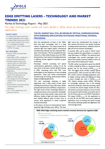
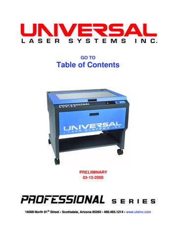
Chapter 1 System overviewCapSure caps and software compatibility1Using the ArcturusXT operating softwareTo start the software, you click the ArcturusXT icon onthe Windows desktop (see Figure 1). The system willdisplay the primary screen, which is shown in Figure 2.This application facilitates the microdissection workflow.Figure 1 ArcturusXT icon.The primary screenThe most prominent feature in the primary screen is the main image window thatshows the live microscope image. To the side of the screen is the tool panel, whichcontains tool panes arranged from top to bottom in the order of the steps for lasermicrodissection:1. Setup2. Inspect3. Select4. MicrodissectBy default, the main image is in the upper-left corner and the tool panel is on the right.You can move the tool panel to the left side by clicking Left-hand Orientation in theView menu.MainimageToolpanelCap and slide handling area, Slide overview image, and QC caps areaFigure 2 The ArcturusXT Instrument primary screen.ArcturusXT Laser Capture Microdissection System User Guide (CapSure LCM MicroCaps)13
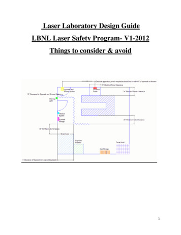
1Chapter 1 System overviewCapSure caps and software compatibilityAt the bottom of the screen are the cap and slide handling areas, the slide overviewimage, and the QC caps area. To move the stage to a particular region and to view thatregion in the main image, you click the slide overview image in that region. Theinformation displayed includes the properties of the currently selected object, such as aslide or a cap.Viewing tool tipsMost items in the ArcturusXT primaryscreen have a tool tip associated with them.The tool tips give you information about theitem.To view tool tips hover the stylus or mouseover the item of in
The ArcturusXT Laser Capture Microdissection (LCM) System provides an automated approach to laser microdissection of individual cells or multi-cellular structures from slides containing tissue sections or cytological samples. The ArcturusXT LCM System consists of the ArcturusXT Instrument, a computer, and the Arcturus XT operating .