Transcription
Circular RNA hsa circ 0006421 inhibitshepatocellular carcinoma by acting as a ceRNAtargeting miR-134-5p/CELF2 pathwayYibin Deng ( 1123049646@qq.com )Center for Clinical Laboratory Diagnosis and Research ;the A liated Hospital of Youjiang MedicalUniversity for NationalitiesLv ZhouYoujiang Medical University for NationalitiesXiaohao ChenYoujiang Medical University for NationalitiesJunxia PuYoujiang Medical University for NationalitiesJunhao ShiYoujiang Medical University for NationalitiesResearch ArticleKeywords: hsa circ 0006421, miR-134-5p, CELF2, hepatocellular carcinomaPosted Date: July 18th, 2022DOI: : This work is licensed under a Creative Commons Attribution 4.0 International License.Read Full License
Circular RNA hsa circ 0006421 inhibits hepatocellular carcinoma by acting as aceRNA targeting miR-134-5p/CELF2 pathwayLv Zhou 1, Xiaohao Chen 1, Junxia Pu 1, Junhao Shi 1and Yibin Deng 2*AbstractBackground: Hepatocellular carcinoma (HCC) has ranked sixth most common cancer innumber of malignancies in the worldwide. There is plenty of evidence indicated thatcircular RNAs (circRNAs) exert vitally important roles in the progression of manymalignancies. Nevertheless, the molecular mechanism and role of hsa circ 0006421 inHCC are still unclear. The aim of our research is to verify the molecular mechanism andeffects of hsa circ 0006421 in HCC.Methods: First, we collected 34 paired HCC tissues and paraneoplastic tissues surgicallyresected from patients and analyzed the expression of hsa circ 0006421 in HCC tissuesand its relationship with clinicopathological characteristics. Then, CCK8, colonyformation, cell migration assay, transwell invasion assay and Annexin-V/PI staining wereused to assess the effects of hsa circ 0006421 on the growth, migration, invasion andapoptosis of HCC cells. Next, quantitative real-time PCR (qRT-PCR) and Western blotswere used to detect the expression of miR-134-5p, CELF2 and hsa circ 0006421. In theend, the targeting interactions of miR-134-5p and hsa circ 0006421 , CELF2 andmiR-134-5p were explored by the dual-luciferase reporter assay.Results: Hsa circ 0006421 was diminished in HCC tissues and its down-regulation wasrelated to cirrhosis history. Knocking down hsa circ 0006421 promoted HCC is,whereasoverexpressing
hsa circ 0006421 had the opposite effects. Moreover, hsa circ 0006421 served as thecompeting endogenous RNA of miR-134-5p. Subsequently, there was a reciprocalrelationship between CELF2 and miR-134-5p. Hsa circ 0006421 could positivelyregulate the protein level of CELF2 in HCC.Conclusion: hsa circ 0006421 inhibits liver cancer by regulating miR-134-5p/CELF2axis.Key Words:hsa circ 0006421; miR-134-5p; CELF2; hepatocellular carcinomaIntroductionHepatocellular carcinoma (HCC) is an incurable disease with high morbidity andmortality rates [1]. According to GLOBOCAN cancer statistics, in 2020, HCC was thesixth most frequently diagnosed malignancy and the number of deaths occupied the thirdplace [2]. China accounts for 47.1% and 45.6% of new deaths and new cases of HCCworldwide, respectively, which seriously threatened the lives and health of Chineseresidents [3]. Therefore, promising biomarkers for early detection and prediction of HCCprognosis need to be identified. The up-regulation or down-regulation of non-codingRNAs (ncRNAs) is related to the pathogenesis of HCC [4]. Therefore, these ncRNAs havepotential to be predictive markers for HCC patients.CircRNAs , a new class of ncRNAs, were first identified in eukaryotic cells in 1979[5].Unfortunately,they received less attention and were deemed as by-products oferroneous splicing in the past [6]. Compared with linear RNAs, circRNAs are stable RNAsbecause of their closed loop structure [7]. The evidence is mounting that the dysregulationof circRNAs expression is related to multiple malignant tumors, such as cervix cancer [8],2
breast tumor [9, 10], colorectal cancer [11], lymphoma [12], GC [13] and HCC [14].Currently, growing evidence revealed that circRNAs could function as sponges ofmiRNAs or modulate RNA-binding proteins (RBPs). For example, hsa circ 0003410facilitates cell proliferation via the miR-139-3p/CCL5 in HCC [15]. Circular RNAcircGLIS3 plays an oncogenic role in bladder cancer through miR-1273f/SKP1/Cyclin D1axis [16].The differentially expressed circRNAs were selected by bioinformatics, afterconsulting the literature, we found that hsa circ 0006421 decelerates cell proliferation instomach cancer through miR-134-5p/CELF2 axis [17], but the biological effects ofhsa circ 0006421 in HCC remains elusive. Therefore, we finally chose hsa circ 0006421as the target, and then further proved whether hsa circ 0006421/miR-134-5p/CELF2pathway also has inhibitory effect in HCC.In this research, hsa circ 0006421 was diminished in HCC tissues and itsdown-regulation was related to cirrhosis history. Then, vitro experiments revealed thathsa circ 0006421 is an oncogene suppressor in HCC. Furthermore, the data demonstratedthathsa circ 134-5p/CELF2 axis. For these reasons, hsa circ 0006421/ miR-134-5p/CELF2 axisis expected to provide a basis for early HCC diagnosis.Materials and MethodsPatients and sample collectionA total of 34 fresh HCC tissue specimens and para cancer tissues were obtained frompatients who underwent surgery. This research was approved by the Medical EthicsCommittee of the Affiliated Hospital of Youjiang Medical University for Nationalities(protocol code: YYFY-LL-2022-13). All of the patients were informed consent. Samples3
receiving chemoradiotherapy prior to surgery before collections were excluded. Thesepatients were pathologically diagnosed as hepatocellular carcinoma and signed thein-formed consents. After surgical excision of the fresh tissues, they were deep frozen withliquid nitrogen and stored at 80ºC for subsequent use.Cell lines and culturesHepG2、MHCC97H cells and human embryotic kidney cells (HEK293T) werepurchased from Chinese Academy of Sciences (Beijing, China). All of the cells werecultured in DMEM high sugar liquid medium (Gibco, USA) supplemented with 10% fetalbovine serum (FBS, Beyotime, Nantong, China), in a 37 C cell incubator containing 5%CO2.Cell transfectionFor sh-hsa circ 0006421 (sh-circ) and its negative control transfection, lentiviraltransfection of HepG2 and MHCC97H cells with a multiplicity of infestation (MOI) of100. And then, purinomycin with concentration of 4g/mL was added to complete culturemedium to screen lentivirus stably transformed cell lines. For hsa circ 0006421overexpression (over-circ), hsa circ 0006421 cDNA was synthesized and cloned intopcDNA3.1. MiR-134-5p mimics, si-hsa circ 0006421 (si-circ) were obtained fromGenePharma Co., Ltd. (Suzhou, China). They are shown in Table 1. Lipofectamine 3000(Invitrogen, ThermoFisher, USA) was utilized to transfect.RNA extraction and qRT-PCR analysisTotal RNA in HCC specimens and cultured cells was extracted by using Trizolrea-gent (Invitrogen, USA). RNA samples were converted into cDNA by using theRevertAid Master Mix (Thermo Fisher Scientific, USA). For miRNA analysis, RT-PCR4
was completed using a miRNA First Strand cDNA Synthesis (Sangon Biotech, China).The synthesized cDNA was detected by qRT-PCR by using SYBR Green (Hieff ,Shanghai, China). The amplification reaction conditions were as follows: 95 C for 10 sand 60 C for 30 s. We used U6 and GAPDH as reference genes for data normalization.The primer sequences are showed in Table 1.Table 1 Sequence of small interfering RNA, short hairpin RNA, mimic, or polymerasechain reaction (PCR) primers.NameSequence (5’-3’)si-circ 0006421: Sense: 5‘AGAGGAGUGGAAGCAGAACTT3’Antisense: 5‘GUUCUGCUUCCACUCCUCUTT3’sh-circ 0006421: Sense: 5’-CAGAGGAGTGGAAGCAGAA-3’miR-134-5p mimic: Sense: 5’-UGUGACUGGUUGACCAGAGGGG-3’Antisense: 5’-CCUCUGGUCAACCAGUCACAUU-3’hsa circ 0006421: Forward: 5’-ACTGGCTTCACGTGGATATGG-3’Reverse: 5’-CCAGTTCTGCTTCCACTCCTC-3’miR-134-5p: Forward: 5’- CCTCTATTCTGTGACTGGTTGACC -3’Reverse: 5’- TATGGTTTTGACGACTGTGTGAT -3’CELF2: Forward: 5'-CTGGCGGGAAACAAACTCTG-3'Reverse: 5'-TCTAAGCCCTTGGCCTCCTC-3'GAPDH: Forward: 5’-CAGGAGGCATTGCTGATGAT-3’Reverse: 5’-GAAGGCTGGGGCTCATTT-3’U6: : 5’-ACGAATTTGCGTGTCATCC-3’Cell proliferation assayAbout three thousand HCC cells (HepG2、MHCC97H) were cultured in 96-wellplates. PBS was added to the other wells around the cells. Before detecting the OD value,we added 10 microliter of Cell Counting Kit-8 (CCK8) (MedChemExpress, China) to each5
well. After 2 hours, the absorbance value was detected by using an enzyme immunoassayanalyzer at 450 nm.Colony formation assayLentiviral stable transfected cell lines were uniformly inoculated with 1.7 103 cellsin a 6-well plate. DMEM medium containing 20% FBS was changed every three days. Onthe ninth day, visible colonies were formed, and cell culture was terminated. Carefully add4% paraformaldehyde along the inner wall to a 6-well plate and fix for 20 minutes, stainedby the crystal violet solution for about fifteen minutes. After washing, the numbers ofclones were then counted and analyzed.Cell migration assayHCC cells (HepG2 and MHCC97H) were seeded into 6-well plates and starved for24h. They were incubated in cell incubator until the cells reached about 95% confluence.Next, we draw several scratches in the cell using a clean 100 µl pipette tip. Finally, aftercultured for 12h and 24h, the moving distances of cells were measured and the cell healingrates were calculated.Transwell invasion assayAfter 24 hours of transfection, the HCC cells (HepG2 and MHCC97H) were digestedand detected by transwell invasion assay. The Matrigel (BD, USA) diluted 10 times withpre-cooled serum-free DMEM medium to obtain a suitable concentration of Matrigelsolution. We added 100μL of the solution to the upper chamber and incubated in theincubator for 1h. After the cells were washed with PBS, they were added to the serum-freemedium and mixed evenly to prepare a cell suspension of 2 105 cells/mL, 200 μL of cellsuspension was seeded into each well of the upper chamber. 600 μL of DMEM complete6
medium was supplied in the lower chamber. After cultured for 24h, the cells in the upperchamber were eliminated. The transmembrane cells were fixed for 15 minutes with 4%paraformaldehyde and 0.5% crystal violet was added to stain. The number of HCC cellinvasions was counted under a microscope in five random visual fields.Western blotAfter transfection for 48 hours, HCC cells were washed with pre-chilled PBS. Proteinsamples were obtained by the RIPA Lysis and Extraction Buffer (Solarbio, China). Theprotein concentrations were detected using a Pierce BCA kit (Beyotime, Shanghai, China).The cell lysates (30µg/lane) were electrophoresed on 10% SDS-PAGE (sodium dodecylsulphate-polyacrylamide gel electrophoresis) and then transferred onto polyvinylidenedifluoride (PVDF) membranes (Millipore, Billerica, MA, USA).After blocking with 5%skim milk for 2h, the membranes were incubated with anti-β-actin (1:3000; ab227387,Abcam), anti-CELF2 (1:3000, ab179447, Abcam) at 4 overnight. Subsequently, washedthese bands with TBST solution and secondary antibodies (1:5000; Invitrogen, USA) wereincubated for one hour.Cell apoptosis analysisAfter transfection, HCC cells (HepG2 and MHCC97H) were collected andcentrifuged for 3 mins with 800 rpm. After washing the HCC cells with pre-chilled PBS,they were stained with an Annexin V-FITC/ PI (Invitrogen, ThermoFisher). Finally, HCCcells were subjected to apoptosis and detected by using a flow cytometer (FACSCanto II,BD Biosciences).Luciferase reporter assayWe obtained the dual-luciferase plasmid from GenePharma Co., Ltd. (Suzhou, China).7
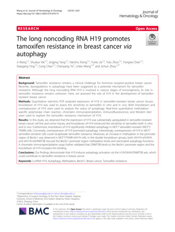
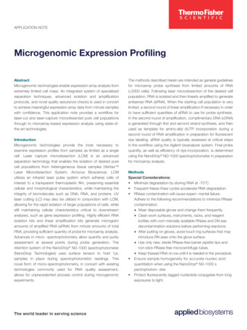
The sequences of hsa circ 0006421 and CELF2 3ʹUTR containing miR-134-5p bindingsites were inserted into the pmirGLO promoter vector. HEK293T cells were cultured in24-well plates overnight and were cotransfected with 1μg of WT or MUT plasmids and1.5μL of miR-134-5p mimics or miR-134-5p NC using the lipofectamine 3000transfection reagent. After 48 hours of transfection, the luciferase activity was quantifiedusing a dual-luciferase reporter assay kit (Promega, USA).Statistical analysisEach experiment was repeated triplicate. All values are expressed as means SD. Weperformed the statistical analysis using R 4.0.2 software. Wilcoxon pair test was used toanalyze the relative expression of hsa circ 0006421 in liver cancer and paracanceroustissue. Student’s t-tests were used to calculate statistical significance. Categorical datawere analyzed using Fisher’s exact test. p 0.05 was considered statistically significant.ResultsHsa circ 0006421 is abnormally low expressed in HCC tissuesWe collected 34 clinical specimens of hepatocellular carcinoma ,as shown byqRT-PCR, judged side by side with paracancerous tissues, hsa circ 0006421 wasdown-regulated in 22 cases and upregulated in 12 cases. After statistical analysis, theexpression level of hsa circ 0006421 was lower in hepatocellular carcinoma compared toparaneoplastic tissues (Fig. 1A). What's more, we analyzed the clinicopathologicalfeatures further of 34 patients and found that the hsa circ 0006421 expression wasassociated with cirrhosis history (p 0.042) (Table 2). To sum up, hsa circ 0006421 isabnormally low expressed in HCC and its low expression is a red flag for patients withcirrhosis, implying that hsa circ 0006421 may be an anti-oncogene in HCC.8
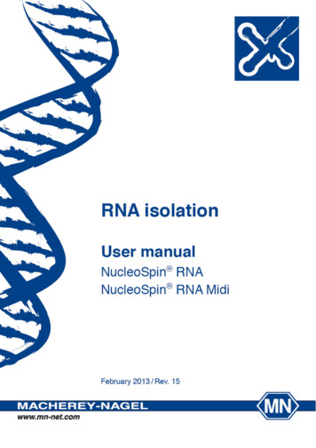
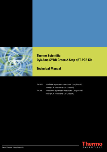
Figure1. Hsa circ 0006421 is lowly expressed in HCC tissues. (A)The expression of hsa circ 0006421 wasexamined in 34 HCC samplesandinhibitornormal tissues.*p 0.05, **p 0.01 and ***p 0.001.Hsa circ 0006421as anof HCCTo further verify the function of hsa circ 0006421 in HCC cells, we used small interferingRNA (si-circ) and lentiviral (sh-circ) prominently reduced the expression ofhsa circ 0006421 in HepG2 and MHCC97H cell lines. Moreover, the hsa circ sshsa circ 0006421.Thehsa circ 0006421 knockdown and overexpression efficiency in HepG2 and MHCC97Hcells were confirmed by qRT-PCR (Fig.2A). After successful construction ofhsa circ 0006421 knockdown and overexpression cell lines, CCK8 assay and cloneformation assay were used to verify cell viability. Migration assay and transwell invasionassay were used to detect cell migration and invasion, respectively. Apoptosis assay wasused to detect cell apoptosis. Results showed that the overexpression of hsa circ 0006421substantially suppressed cell viability (Fig. 2B, 2C), cell migration (Fig. 2D), cell invasion9
(Fig. 2E) and promoted apoptosis (Fig. 2F), whereas knocking down hsa circ 0006421had the opposite effects in HepG2 and MHCC97H cells. To sum up, We came to theconclusion that hsa circ 0006421 inhibits invasion and proliferation of hepatocellularcarcinoma cells.Table 2 Correlation between hsa circ 0006421 expression and clinicopathologicalcharacteristics in HCCCharacteristicsN 34Expression of hsa circ 0006421Low(n 22)High(n 12)Agep0.462 50y tiveNegativeCirrhosis historyPositiveNegativeTumor size 5cm 5cmAFP 20ng/mL 20ng/mLT classificationT3-T4T1-T2Tumor number 1166750.082113166570.6101123715.1048
Figure2. Knocking down hsa circ 0006421 promoted HCC cells growth, migration, invasion and inhibited apoptosis,whereas overexpressing hsa circ 0006421 had the opposite effects. (A)The expression of hsa circ 0006421 wasdetermined by qRT-PCR in hsa circ 0006421 overexpression or knockdown HCC cells. The effect ofhsa circ 0006421 on HCC cell proliferation was determined by CCK-8(B) and colony formation(C). (D)The effectof hsa circ 0006421 on HCC cell migration was evaluated by migration assay .(E) The effect of hsa circ 0006421 onHCC cell invasion was evaluated by transwell assays. (F) The apoptosis of HCC cells was measured by staining withAnnexinV/PI, followed by FACS analysis. The scale bars were 100 μm. Data were expressed as mean SD.11
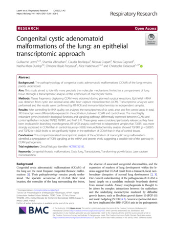
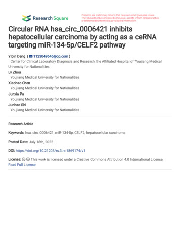
Hsa circ 0006421 inhibits HCC progression via miR-134-5p/CELF2 pathwayA new study reveals that hsa circ 0006421 inhibits stomach cancer throughregulating miR-134-5p/CELF2 pathway [17]. To investigate whether hsa circ 0006421could inhibit hepatocellular carcinoma through this pathway, we overexpressedhsa circ 0006421 in HepG2 and MHC97H cell lines, the result of qRT-PCR showed thatmiR-134-5p was significantly downregulated (Fig. 3A), implying that they werenegatively regulated. Then, the interaction relationship between hsa circ 0006421 andmiR-134-5p was further verified by luciferase reporter assay, the binding sites betweenmiR-134-5p and hsa circ 0006421 are shown in (Fig. 3B). Results showed that theluciferase intensities were reduced in miR-134-5p mimics and WT-hsa circ 0006421co-transfected group in HEK293T cell lines. However, the luciferase intensities ofMUT-hsa circ 0006421 were nearly unchanged (Fig. 3C). Thus, hsa circ 0006421 andmiR-134-5p have a targeting relationship in hepatocellular carcinoma.Next, we validated the targeting relationship between miR-134-5p and CELF2 inHCC and we found that the overexpression of miR-134-5p significantly reduced CELF2expression in HCC cells according to the qRT-PCR results (Fig. 3D). We demonstrated abinding target between CELF2 and miR-134-5p shown in Fig. 3E. Dual-luciferase assaysrevealed that the luciferase activity of WT CELF2 3’UTR reduced in miR-134-5p mimicsgroup, but the activity of the MUT CELF2 3’-UTR remained unchanged (Fig. 3F).Therefore, CELF2 and miR-134-5p have a targeting relationship in hepatocellularcarcinoma.Finally, we verified whether hsa circ 0006421 could positively regulate theexpression of CELF2. We used small interfering RNA and plasmid to knock down and12
overexpress hsa circ 0006421 in HepG2 and MHCC97H cells respectively, and thendetected the expression of CELF2 protein by Western blot. The results display thathsa circ 0006421 could significantly positively regulate the expression of CELF2 proteinin HCC cells (Fig. 3G). These data are sufficient to support our view thathsa circ 0006421 inhibits HCC progression via miR-134-5p/CELF2 pathway.Figure3. Hsa circ 0006421 inhibits HCC by regulating miR-134-5p/CELF2 axis. (A)The qRT-PCR results showedthat the overexpression of hsa circ 00006421 significantly reduced miR-134-5p expression in HepG2 cells andMHCC97H cells. (B) The potential binding site of hsa circ 0006421 and miR-134-5p.( C) Dual-luciferase assaysrevealed that the miR-134-5p mimics could reduce the luciferase activity of WT hsa circ 0006421 3’UTR. (D) TheqRT-PCR results showed that miR-134-5p could negatively regulate the expression of CELF2 in HepG2 cells andMHCC97H cells. (E) The potential binding site of miR-134-5p and CELF2. (F) Dual-luciferase assays revealed thatthe miR-134-5p mimics could reduce the luciferase activity of WT CELF2 3’UTR. (G) Western blot analysis showedthat hsa circ 00006421 could positively regulate the expression of CELF2 protein in HCC cells.13
DiscussionHepatocellular carcinoma has become one of the most refractory diseases in theworld , due to its complexity and high recurrence [18]. With the high burden ofhepatocellular carcinoma in China, it is an urgent thing to find specific biomarkers andmore effective treatments for the diagnosis and treatment of early stage HCC to reducepatient mortality and improve their prognosis [19]. HCC occurs because the genes of livercells have changed, and the normal regulation of hepatocyte proliferation and apoptosis islost [20].CircRNA is a non-coding RNAs that have attracted attention for their extensiveinvolvement in tumor development [21]. Many surveys show that the up-regulation anddown-regulation of circRNA expression were closely related to many types of incurablediseases. For example, hsa circ 0002232 suppresses malignant progression of coloncancer through inhibition of TGF-β/Smad signaling [22]. CircKDM4B inhibits way[23].ThecircROBO1/KLF5/FUS feedback loop promotes malignant progression and livermetastasis of breast cancer [24]. CircVAMP3 inhibits the progression of HCC bysuppressing c-Myc translation [25]. In the present study, our results indicated thathsa circ 0006421 is decreased in HCC tissues, and the down-regulation ofhsa circ sthathsa circ 0006421 is a tumor suppressor in hepatocellular carcinoma. Actually, wedemonstrated that hsa circ 0006421 overexpression decelerated the growth and migrationof HCC in vitro. Furthermore, we identified hsa circ 0006421 suppressed miR-134-5pexpression in HCC cells. In addition, we also proved their interaction by luciferase14
reporter assay. A previous study showed that high expression of miR-134-5p promotes therelapse of early-stage lung adenocarcinoma, leading to poor survival [26]. Other studiesalso indicated that miR-134-5p suppresses the aggressiveness and growth of AML cells[27]. However, the role of miR-134-5p in HCC remains elusive. In this work, we foundthat miR-134-5p is a potential cancer-promoting factor in HCC.CELF2 belongs to the CUGBP Elav-like family and is a RNA-binding protein [28].High expression of CELF2 means a better prognosis in various tumors [29]. For example,CELF2 inhibits the progression of breast cancer by downregulating NFATc1 [30]. CELF2inhibits ovarian cancer cell invasion by Stabilizing FAM198B [31]. LncRNA CRNDEpromotes hepatocellular carcinoma development by facilitating epigenetic repression ofCELF2 and LATS2 [32]. STYXL1 promotes hepatocellular carcinoma cell invasion bydownregulating CELF2 through the PI3K/Akt axis [33]. We found that miR-134-5pnegatively regulates CELF2 in HCC. Moreover, the dual-luciferase activity showed thatthe luciferase activity of WT CELF2 3’UTR reduced in miR-134-5p mimics group, whichmeans that there was a targeting relationship between CELF2 and miR-134-5p.Mean-while, overexpression of hsa circ 0006421 promotes the CELF2 protein levels inHCC cells, whereas knocking down hsa circ 0006421 has the opposite effects. For theserea-sons, hsa circ 00006421 could positively regulate the expression of CELF2 protein inHepG2 and MHCC97H cells. Last but not least, hsa circ 0006421 inhibits HCC byregu-lating miR-134-5p/CELF2 pathway.In conclusion,we first found that hsa circ 0006421 acts as an anti-oncogene in HCC,and its low expression may increase the possibility of canceration for patients withcirrhosis of liver. Our studies indicated hsa circ 0006421 inhibits to HCC progression via15
increasing CELF2 expression by sponging miR-134-5p, suggesting that hsa circ 0006421might be a promising target for HCC therapy.ConclusionsIn summary, our results indicate that hsa circ 0006421 inhibits hepatocellularcarcinoma by acting as a ceRNA targeting miR-134-5p/CELF2 pathway, which mayrepresent a vital strategy for inhibiting the proliferation of liver cancer.AcknowledgementsNot applicable.Author Contributionswriting—review and editing, project administration:Y.D.;data curation, writing—originaldraft preparation, formal analysis: L.Z.; validation, supervision: X.C.; methodology: J.P.;software: J.S.; All authors have read and agreed to the published version of the eclarationsInstitutional Review Board StatementThe study was conducted in accordance with the Declaration of Helsinki, and approved bythe Ethics Committee of the Affiliated Hospital of Youjiang Medical University forNationalities (protocol code: YYFY-LL-2022-13 and date of approval:27 June 2022 ).Consent for publicationNot applicable in the declarations section.16
Competing interestsThe authors declare that they have no competing interests.Author details1Youjiang Medical University for Nationalities, Baise, Guangxi 533000, China.2AffiliatedHospital of Youjiang Medical University for Nationalities, Baise, Guangxi 533000, China.References1.HUANG X Y, HUANG Z L, ZHANG P B, et al. CircRNA-100338 Is Associated With mTOR Signaling Pathwayand Poor Prognosis in Hepatocellular Carcinoma [J]. Front Oncol, 2019, 9: 392.2.FERLAY J, COLOMBET M, SOERJOMATARAM I, et al. Cancer statistics for the year 2020: An overview [J]. IntJ Cancer, 2021.3.SUNG H, FERLAY J, SIEGEL R L, et al. Global Cancer Statistics 2020: GLOBOCAN Estimates of Incidence andMortality Worldwide for 36 Cancers in 185 Countries [J]. CA Cancer J Clin, 2021, 71(3): 209-249.4.HUANG Z, ZHOU J K, PENG Y, et al.The role of long noncoding RNAs in hepatocellular carcinoma [J]. MolCancer, 2020, 19(1): 77.5.ZHOU H, ZHENG X D, LIN C M, et al. Advancement and properties of circular RNAs in prostate cancer: Anemerging and compelling frontier for discovering [J]. Int J Biol Sci, 2021, 17(2): 651-669.6.LIU C, LI N, DAI G, et al.A narrative review of circular RNAs as potential biomarkers and therapeutic targets forcardiovas-cular diseases [J]. Ann Transl Med, 2021, 9(7): 578.7.SHEN H, LIU B, XU J, et al. Circular RNAs: characteristics, biogenesis, mechanisms and functions in liver cancer[J]. J Hema-tol Oncol, 2021, 14(1): 134.8.SONG T F, XU A L, CHEN X H, et al. Circular RNA circRNA 101996 promoted cervical cancer development byregulating miR-1236-3p/TRIM37 axis[J]. Kaohsiung J Med Sci, 2021, 37(7): 547-561.17
9.ZHENG X, HUANG M, XING L, et al. The circRNA circSEPT9 mediated by E2F1 and EIF4A3 facilitates thecarcinogenesis and development of triple-negative breast cancer [J]. Mol Cancer, 2020, 19(1): 73.10. LI J, MA M, YANG X, et al. Circular HER2 RNA positive triple negative breast cancer is sensitive toPertuzumab[J]. Mol Cancer, 2020, 19(1): 142.11. XU H, LIU Y, CHENG P, et al. CircRNA 0000392 promotes colorectal cancer progression through themiR-193a-5p/PIK3R3/AKT axis [J]. J Exp Clin Cancer Res, 2020, 39(1): 283.12. ZHAO C X, YAN Z X, WEN J J, et al. CircEAF2 counteracts Epstein-Barr virus-positive diffuse large B-celllymphoma pro-gression via miR-BART19-3p/APC/beta-catenin axis [J]. Mol Cancer, 2021, 20(1): 153.13. WANG G, SUN D, LI W, et al. CircRNA 100290 promotes GC cell proliferation and invasion via themiR-29b-3p/ITGA11 axis and is regulated by EIF4A3[J]. Cancer Cell Int, 2021, 21(1): 324.14. XU J, JI L, LIANG Y, et al. CircRNA-SORE mediates sorafenib resistance in hepatocellular carcinoma bystabilizing YBX1 [J]. Signal Transduct Target Ther, 2020, 5(1): 298.15. CAO P, MA B, SUN D, et al. hsa circ 0003410 promotes hepatocellular carcinoma progression by increasing theratio of M2/M1 macrophages through the miR-139-3p/CCL5 axis [J]. Cancer Sci, 2022, 113(2): 634-647.16. WU S, YANG J, XU H, et al. Circular RNA circGLIS3 promotes bladder cancer proliferation via themiR-1273f/SKP1/Cyclin D1 axis[J]. Cell Biol Toxicol, 2022, 38(1): 129-146.17. FAN H N, ZHAO X Y, LIANG R, et al. CircPTK2 inhibits the tumorigenesis and metastasis of gastric cancer bysponging miR-134-5p and activating CELF2/PTEN signaling [J]. Pathol Res Pract, 2021, 227: 153615.18. LIU D, LIU W, CHEN X, et al. circKCNN2 suppresses the recurrence of hepatocellular carcinoma at least partiallyvia regu-lating miR-520c-3p/methyl-DNA-binding domain protein 2 axis [J]. Clin Transl Med, 2022, 12(1): e662.19. LUO Z, LU L, TANG Q, et al. CircCAMSAP1 promotes hepatocellular carcinoma progression throughmiR-1294/GRAMD1A pathway [J]. J Cell Mol Med, 2021, 25(8): 3793-3802.20. ZHANG P F, WEI C Y, HUANG X Y, et al. Circular RNA circTRIM33-12 acts as the sponge of MicroRNA-191 to18
suppress hepatocellular carcinoma progression[J]. Mol Cancer, 2019, 18(1): 105.21. WANG Z, LEI X, WU F X. Identifying Cancer-Specific circRNA-RBP Binding Sites Based on Deep Learning[J].Molecules,2019, 24(22).22. ZHENG L, LIANG H, ZHANG Q, et al. circPTEN1, a circular RNA generated from PTEN, suppresses cancerprogression through inhibition of TGF-beta/Smad signaling [J]. Mol Cancer, 2022, 21(1): 41.23. GUO X Y, LIU T T, ZHU W J, et al. CircKDM4B suppresses breast cancer progression via the miR-675/NEDD4Laxis[J]. Onco-gene, 2022.24. WANG Z, YANG L, WU P, et al. The circROBO1/KLF5/FUS feedback loop regulates the liver metastasis of breastcancer by inhibiting the selective autophagy of afadin[J]. Mol Cancer, 2022, 21(1): 29.25. CHEN S, CAO X, ZHANG J, et al. circVAMP3 Drives CAPRIN1 Phase Separation and Inhibits HepatocellularCarcinoma by Suppressing c-Myc Translation [J]. Adv Sci (Weinh), 2022: e2103817.26. ZHANG L, HUANG P, LI Q, et al. miR-134-5p Promotes Stage I Lung Adenocarcinoma Metastasis andChemoresistance by Targeting DAB2 [J]. Mol Ther Nucleic Acids, 2019,18: 627-637.27. LIU Y, CHEN X, LIU J, et al. Circular RNA circ 0004277 Inhibits Acute Myeloid Leukemia Progression ThroughMi-croRNA-134-5p / Single stranded DNA binding protein 2[J]. Bioengineered, 2022, 13(4): 9662-9673.28. LIAO C, CHEN W, WANG J. MicroRNA-20a Regulates Glioma Cell Proliferation, Invasion, and Apoptosis byTargeting CUGBP Elav-Like Family Member 2[J]. World Neurosurg, 2019, 121: e519-e527.29. WANG L, LIU Z, LIU L, et al. CELF2 is a candidate prognostic and immunotherapy biomarker in triple-negativebreast cancer and lung squamous cell carcinoma: A pan-cancer analysis [J]. J Cell Mol Med, 2021, 25(15): 7559-7574.30. ZHOU L, XIE X. RNA-binding protein CELF2 inhibits breast cancer cell invasion and angiogenesis bydownregulating NFATc1 [J]. Exp Ther Med, 2021, 22(2): 898.31. GUO Q, WU Y, GUO X, et al. The RNA-Binding Protein CELF2 Inhibits Ovarian Cancer Progression byStabilizing FAM198B [J]. Mol Ther Nucleic Acids, 2021, 23: 169-184.19
32. XIE S C, ZHANG J Q, JIANG X L, et al. LncRNA CRNDE facilitates epigenetic suppression of CELF2 andLATS2 to promote proliferation, migration and chemoresistance in hepatocellular carcinoma[J]. Cell Death Dis, 2020,11(8): 676.33. WU J Z, JIANG N
RNA samples were converted into cDNA by using the RevertAid Master Mix (Thermo Fisher Scientific, USA). For miRNA analysis, RT-PCR . 5 was completed using a miRNA First Strand cDNA Synthesis (Sangon Biotech, China). The synthesized cDNA was detected by qRT-PCR by using SYBR Green (Hieff , . we added 10 microliter of Cell Counting Kit-8 (CCK8 .