Transcription
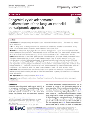
Lezmi et al. Respiratory Research(2020) ARCHOpen AccessCongenital cystic adenomatoidmalformations of the lung: an epithelialtranscriptomic approachGuillaume Lezmi1,2,3, Shamila Vibhushan2, Claudia Bevilaqua4, Nicolas Crapart4, Nicolas Cagnard5,Naziha Khen-Dunlop3,6, Christine Boyle-Freyssaut5, Alice Hadchouel1,2,3 and Christophe Delacourt1,2,3*AbstractBackground: The pathophysiology of congenital cystic adenomatoid malformations (CCAM) of the lung remainspoorly understood.Aim: This study aimed to identify more precisely the molecular mechanisms limited to a compartment of lungtissue, through a transcriptomic analysis of the epithelium of macrocystic forms.Methods: Tissue fragments displaying CCAM were obtained during planned surgical resections. Epithelial mRNAwas obtained from cystic and normal areas after laser capture microdissection (LCM). Transcriptomic analyses wereperformed and the results were confirmed by RT-PCR and immunohistochemistry in independent samples.Results: After controlling for RNA quality, we analysed the transcriptomes of six cystic areas and five control areas. In total,393 transcripts were differentially expressed in the epithelium, between CCAM and control areas. The most highlyredundant genes involved in biological functions and signalling pathways differentially expressed between CCAM andcontrol epithelium included TGFB2, TGFBR1, and MAP 2 K1. These genes were considered particularly relevant as they havebeen implicated in branching morphogenesis. RT-qPCR analysis confirmed in independent samples that TGFBR1 was morestrongly expressed in CCAM than in control tissues (p 0.03). Immunohistochemistry analysis showed TGFBR1 (p 0.0007)and TGFB2 (p 0.02) levels to be significantly higher in the epithelium of CCAM than in that of control tissues.Conclusions: This compartmentalised transcriptomic analysis of the epithelium of macrocystic lung malformationsidentified a dysregulation of TGFB signalling at the mRNA and protein levels, suggesting a possible role of this pathway inCCAM pathogenesis.Trial registration: ClinicalTrials.gov Identifier: NCT01732185.Keywords: Congenital thoracic malformations, Cystic lung, Transcriptome, Transforming growth factor, Laser capturemicrodissectionBackgroundCongenital cystic adenomatoid malformations (CCAM) ofthe lung are the most frequent congenital thoracic malformations [1]. Their pathophysiology remains poorly understood. The sporadic occurrence of CCAM, their focallocation, the normality of the lung surrounding the lesion,* Correspondence: christophe.delacourt@aphp.fr1Service de Pneumologie et d’Allergologie Pédiatriques, AP-HP, HôpitalUniversitaire Necker-Enfants Malades, 75743 Cedex 15 Paris, France2INSERM, U955, Institut Mondor de Recherche Biomedicale (IMRB), Equipe 4,94000 Créteil, FranceFull list of author information is available at the end of the articlethe absence of associated congenital abnormalities, and theexpression of markers of lung development within the lesions suggest that CCAM result from a transient, focal, nonhereditary disruption of normal lung development [2, 3].Our current understanding of the pathogenesis of CCAM isbased largely on a candidate molecule hypothesis derivedfrom animal models. Airway morphogenesis is thought tobe driven by complex interactions between the epitheliumand the underlying mesenchyme mediated by diffusiblegrowth factors, such as fibroblast growth factor 10 (FGF-10)and sonic hedgehog (SHH) [4, 5]. Several experimental studies have implicated the SHH-FGF10 axis in the pathogenesis The Author(s). 2020 Open Access This article is distributed under the terms of the Creative Commons Attribution 4.0International License (http://creativecommons.org/licenses/by/4.0/), which permits unrestricted use, distribution, andreproduction in any medium, provided you give appropriate credit to the original author(s) and the source, provide a link tothe Creative Commons license, and indicate if changes were made. The Creative Commons Public Domain Dedication o/1.0/) applies to the data made available in this article, unless otherwise stated.
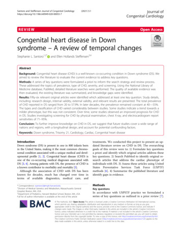
Lezmi et al. Respiratory Research(2020) 21:43of cystic abnormalities during lung development [3]. FGF10and its receptor, FGFR2b, are strongly expressed in the mesenchyme and epithelium of human CCAM, differentiatingthese abnormalities from other cystic abnormalities, such astype 1 pleuropneumoblastoma [6]. However, the levels ofexpression of these molecules do not differ between CCAMand control tissues [6]. A recent analysis of the human congenital lung lesion transcriptome revealed an upregulationof epithelium-related genes, and a dysregulation of the expression of genes related to the Ras and PI3K-AKT-mTORsignalling pathways [7]. Neither FGF10 nor FGFR2b wasfound to be dysregulated. However, the use of whole lungsin this study may have biased gene identification againstgenes with expression limited to a particular compartmentof the lung parenchyma. Furthermore, only two macrocysticlesion transcriptomes were analysed. These lesions appearedto cluster separately from microcystic and hybrid lesions,and were excluded from the analysis. It this study, we performed a transcriptomic analysis of the epithelium ofmacrocystic forms. The tissues were isolated by laser capturemicrodissection, and the transcriptomes of the epithelialcells in cystic zones and adjacent healthy tissue areas wereanalysed. Our results suggested a potential role for TGFbeta signalling. We validated these results by PCR and immunohistochemistry, which confirmed the differences in expression of these molecules between zones of malformationand healthy tissue.Materials and methodsThis study was approved by the institutional ethics committee (Comité de Protection des Personnes-Ile de FranceVII (N 12–025)), and by the French national drug safetyagency, the Agence Nationale de Sécurité du Médicament (B120601–10). Written informed consent was obtained from the parents of the patients.Patient selectionChildren with congenital cystic pulmonary malformationwere recruited during follow-up visits at the PaediatricPulmonology Department of Necker Hospital. Tissue fragments were collected prospectively during programmedsurgical removal of the malformation, usually at about 6months of age. The diagnosis of CCAM was confirmedafter surgical resection, by histological analysis, andCCAM were classified according to the Stocker classification [8]. Non-cystic pulmonary malformations or hybridlesions were excluded from the study. Children who hadan infection of their malformation before surgery werealso excluded. Infection was defined by fever and a newradiological infiltrate at the location of the malformation.Tissue acquisitionAfter resection, the explant was immediately cooled fora few seconds in PBS buffer at 4 C to reduce the activityPage 2 of 10of endogenous RNAases. Several fragments were rapidlycollected on ice, placed in cryomolds and cryotubes andimmersed in liquid nitrogen for storage at 80 C. Thesefragments were used for laser microdissection and RTqPCR analysis. For each CCAM, we collected tissuesfrom two areas, defined on the basis of their macroscopic appearance: a cystic area and an area of normalappearance. The macroscopically normal tissue was collected from the area surrounding the lesion, and wasused as a control if this normal appearance was alsoconfirmed microscopically (see below). The remainderof the explant was used for histological analysis by apathologist to confirm the diagnosis of CCAM.Laser capture microdissectionWe cut 8 μm-thick sections, which were then stainedwith cresyl violet for 30 s, rinsed in 95% ethanol anddehydrated in 100% ethanol. The sections wereimmersed in xylene to eliminate the ethanol, dried inroom air and placed in a vacuum chamber until use.The sections were then microdissected with a VERITASlaser dissecting microscope (Arcturus, Life Technologies) at the ICE core facility (INRA, Jouy-en-Josas).Slides were viewed at 20 or 40 magnification, andthe epithelium was delineated on both the cystic andcontrol slides. Normal and abnormal structures are difficult to differentiate in CCAM. This first step was therefore also used to confirm that the macroscopicallynormal tissues did not contain any abnormal structures.The epithelium tissues from cystic and normal areaswere then dissected out and collected (Fig. 1). They wereimmediately placed in RNA extraction buffer (ArcturusPicopure RNA isolation kit) and stored at 80 C untilextraction. Total RNA was isolated with the ArcturusPicopure kit (Life Technologies), according to the manufacturer’s instructions.Assessment of the quality of the extracted RNARNA quality was assessed with an Agilent Model 2100Bioanalyser (Agilent Technologies). An RNA integritynumber (RIN) greater than 7 was considered acceptablefor the analysis. The quality of the RNA extracted fromthe first cut (control block) was used to assess the quality of the RNA in the work piece. The RIN of the RNAextracted from the control blocks ranged from 7.5 to8.5, reflecting partial degradation of the RNA during thesurgical procedure. As recommended [9], Agilent analysis was also systematically performed on the RNA obtained from the tissue remaining on the slides aftermicrodissection (control slide), to assess the degradationof the RNA by the procedure. For small amounts ofextracted RNA ( 150 μg/μl), the RIN obtained was artificially low. In such cases, if the RIN on the
Lezmi et al. Respiratory Research(2020) 21:43Page 3 of 10Fig. 1 Laser capture microdissection of epithelial samplescorresponding control slide exceeded 7, the sample wasconsidered to be of sufficient quality for further analysis.There is no broad consensus RIN threshold for the inclusion of samples in transcriptomic analyses, but RINscores higher than 6 were required for the GenotypeTissue Expression (GTEx) project anscriptomic analysisAn expression analysis was performed with the GeneChip Human Exon 1.0 ST Array (Affymetrix), investigated more than one million exon clusters within theknown and predicted transcribed regions of the entiregenome (the genomics core facility of the Imagine Institute, Paris). Two kits from NuGEN (NuGEN Technologies) were used for the amplification and postamplification treatment of samples before hybridisation.Using 200 pg of total RNA as the starting material, wesubjected samples to Ribo-SPIA RNA Amplification(SPIA, for single primer isothermal amplification). Theamplicons obtained were fragmented and labelled withbiotin, with the One-Direct System and the Encore Biotin Module, as recommended by the manufacturer. Thebiotinylated DNA fragments obtained were hybridisedwith the Human Exon 1.0 ST Array, with theHybridization Wash Stain kit (Affymetrix). The arrayswere washed, stained, and scanned with an AffymetrixModel 450 Fluidics Station, an Affymetrix Model 3000scanner and Command Console software, which pilotedthe GeneChip systems.Fluorescence data were imported into two softwaresuites for analysis: Affymetrix Expression Console andR Bioconductor. Gene expression was evaluated by calculating the RMA algorithm in Expression Console, andflags were computed with a custom algorithm in R. Assuming that no more than 80% of genes are expressed,we selected the lowest 20% of values obtained for eachmicroarray for use as the background. The thresholdwas fixed at two standard deviations above the mean ofthe background values. All probes with normalised intensity values below this threshold were flagged as 0rather than 1. Lists were created by filtering for probesflagged as “Present” (flag 1) for at least half the chips.Groups were compared in Student’s t tests. False discovery rates were estimated by filtering the resulting pvalues at 5%. Hierarchical clustering was performed withSpearman’s rho valus and the average link algorithm.Data were subjected to Ingenuity Pathway Analysis(IPA) for the modelling of networks, to identify the relevant pathways and biological processes. The filters pvalue 0.05 and a 1.2-fold change in probe expressionlevel were used to identify transcripts differentiallyexpressed in the epithelium, between CCAM and controlareas. Gene-set enrichment analysis was performed withIngenuity software.RT-qPCR analysisFor confirmation of the results of transcriptomic analysis,we used RT-PCR on small amounts of preamplified cDNA(QuantiTect Whole Transcriptome, Qiagen) to assessgene expression levels. We used the following primers:TGFb2 (NM 003238.3) F: ATGCCAACTTCTGTGCTGGA R: AAATCTTGGGACACGCAGCA, TGFbR1(NM 004612,3) F: CTCCAAACCACAGAGTGGGAA R:ATCATCGAGAACTTCAGGGGC, MAP 2 K1 (NM002755,3) F: CGCCCATCCAGCTGAACC, R: TCATCAAGCTCTAGCTCCTCCA.ImmunohistochemistryAfter resection, the tissues were embedded in paraffinand 5 μm sections were cut for immunohistochemistry.Sections were processed as previously described [6], andincubated with the primary antibodies for TGFb2 (1:200,PA5–32629, Thermo Fisher Scientific Inc., Waltham,MA), TGFbR1 (abcam, ab31013, 1:200); MEK1/MAP 2K1, Thermo Fisher Scientific, MA1–095, 1:10000) andwith isotype control rabbit IgG (02–6102, Life Technologies, Carlsbad, USA) or mouse IgG, (Dako, X0931), with1.5% normal horse serum (PK-7800, RTU Vectastain,Vector Laboratories, Burlingame, USA) in PBS. For eachpatient, and for each immunostaining reaction, wephotographed 10 zones of epithelium selected at
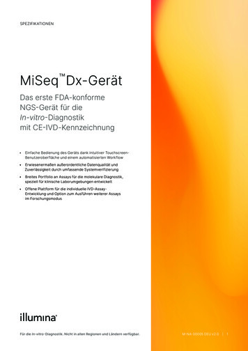
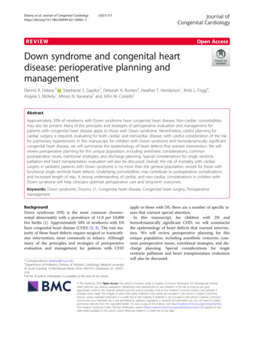
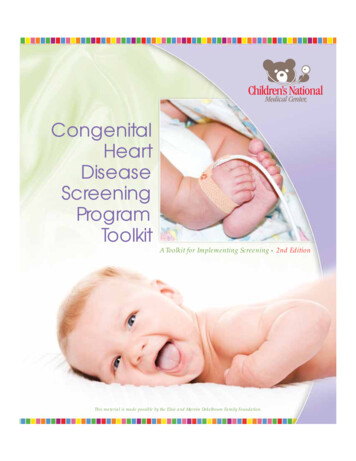
Lezmi et al. Respiratory Research(2020) 21:43random, at a magnification of 20. Analyses were performed with Image J analysis software (http://rsb.info.nih.gov/ij/). Immunostaining in epithelial cells (brown)and nucleus counterstaining (purple) signals were separated and converted into greyscale intensities with theColour Deconvolution plugin of Image J software. Twoof the authors (GL and SV) analysed the staining blindand independently of each other. The ratio of immunostaining to counterstaining intensities was calculated foreach Image.AnalysisThe values obtained in RT-PCR and immunostaininganalyses were compared in Mann-Whitney tests, with pvalues 0.05 considered significant.ResultsTranscriptomic analysisFollowing LCM, RNA of sufficient quality was isolatedfrom 10 patients (Additional file 1: Table S1). For onepatient, mRNA was obtained from both cystic and control areas. For five patients, mRNA was obtained exclusively for the cystic area, and, for another four patients,mRNA was obtained only from the control area. In total,we analysed six cystic areas and five control areas. Themedian age of the six patients for whom cystic areas (3type I, 3 type II CCAM) were analysed was 5.7 months(range: 4.2–7.8 months), and did not differ from that ofthe patients in whom control areas were analysed (8months (range: 4.2–13.8 months)), p 0.88.We observed marked differences in the global profileof epithelial transcripts between CCAM and controlareas (Fig. 2). Using Ingenuity software with filtering ona p value 0.05 and a 1.2-fold difference in probe expression, we identified 393 genes as differentially expressedin the epithelium, between CCAM and control areas:203 were downregulated (Additional file 1: Table S2),and 190 upregulated (Additional file 1: Table S3). Geneontology analysis of the genes differentially expressedbetween CCAM epithelium and control epitheliumshowed that several of the significantly dysregulatedgenes were related to cancer and developmental processes (Fig. 3). Gene-set enrichment analysis identifiedgroups of genes that were significantly enriched ordepleted as belonging to signalling pathways (Fig. 4,Additional file 1: Table S4). To select candidate genesfor confirmation by qPCR and immunohistochemistry,we considered redundant genes within the 20 signallingpathways differing most significantly between CCAMand controls (Fig. 4). Ninety seven genes were identifiedwithin these 20 signalling pathways (Additional file 1:Table S4). Only 6 genes were highly redundant: PLCD4(11 pathways), PRKCA (11 pathways), MAP 2 K1 (10pathways), PLCE1 (10 pathways), TGB2 (10 pathways),Page 4 of 10and TGFBR1 (9 pathways). We selected TGFB2,TGFBR1, and MAP 2 K1 for further analysis, becausethese genes had already been implicated in branchingmorphogenesis [10, 11]. Predicted upstream pathwayanalysis identified genes involved in lung development,such as FGF1, WNT11, CEBPA, CREB1, and HIF1A, aspotential upstream regulators of groups of genes including TGFB2, TGFBR1 or MAP 2 K1 (Additional file 1:Table S5).Confirmation of the dysregulated expression of candidategenes in independent samplesWe then analysed the levels of mRNA and protein forTGFB2, TGFBR1 and MAP 2 K1 in tissue samples from17 new patients. The RT-qPCR data for each gene werenormalised against the levels of 18S rRNA, beta 2 microglobulin and RPLP0. TGFBR1 mRNA levels were significantly higher in CCAM than in control tissues (p 0.03),whereas no significant difference was found in the levelsof mRNA for TGFB2 or MAPK21 (Fig. 5a).Immunohistochemistry showed the levels of TGFBR1(p 0.0007) and TGFB2 (p 0.02) in the epithelium tobe significantly higher in CCAM than in control tissues.No difference in MAP 2 K1 protein levels was observedbetween CCAM and control tissues (Fig. 5b). TGFB2and TGFBR1 were expressed in a diffuse mannerthroughout the epithelium of CCAM tissues, but wereexpressed only sparsely in the control epithelium (Fig. 6).No expression of TGFB2 was observed in the mesenchyme. A low expression of TGFBR1 was observed inthe mesenchyme, with no difference between controlsand CCAM.DiscussionWe used a laser microdissection-based protocol to separate out CCAM and control areas of the epithelium. Wethen demonstrated differences in the gene expression profiles of the CCAM and control tissues. TGFB2 andTGFBR1 were among the genes identified as belonging tothe epithelial biological processes and signalling pathwaysdiffering most significantly between CCAM and controltissues. The overexpression of these genes was confirmedin analyses of CCAM and control samples from additionalpatients. This study therefore provides the first evidencefor a role of TGF in the pathophysiology of congenitalmacrocystic lung malformations.During lung development, the epithelium and surrounding mesenchyme display specific patterns of geneexpression [12]. Analyses on whole-tissue homogenatesmay therefore be inconclusive or misleading, as the contribution of a specific tissue compartment may bemasked. In addition, the term “CCAM” covers a numberof different malformation phenotypes, including microcystic and macrocystic forms, with or without systemic
Lezmi et al. Respiratory Research(2020) 21:43Page 5 of 10Fig. 2 Unbiased clustering of genes based on their levels of expression in microarray analyses of epithelial samples from six cystic and fivecontrol areas. Samples CCAM 2 E, CCAM 6 E and CCAM 19 E are type I CCAM; samples CCAM 5 E, CCAM 14 E and CCAM 16 E are type IICCAM. The cluster correlation dendrogram showed a clear separation of CCAM samples from control samples. No clear clustering differentiatedtype I CCAM from type II CCAMvascularisation, and it remains unknown whether thesedifferent phenotypes share the same molecular dysfunctions. We aimed to homogenise our analysis as much aspossible, by focusing on a single compartment (theepithelium) in a single malformation phenotype (macrocystic forms). LCM was used for the epithelium-specificmolecular profiling of areas of malformation and adjacent healthy zones. The LCM procedure itself and the
Lezmi et al. Respiratory Research(2020) 21:43Page 6 of 10Fig. 3 Gene ontology analysis of genes differentially expressed between CCAM epithelium and control epithelium. TGFB2 was identified in 17 ofthe top 20 biological processes, and TGFbR1 in 12 of the top 20 biological processesanalysis of microdissected material can be challenging,particularly due to the small amounts of nucleic acidsisolated and the loss of RNA due to degradation duringthe process. With the most recent guidelines for LCM,we were able to obtain sufficient amounts of RNA withlimited degradation [9, 13].Our results indicate significant differences in geneexpression between the epithelia of CCAM andhealthy tissues. Dysregulated lung epithelium was recently suggested to play a key role in branching morphogenesis in congenital lung malformations, in astudy on whole lung tissues corresponding to different malformation phenotypes [7]. Our findings confirm that epithelial dysfunction makes a significantcontribution to the pathogenesis of pulmonary malformations. Furthermore, our findings identify TGB2and TGFBR1 as key molecules in epithelial pathwaydysregulation, and confirm their overexpression inadditional samples. These findings are consistent witha causal role for TGFβ signalling in CCAM. TheTGFβ signalling pathway is involved in various biological processes, including development and extracellular matrix production, and is crucial for epithelial-mesenchymal interactions during lung branching morphogenesis and alveolarisation [11, 14]. A dimericform of activated TGFβ binds to the transmembraneTGFBR1 and TGFBR2 receptors [11]. TGFBR2 phosphorylates TGFBR1, which activates the canonicalSmad-dependent pathway [11, 15]. During airwaymorphogenesis, different expression patterns havebeen described for the different isoforms of TGFB,TGFB2 being localised in the distal epithelium,whereas TGFB1 and TGFBR3 are expressed in themesenchyme [16]. Several experimental studies haveshown TGF signalling to be crucial for lung branching [11]. Branching morphogenesis is disrupted bylung mesenchymal knockouts of the TGFB1 gene[17]. More specifically, TGFB2-knockout mice developa number of specific developmental defects, includingcollapsed distal airways [18]. The epithelium-specificdeletion of TGFBR1 in mouse lungs leads to an enlargement of the airways, with disorganisation of theepithelium [19]. In an organ culture model, TGFB2was found to inhibit lung explant branching 50 timesmore potently than TGFB1 [20]. Based on theseexperimental data, our results suggest that the
Lezmi et al. Respiratory Research(2020) 21:43Page 7 of 10Fig. 4 Gene-set enrichment analysis of genes differentially expressed between cystic and control areas of epithelium. TGFB2 was identified in 10of the top 20 signalling pathways, and TGFbR1 in 9 of the top 20 signalling pathwaysdysregulation of TGF signalling in the pulmonary epithelium plays a direct role in CCAM pathogenesis.However, one limitation of our study, as in all humanstudies on these malformations, was the postnatal collection of the tissues, at a time point well after thetriggering molecular event in the pseudoglandularphase of development. The observed signalling anomalies may, therefore, be no more than a consequenceof the initial molecular dysregulation, rather than theactual triggering event.The hypothesis of a direct role for TGF signalling inthe pathogenesis of CCAM is also supported by our upstream transcriptomic study, which identified several developmental genes as potential upstream regulators ofgroups of genes including TGFB2 or TGFBR1 alreadyshown to be involved in the branching process. CEBPAis crucial for the proximo-distal differentiation of thepulmonary epithelium [21]. The relationship betweenTGFβ signalling and CEBPA-mediated transcriptionalregulation has already been demonstrated during liverdevelopment [22]. HIF1A plays a major role in early pulmonary vasculogenesis, and its signalling is amplifiedthrough interactions between the mammalian target ofrapamycin (mTOR) andFGF-10 signaling [23], both ofwhich are dependent on TGF for activation [17, 24].WNT11 has been shown to interact directly with TGFsignalling to induce the production of smooth musclealpha-actin [25]. FGF1 is expressed very early in themouse lung epithelium, during branching, and has beenshown to induce branching morphogenesis in Matrigel-
Lezmi et al. Respiratory Research(2020) 21:43Page 8 of 10Fig. 5 RT-PCR (a) and immunostaining (b) results for TGFb2, TGFbR1 and MAP 2 K1 in control and CCAM samples. TGFB2: 5 controls vs. 17 CCAMsamples and 9 controls vs. 9 CCAM samples for RT-PCR and immunostaining, respectively; TGFBR1: 4 controls vs. 11 CCAM samples and 13controls vs. 13 CCAM samples for RT-PCR and immunostaining, respectively; MAP 2 K1: 4 controls vs. 17 CCAM samples and 13 controls vs. 11CCAM samples for RT-PCR and immunostaining, respectively. * p 0.05; ***p 0.001Fig. 6 Immunostaining for TGFB2 (A, A', B, B') and TGFBR1 (C, C', D, D') in CCAM (B, B', D, D') and control samples (A, A', C, C') , at magnificationsof 20 (A, C, B, D) and 40 (A', B', C', D'). Diffuse expression is observed in CCAM epithelium
Lezmi et al. Respiratory Research(2020) 21:43embedded E11 epithelium [26]. FGF1 has also beenshown to interact with TGF-driven fibroblast-tomyofibroblast transdifferentiation [27].In conclusion, this transcriptomic analysis of the epithelium of CCAM tissues reveals a dysregulation ofTGFB signalling at the mRNA and protein levels. Thesedata are consistent with previous studies demonstratingthe involvement of TGFB signalling in airway morphogenesis, and thus identify a possible role for this signalling pathway in CCAM pathogenesis.Supplementary informationSupplementary information accompanies this paper at al file 1: Table S1. RIN and mRNA concentrations fromsamples used for transcriptome analysis. Table S2. List of downregulatedgenes in CCAM epithelium compared to control zones. These genes wereselected using Ingenuity software with a filter p value 0.05 and a 1.2-foldalteration of probe expression. Table S3. List of upregulated genes in CCAMepithelium compared to control zones. These genes were selected usingIngenuity software with a filter p value 0.05 and a 1.2-fold alteration of probeexpression. Table S4. Pathway analysis. Table S5. Upstream analysis.AbbreviationsCCAM: Congenital cystic adenomatoid malformation; FGF: Fibroblast GrowthFactor; LCM: Laser capture microdissection; SHH: Sonic hedgehog;TGF: Transforming Growth FactorAcknowledgementsWe thank the Unité de Recherche Clinique (URC) from Necker Hospital for itssupport.Authors’ contributionsEach author significantly contributed to the conception (GL, CD), to thedesign (GL, CB-F, AH, CD), to the acquisition, analysis, and interpretation ofdata (GL, SV, CB, NC, NG, NK-D, CB-F, CD), to the draft of the work (GL, CD)and to its revision (All authors). All authors have approved the submitted version. All authors agree both to be personally accountable for the author’sown contributions and to ensure that questions related to the accuracy orintegrity of any part of the work, even ones in which the author was not personally involved, are appropriately investigated, resolved, and the resolutiondocumented in the literature.FundingThis study was funded by APHP-Programme Hospitalier de Recherche Clinique (PHRC). 2012-A00538–35- Funding was obtained for collection and analysis of data.Availability of data and materialsThe datasets used and/or analysed during the current study are availablefrom the corresponding author on reasonable request.Ethics approval and consent to participateThis study was approved by the institutional ethics committee (Comité deProtection des Personnes-Ile de France VII (N 12–025)), and by the Frenchnational drug safety agency, the Agence Nationale de Sécurité du Médicament(B120601–10). Written informed consent was obtained from the parents ofthe patients.Consent for publicationNot applicable.Competing interestsThe authors declare that they have no competing interests.Page 9 of 10Author detailsService de Pneumologie et d’Allergologie Pédiatriques, AP-HP, HôpitalUniversitaire Necker-Enfants Malades, 75743 Cedex 15 Paris, France. 2INSERM,U955, Institut Mondor de Recherche Biomedicale (IMRB), Equipe 4, 94000Créteil, France. 3Paris Descartes University, Paris, France. 4Institut National dela Recherche Agronomique (INRA), AgroParisTech, Université Paris-Saclay,Jouy en Josas, France. 5Inserm UMR1163, Imagine Institute, Genomics CoreFacility, Paris, France. 6Service de Chirurgie Pédiatrique, AP-HP, HôpitalUniversitaire Necker-Enfants Malades, 75743 Cedex 15 Paris, France.1Received: 5 November 2019 Accepted: 27 January 2020References1. European surveillance of congenital anomalies (EUROCAT). 2019. /prevalencetables Freeregistration needed for access to national prevalence tables.2. Alt B, Shikes RH, Stanford RE, Silverberg SG. Ultrastructure of congenitalcystic adenomatoid malformation of the lung. Ultrastruct Pathol. 1982;3:217–28.3. Boucherat O, Jeannotte L, Hadchouel A, Delacourt C, Benachi A.Pathomechanisms of congenital cystic lung diseases: focus on congenitalcystic adenomatoid malformation and pleuropulmonary blastoma. PaediatrRespir Rev. 2016;19:62–8.4. El Agha E, Bellusci S. Walking along the fibroblast growth factor 10 route: akey pathway to understand the control and regulation of epithelial andmesenchymal cell-lineage formation during lung development and repairafter injury. Scientifica (Cairo). 2014;2014:538379.5. Yuan T, Volckaert T, Chanda D, Thannickal VJ, De Langhe SP. Fgf10 signalingin lung development, homeostasis, disease, and repair after injury. FrontGenet. 2018;9:418.6. Lezmi G, Verkarre V, Khen-Dunlop N, Vibhushan S, Hadchouel A, RambaudC, Copin MC, Rittie JL, Benachi A, Fournet JC, Delacourt C. FGF10 signalingdifferences between type I pleuropulmonary blastoma and congenital cysticadenomatoid malformation. Orphanet J Rare Dis. 2013;8:130.7. Swarr DT, Peranteau WH, Pogoriler J, Frank DB, Adzick NS, Hedrick HL,Morley M, Zhou S, Morrisey EE. Novel molecular and phenotypic insightsinto congenital lung malformations. Am J Respir Crit Care Med. 2018;197:13
immediately placed in RNA extraction buffer (Arcturus Picopure RNA isolation kit) and stored at 80 C until extraction. Total RNA was isolated with the Arcturus Picopure kit (Life Technologies), according to the manu-facturer's instructions. Assessment of the quality of the extracted RNA RNA quality was assessed with an Agilent Model 2100