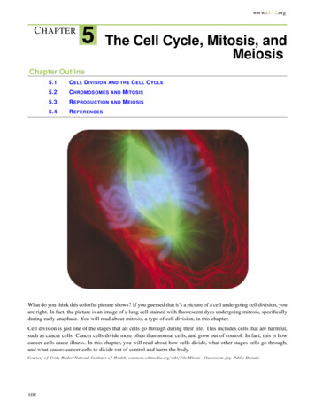
Transcription
Introduction to single-cell RNAseq analysisBaRC Hot TopicsBioinformatics and Research ComputingWhitehead InstituteMarch 7th 2019http://barc.wi.mit.edu/hot topics/
Outline Introduction to single-cell RNA-seq data analysis– Overview of scRNA-seq technology, cell barcoding, UMIs– Experimental design– Analysis pipeline Preprocessing and quality controlNormalizationDimensionality reductionClustering of cellsTrajectory inferenceDifferential expression and functional annotation Hands-on analysis using the package Seurat2
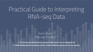
Why do single cell RNA-seq? Identify expression profiles ofindividual cells (that may bemissed with bulk RNA-seq) Discover of new cell states/types Order cells within adevelopmental trajectoryLummertz da Rocha, Nature Communications 2018Etzrodt, Cell Stem Cell 20143
Advances on scRNA-seq technologySvensson, Vento-Tormo, and Teichmann, arXiv:1704.01379v24
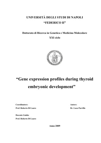
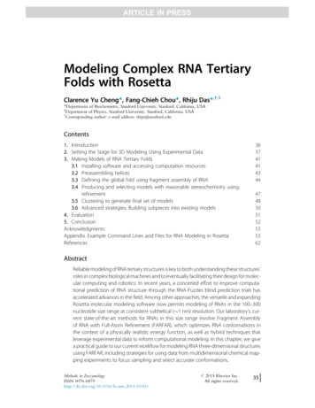
Library preparation stepsComparative Analysis of Single-Cell RNA Sequencing MethodsZiegenhain et. al, Molecular CellVolume 65, Issue 4, 16 February 2017,5
Features of scRNA-seq methodsNameTranscript coverageStrand specificityPositional biasUMI possible?Tang methodNearly full-lengthNoStrongly 3 NoSmart-seqFull-lengthNoMedium 3 NoSmart-seq2Full-lengthNoWeakly 3 NoSTRT-seq & STRT/C15 -onlyYes5 -onlyYesCEL-seq3 -onlyYes3 -onlyNoCEL-seq23 -onlyYes3 -onlyYesMARS-seq3 -onlyYes3 -onlyYesCytoSeqPre-defined genesonlyYes3 -onlyYesDrop-seq/InDrop3 -onlyYes3 -onlyYesSingle-cell RNA-sequencing: The future of genome biology is nowSimone Picelli, RNA Biology, Volume 14, 2017 - Issue 56
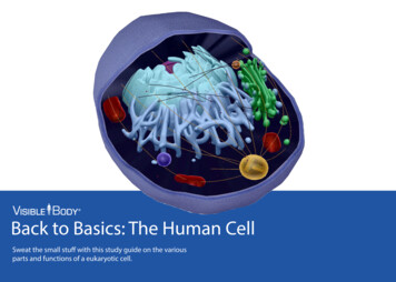
Sensitivity of scRNA-seq methodsComparative Analysis of Single-CellRNA Sequencing MethodsZiegenhain et. al, Molecular CellVolume 65, Issue 4, 16 Feb 20177
s/
s/
Goals of scRNA-seq analysis methodsLummertz da Rocha, Nature Communications 201810
Goals of scRNA-seq analysis methodsComputationalapproaches forinterpreting scRNA‐seqdata, Rostom et al. FEBSLetters, Volume: 591,Issue: 15.11
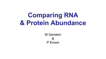
Analysis pipelinePre-ProcessingClusteringBiologyExpression Matrix(GENES x CELLS)1. IdentifyVariable Genes5. DifferentiallyExpressed Genes2. DimensionalityReductionFilter Cells/QualityControl6. Assigning CellType3a. ClusteringNormalization4a. ExploringKnown MarkerGenes7. FunctionalAnnotationPseudotime analysisAdapted cuits-computational-genomicsworkshop3b. Trajectorymodeling4b. Geneexpressiondynamics12
Technical challenges Data is noisy due to– cDNA amplification bias– mRNA capture efficiency– drop outs: large number of genes with 0 countsdue to limiting mRNA. Zero expression doesn'tmean the gene isn’t on. Cells can change or die during isolation.13
Experimental design Process your samples in a way that the condition cannot be confounded with a batch effect, like processingdate, facility, or reagents used.– i.e. If you have to process your cells in several batches,each batch should contain an equal number of cells fromeach condition. If you are comparing your data to published data youmay have to remove batch effects.– R packages like Combat can be used for ersions/3.20.0/topics/ComBat)– See “Dealing with confounders” section of the "Analysisof single cell RNA-seq data" course (Hemberg Group).14
Preprocessing for Smart-seq2 Demultiplexing: assign all the reads with thesame cell barcode to the same cell. Done atthe sequencing facility. We can check the quality of the reads withFastQC and the library composition with FastQScreen as we would do with bulk RNA-seq.15
Preprocessing for technologies using UniqueMolecular Identifiers (UMIs) Demultiplexing: assign all the reads with the samecell barcode to the same cell. Remove PCR duplicates: if several reads have thesame UMI and map to the same location in thegenome, keep only one.– Cell range software for 10x data (run by the genometechnology core)– Drop-seq tools for drop-seq and seq-well modules/
Demultiplexing and counting 10x dules/17
CellRanger web summary18
Demultiplexing and counting Drop-seq orSeq-well dataFASTQ read1FASTQ read2Unmapped BAM1. Extract cell-barcodeand UMIUnmapped BAMWith barcode andUMI infoFASTQ2. Map readsAlignedBAM3. Merge bam filesAlignedBAM withcell barcode andUMI info3. Tag reads with gene4. Count UMIs, select cellbarcodesCount matrix19
Analysis pipelinePre-ProcessingClusteringBiologyExpression Matrix(GENES x CELLS)1. IdentifyVariable Genes5. DifferentiallyExpressed Genes2. DimensionalityReductionFilter Cells/QualityControlNormalization3. ExploringKnown MarkerGenes4. Clustering6. Assigning CellType7. FunctionalAnnotationAdapted cuits-computational-genomics-workshop20
Quality control and filtering Quality control– Number of reads per cell– Number of genes detected per cell– Proportion of reads mapping to mitochondrial reads Remove cells with poor quality– Filter out cells with percentage of mitochondrial reads higher than a cut off– Filter out cells with less than a lower threshold on the number of genes orcounts per cell Remove doublets (two cells captured with one bead in the droplet)– Filter out cells with more than an upper threshold on the number of genes orcounts per cell in your data– More sophisticated way of removing doublets https://github.com/JonathanShor/DoubletDetection https://github.com/AllonKleinLab/scrublet 5248421
NormalizationCorrect for sequencing depth (i.e. library size) of eachcell so we can compare across cells1. Normalize gene expression for each cell bytotal expression2. Multiply by a scale factor (i.e. 10,000).3. Log transform the scaled countsThis is the log normalization implemented in Seurat22
Clustering and Biology: What do you want tolearn from the experiment? Classify cells and discover new cellpopulations Compare gene expression between differentcell populations Reconstruct developmental 'trajectories' toreveal cell fate decisions of distinct cellsubpopulations23
Lots of software available to analyze singlecell RNA-seq data andavi/awesome-single-cell24
Seurathttps://satijalab.org/seurat/ Seurat is an R package designed for QC, analysis,and exploration of single cell RNA-seq data. Developed and by the Satija Lab at the New YorkGenome Center. It is well maintained and well documented. It has a built in function to read 10x Genomicsdata. It has implemented most of the steps needed incommon analyses.25
Read data and explore QC metrics plots Read data– Read10X()– read.table() Create Seurat object: CreateSeuratObject() Calculate the % mitochondrial genes Plot nUMI, nGenes and % mito to decide on cut offshttps://satijalab.org/seurat/pbmc3k tutorial.html26
Select cells, normalize and scale data. Filter cells based on number of genes detected and percent ofmitochondrial genesSObj - FilterCells(object SObj,subset.names c("nGene","percent.mito"),low.thresholds c(4000, -Inf),high.thresholds c(11000, 0.06)) Normalize countsSObj - NormalizeData(object SObj,normalization.method "LogNormalize",scale.factor 1e4) Scaling the data and removing unwanted sources of variationSObj - ScaleData(object SObj) # just scale genes acrosssamplesSObj - ScaleData(object SObj, vars.to.regress c(“batch"))# remove cell-cell variation in gene expression driven bythe batch/day samples were processed.27
Select variable genes that will be used fordimensionality reduction“FindVariableGenes” calculates the average expression and dispersion foreach gene, places these genes into bins, and then calculates a z-score fordispersion within each bin. This helps control for the relationship betweenvariability and average expression.pbmc - FindVariableGenes(object pbmc, mean.function ExpMean, dispersion.function LogVMR, x.low.cutoff 0.0125,x.high.cutoff 3, y.cutoff 0.5)length(x pbmc@var.genes)## gives you the number of genes selected, 1838 in this example28
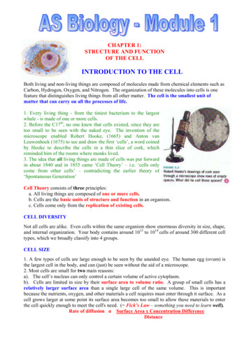
PC 2Principal component analysisPC 1Cells in 20000 (genes)dimensional spacePCACells in 10-50 principalcomponents spaceSome genes have low expressionMany genes are co-regulatedWikipedia and adapted from Hojun Li
Other dimensionality reductionmethodsCells in 20000 (genes)dimensional spacePCACells in 10-50 principalcomponents spaceHow can we further summarize these multiplePCAs into just 2 dimensions?Cells in 10-50 principalcomponents spacetSNE, UMAP, otherCells in 2D space30
t-Distributed Stochastic NeighborEmbedding (tSNE) Takes a set of points in a high-dimensional space andfinds a faithful representation of those points in alower-dimensional space, typically the 2D plane. The algorithm is non-linear and adapts to theunderlying data, performing different transformationson different regions. The t-SNE algorithm adapts its notion of “distance” toregional density variations in the data set. As a result, itnaturally expands dense clusters, and contracts sparseones, evening out cluster sizes. Distances between clusters might not mean anything.https://distill.pub/2016/misread-tsne/31
UMAPUniform manifold approximation and projection It is a non linear dimensionality reductionalgorithm. Preserves the local structure but also theglobal structure and the continuity of the cellsubsets better. See PMID: 30531897 for comparison of Seuratand UMAP.32
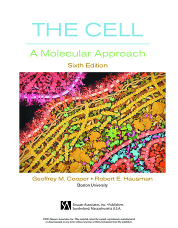
Dimensionality reduction and clustering Linear dimensionality reduction: PCApbmc - RunPCA(object pbmc, pc.genes pbmc@var.genes, do.print TRUE, pcs.print 1:5, genes.print 5) Cluster the cells and run non-lineardimensional reduction (tSNE)pbmc - FindClusters(object pbmc, reduction.type "pca", dims.use 1:10, resolution 0.6, print.output 0, save.SNN TRUE)pbmc - RunTSNE(object pbmc, dims.use 1:10, do.fast TRUE)33
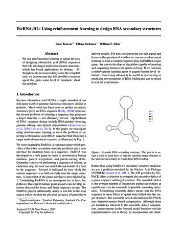
Visualize the tSNE plotTSNEPlot(object pbmc)Location of cells on the plot is coming from the tSNE plot, color is comingfrom the “FindClusters” the algorithm.34
Differential expression and visualization Finding differentially expressed genes (clusterbiomarkers)# find all markers distinguishing cluster 5 from clusters 0 and 3cluster5.markers - FindMarkers(object pbmc, ident.1 5, ident.2 c(0, 3), min.pct 0.25) Visualize DE genesVlnPlot(object pbmc, features.plot c("MS4A1", "CD79A"))FeaturePlot(object pbmc, features.plot c("MS4A1", "GNLY","CD14", "FCER1A"), cols.use c("grey", "blue"),reduction.use "tsne")35
Reconstructing 'trajectories‘Pseudotime analysis Applicable when studying a process where cells changecontinuously. For example cell differentiation duringdevelopment, or cell response to a stimulus.MonocleTSCANSlicerSlingshotDiffusion maps– Scanpy,– Seurat– density36
References and resources A practical guide to single-cell RNAsequencing for biomedical research and clinical applications. PMID: 28821273 "Analysis of single cell RNA-seq data" course (HembergGroup). Single cell RNA sequencing - NGS Analysis - NYU 2017/2018 Single Cell RNA Sequencing AnalysisWorkshop (UCD,UCB,UCSF) seandavi/awesome-single-cell Broad Institute single cell portal Tabula Muris (https://tabula-muris.ds.czbiohub.org/)37
Exercises Goal:– To walk you through an example analysis of scRNA-seqdata. Exploring the data Performing quality control Identifying cell type subsets.– To introduce you to scRNA-seq analysis using the Seuratpackage. We will be analyzing the a dataset of Non-Small CellLung Cancer Cells (NSCLC) freely available from 10XGenomics tasets/2.2.0/vdj v1 hs nsclc 5gex)38
Simone Picelli, RNA Biology, Volume 14, 2017 - Issue 5 . Sensitivity of scRNA-seq methods . 7 . Comparative Analysis of Single-Cell RNA Sequencing Methods . Ziegenhain et. al, Molecular Cell . . "Analysis of single cell RNA-seq data" course (Hemberg Group). Single cell RNA sequencing - NGS Analysis - NYU