Transcription
Amaral et al. BMC Cancer (2016) 16:602DOI 10.1186/s12885-016-2629-yRESEARCH ARTICLEOpen AccessS6Ks isoforms contribute to viability,migration, docetaxel resistance and tumorformation of prostate cancer cellsCamila L. Amaral1†, Lidia B. Freitas1†, Rodrigo E. Tamura2, Mariana R. Tavares1, Isadora C. B. Pavan1,Marcio C. Bajgelman3 and Fernando M. Simabuco1*AbstractBackground: The S6 Kinase (S6K) proteins are some of the main downstream effectors of the mammalian TargetOf Rapamycin (mTOR) and act as key regulators of protein synthesis and cell growth. S6K is overexpressed in avariety of human tumors and is correlated to poor prognosis in prostate cancer. Due to the current urgency toidentify factors involved in prostate cancer progression, we aimed to reveal the cellular functions of three S6Kisoforms–p70-S6K1, p85-S6K1 and p54-S6K2–in prostate cancer, as well as their potential as therapeutic targets.Methods: In this study we performed S6K knockdown and overexpression and investigated its role in prostatecancer cell proliferation, colony formation, viability, migration and resistance to docetaxel treatment. In addition,we measured tumor growth in Nude mice injected with PC3 cells overexpressing S6K isoforms and tested theefficacy of a new available S6K1 inhibitor in vitro.Results: S6Ks overexpression enhanced PC3-luc cell line viability, migration, resistance to docetaxel and tumorformation in Nude mice. Only S6K2 knockdown rendered prostate cancer cells more sensitive to docetaxel.S6K1 inhibitor PF-4708671 was particularly effective for reducing migration and proliferation of PC3 cell line.Conclusions: These findings demonstrate that S6Ks play an important role in prostate cancer progression,enhancing cell viability, migration and chemotherapy resistance, and place both S6K1 and S6K2 as a potentialtargets in advanced prostate cancer. We also provide evidence that S6K1 inhibitor PF-4708671 may be consideredas a potential drug for prostate cancer treatment.Keywords: mTOR, S6K, CancerBackgroundProstate cancer is the second most frequently diagnosedcancer among men worldwide and the first in developedcountries [1]. Although prostate cancer has a goodprognosis in its early stages, with nearly all men living atleast five years after diagnosis, the 5-year survival ratedecreases drastically, to less than 30 %, when it reachesadvanced and metastatic stages. This reveals the currenturgency to identify factors involved in prostate cancerprogression [2].* Correspondence: simabuco@gmail.com†Equal contributors1Laboratory of Disorders of Metabolism, School of Applied Sciences,University of Campinas, R. Pedro Zaccaria, 1300, sala LA 421, 13484-350Limeira, São Paulo, BrazilFull list of author information is available at the end of the articleThe S6K proteins are members of the AGC family ofserine/threonine kinases and one of the main downstream effectors of the mammalian Target Of Rapamycin(mTOR) protein. In mammals, the S6K family is composed of several proteins encoded by two differentgenes: RPS6KB1 and RPS6KB2. Due to the alternativeuse of AUG start codons, each S6K gene generates twodistinct isoforms: p70-S6K1, p85-S6K1, p54-S6K2 andp56-S6K2 [3, 4]. More recently, it has been discoveredthat the splicing factor SF2/ASF acts on S6K1 genepromoting the expression of a novel isoform, p31-S6K1,that lacks most of its catalytic domain [5]. Once activated by mTOR, the S6K proteins are able to phosphorylate targets as rpS6 (ribosomal protein S6), eIF4B(eukaryotic translation Initiation Factor 4B) and eEF2K 2016 The Author(s). Open Access This article is distributed under the terms of the Creative Commons Attribution 4.0International License (http://creativecommons.org/licenses/by/4.0/), which permits unrestricted use, distribution, andreproduction in any medium, provided you give appropriate credit to the original author(s) and the source, provide a link tothe Creative Commons license, and indicate if changes were made. The Creative Commons Public Domain Dedication o/1.0/) applies to the data made available in this article, unless otherwise stated.
Amaral et al. BMC Cancer (2016) 16:602(eukaryotic Elongation Factor 2 Kinase), promoting protein synthesis and cell growth [3].Due to their key role in regulating cell growth and proliferation, several studies have shown that S6K genes areamplified in a variety of human tumors, including prostate cancer [6–9]. In fact, S6K is not only overexpressedin prostate cancer, but also is related to its progression[10], making it a potential target for prostate cancertreatment. Despite the high homology shared betweenS6K1 and S6K2, evidence shows that they might playsome distinct cellular functions [11]. Global expressionprofiles for breast tumors harboring high levels of S6Ksrecently revealed that only a few set of genes stronglycorrelated to both S6K1 and S6K2, suggesting that eachprotein play different functions in tumorigenesis andcancer progression [12]. However, these differences havebeen poorly investigated and the major understandingabout S6Ks roles in cancer is from studies restricted top70-S6K1 [13–19].Here, we aimed to reveal the cellular functions of threeS6K isoforms–p70-S6K1, p85-S6K1 and p54-S6K2–inprostate cancer, as well as their potential as therapeutictargets. We show that all isoforms were important forincreasing prostate cancer cells proliferation, migrationand resistance to docetaxel in vitro. Moreover, S6Ks presented an important effect for tumor progression in vivo.Finally, we demonstrate the potential use of an availableS6K1 inhibitor.MethodsCell cultureHuman metastatic prostate cancer cell line PC-3 and theluciferase expressing cell line PC3-luc were cultured inHam’s F12 (Thermo Scientific) supplemented with 10 %FBS (fetal bovine serum) and 1 % penicillin/streptomycin(Thermo Scientific). Human metastatic prostate cancercell line DU-145 was cultured in Dulbecco’s ModifiedEagle Medium (Thermo Scientific) supplemented with10 % FBS and 1 % penicillin/streptomycin (Thermo Scientific). Cells were maintained at 37 C in a humidified atmosphere containing 5 % carbon dioxide.Transfection of human cellsCells were seeded 24 h before transfection. Transfectionwas performed with Lipofectamine and PLUS reagents(Thermo Scientific). Briefly, DNA and PLUS reagentwere diluted in serum free medium and incubated for15 min at room temperature. Lipofectamine was thendiluted in serum free medium, mixed to the DNA solution and incubated for 15 min at room temperature. Thecells were washed with serum free medium and theDNA/Lipofectamine complexes were added. After 3 h,the medium was exchanged for medium containing 10 %FBS and the cells incubated for 24 to 72 h.Page 2 of 9Virus productionLentivirus and retrovirus preparations were generated atViral Vector Laboratory, Brazilian National Laboratory forBiosciences, Brazilian Center for research in Energy andMaterials. Virus were titrated by puromycin selection andcounting forming colonies.S6Ks RNAi knockdown and overexpressionLentiviruses expressing shRNA targeting S6K1 and S6K2human mRNA were produced using pLK0.1 vector (SigmaAldrich). S6K1 shRNA sequence (TRCN0000022904) hasbeen published before [20]. For S6K2 knockdown, wetested two different shRNA sequences: TRCN0000010539and TRCN0000199878, identified respectively as shRNAS6K2-1 and shRNA-S6K2-2. For the control, we used apLKO.1 plasmid (SHC002) that targets no mammaliangenes and was identified as shC.The retroviral plasmid pBABE-puro was used to performS6K overexpression. Fragments of S6K isoforms werecloned into BamHI/SalI restriction sites and retroviralmediated gene transfer was performed as described previously [21, 22].Viral transductionPC3-luc cells were seeded at a density of 8 103 cells/wellin 96-well plates and incubated for 24 h. Virus particleswere added at a low multiplicity of infection (MOI) of 0.3for lentiviruses and 0.1 for retroviruses in the presence of8 μg/ml of polybrene. Cell culture medium was changed24 h after transduction and cells were then selected with1 μg/mL of puromycin until complete death of controlcells.Western blottingProteins were separated by SDS-PAGE and transferredonto nitrocellulose membranes. Nitrocellulose membraneswere blocked in a solution of TBS containing 5 % nonfatdry milk and 0,1 % Tween-20 for 2 h with constant agitation. After blocking, the membranes were incubated withanti-p70-S6K1 (Cell Signaling), anti-S6K2 (Bethyl) or antiα-tubulin (Calbiochem) antibodies overnight at 4 C. Membranes were washed with TBS-T (3 times for 15 min) andincubated with horseradish peroxidase-conjugated secondary antibodies (Millipore) for 1 h at room temperature withconstant agitation. Bands were visualized using the ECL kit(GE Healthcare). Band densitometry was measured usingImageJ software.MTT viability assayPC3-luc cells with stable S6K knockdown or overexpression were seeded at a density of 104 cells/well in 96-wellplates and incubated for 24, 48 and 72 h. Following eachperiod of incubation, 12 mM of 3-(4, 5-methylthiazol2-yl)-2, 5-diphenyl-tetrazolium bromide (MTT) was
Amaral et al. BMC Cancer (2016) 16:602added to each well for 4 h. The culture medium was aspirated and the formazan crystals were solubilized with asolution of HCl 1 N:isopropanol (1:25) for 15 min. Theoptical density of the plates was measured at 570 nm.Migration assayPC3-luc cells with stable S6K knockdown or overexpression were seeded at a density of 5 105 cells/well in 24well plates and incubated until confluence. After that, cellmonolayers were scratched in the middle of the wells witha p200 pipette tip and the culture medium was replacedby serum free media [23]. Scratch area was analyzed underlight microscope and images were captured right after thescratch (0 h) and after incubation for 24 and 48 h. Thescratch area was quantified using ImageJ software.Colony-forming assayCells were plated at low density (5 102 cells / plate) in60 mm plates and transfected with plasmids pFLAGp70-S6K1, - p85-S6K1, - p54-S6K2 or pFLAG (emptyvector). The cells were incubated at 37 C for 10 days.For staining, the cells were washed with PBS and stainedwith 1 mL of methylene blue dye (3 %) for 30 min. Theplates were washed and colonies were counted, excluding colonies smaller than 1 mm in diameter.Proliferation assayCells were plated in 24-well plates, transfected with plasmids pFLAG- p70-S6K1, - p85-S6K1, - p54-S6K2 orpFLAG (empty vector) and then incubated for up to6 days with 10 % FBS at 37 C. Counts were performedusing automated cell counter on days 2, 4 and 6 aftertransfection.Docetaxel resistance assayDocetaxel resistance assay was performed as previouslydescribed by Uzoh et al. [24], with slight modifications.PC3-luc cells with stable S6K knockdown or overexpression were seeded at a density of 8 105 cells/well in 24well plates and incubated for 24 h. The culture mediumwas then replaced by serum free media and the plateswere incubated for 24 h. Afterwards, cells were treatedwith 30 nM of docetaxel (Sigma Aldrich) for 48 h, whichwas firstly dissolved in DMSO and then diluted in serumfree media. Control cells received serum free media andthe vehicle. The number of living cells was counted usingan automated cell counter (Thermo Scientific) after Trypan Blue staining.Page 3 of 9knockdown were injected subcutaneously into 7 maleathymic Nude mice per group. Tumor growth was measured about three times per week using a caliper rule andcalculated according to the formula: ½ (larger diameter) (smaller diameter) 2 until they reached the maximum volume of 1000 mm3.Treatment of cells with PF4708671PC3 and DU145 cells were treated with 10 μM of S6K1inhibitor PF4708671 [25], by changing medium containingthe drug every day, until 6 days. The drug was firstdissolved in DMSO and then diluted in medium withserum.Statistical analysisValues presented are means standard deviation (SD).Statistical analyzes were assessed by one-way and two-wayanalysis of variance (ANOVA) followed by Bonferroni’spost-test and performed using GraphPadPrism 5 software.p-values 0.05 were considered significant (*p 0.05;**p 0.01; ***p 0.001).ResultsS6K knockdown inhibits cell viabilityS6Ks stable knockdown and overexpression in PC3-luccells were confirmed by western blotting (Fig. 1 a-b).Although some reports indicate that the knockout of oneS6K isoform in vivo may cause a compensatory upregulation of the other [26, 27], we observed a slight compensatory effect by S6K1 in the S6K2-1 knockdown in vitro. Toinvestigate the effects of S6Ks knockdown or overexpression in PC3-luc prostate cancer cell line viability, weperformed the MTT assay. S6K2 knockdown significantlyreduced cell viability in all time periods (Fig. 1c). Similarinhibition was observed in both S6K2 shRNA sequencestested. S6K1 knockdown showed significant reductiononly on the second day and appeared to be less relevantthan S6K2. However, the overexpression of all three S6Kisoforms significantly raised cell viability (Fig. 1d).S6K knockdown inhibits cell migrationNext we investigated if S6K isoforms also impacted themigration capacity of prostate cancer cells (Fig. 2). BothS6K1 and S6K2 knockdown resulted in a significant decrease in cell migration. Again, no difference was observedbetween both S6K2 shRNA sequences. In contrast, asexpected, the overexpression of S6K isoforms significantlyincreased scratch area closure.In vivo tumor formation assayS6K2 expression is related to docetaxel sensitivityAll animal experiments were performed in accordance tothe internal committee of ethics in animal research of theFaculty of Medicine of the University of São Paulo. Onemillion PC3-luc cells with stable S6K overexpression orWe investigated whether S6Ks might play a role in prostate cancer chemotherapy resistance. We treated PC3-lucwith docetaxel, a chemotherapeutic drug widely used inhormone-refractory prostate cancer treatment [28]. As
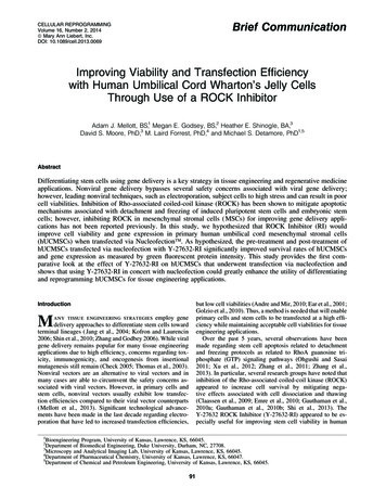
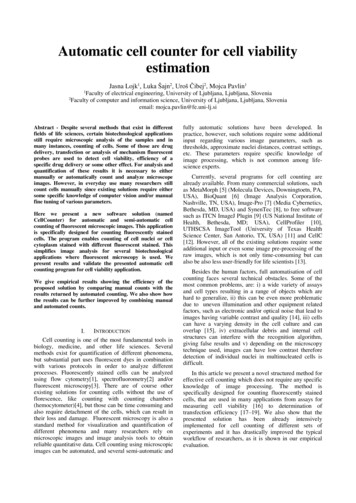
Amaral et al. BMC Cancer (2016) 16:602Page 4 of 9Fig. 1 S6Ks increase cell viability in PC3-luc cells. Western blotting analyses of S6K isoforms expression modulation in human prostate cancer cellline PC3-luc transduced with a lentiviruses pLKO.1-shRNAs against S6K1 and S6K2 and b with retroviruses carrying pBABE construction. S6K1antibody detects both p85-S6K1 and p70-S6K1 proteins. c Relative cell viability in PC3-luc cells with knockdown of S6Ks isoforms and d inPC3-luc cells overexpressing S6Ks isoforms. Cells were seeded in 96-well plates at a density of 104 cells/well. After 24, 48 and 72 h, cellswere treated with MTT (12 mM) for 4 h and absorbance was measured at 570 nm. *p 0.05, **p 0.01, ***p 0,001, n 3Fig. 2 S6Ks increase migration of PC3-luc cells. Cells were seeded in 24-well plates at a density of 5x10⁴ cells/well and incubated until confluence.A scratch was made in the cells monolayer with a pipette tip and cells were washed and incubated in serum free media. The scratch area wasmeasured at 0 (time of scratch), 24 and 48 h. a Migration assay in PC3-luc cells with knockdown of S6Ks isoforms. a, b Scratch relative area ofPC3-luc cells overexpressing S6Ks isoforms. c, d Representative images of scratch area taken at 0, 24 and 48 h of PC3-luc cells with S6Kknockdown and overexpression, respectively. *p 0.05, **p 0.01, ***p 0,001, n 3
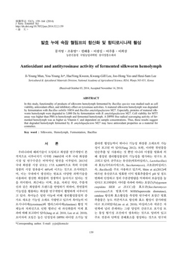
Amaral et al. BMC Cancer (2016) 16:602expected, docetaxel treatment reduced the number of living cells in all groups when compared to untreated cells(Fig. 3). However, the reduction was significantly greaterin cells with S6K2 knockdown (Fig. 3a). When overexpressed, all S6Ks isoforms showed significantly less reduction in the number of living cells than the GFP control(Fig. 3b).S6K overexpression enhances tumor growth in Nude miceAfter having determined that S6Ks are involved in someprostate cancer features in vitro, we investigated theirtumorigenic ability in vivo, by injecting PC3-luc cells overexpressing S6K isoforms or knocked down for S6K1 orS6K2 subcutaneously into the flanks of male athymic Nudemice and measuring tumor growth. Tumors started growing about 10 days after injection and from this momenttumor volume was assessed for about 30 days. Animalsinjected with PC3-luc cells overexpressing S6K isoforms,particularly p70-S6K1, showed significantly greater tumorgrowth than those injected with cells harboring GFP control (Fig. 4a). Conversely, animals injected with PC3-luccells containing the knockdown of S6K1 or S6K2 presentedsignificantly reduced tumor growth in vivo (Fig. 4b).S6K1 inhibitor PF47086701 decreases proliferation andmigration of PC3 prostate cancer cell lineTo check the efficiency of S6K1 inhibitor in prostatecancer cells DU145 and PC3, western blotting was performed in order to evaluate the S6 phosphorylation status(Fig. 5a). Rapamycin was used in this experiment as control. PF47086701 was able to reduce S6 phosphorylationin both DU145 and PC3 cells lines, although its effect wasmore discrete when compared to rapamycin, which completely abolished S6 phosphorylation (Fig. 5a). In a proliferation assay, DU145 and PC3 cells were plated andcounted after treatment with PF47086701 (10 μM). OnlyPC3 cells showed a decrease in the number of cells whentreated with the inhibitor of S6K1 (Fig. 5b). In a migrationassay, the scratched monolayers were treated with serumfree medium and PF4708671 (10 μM) daily. The areasPage 5 of 9were measured within 48 h after the scratch. PC3 cellsshowed a significant decrease in migration compared tocontrol in the presence of S6K1 inhibitor (Fig. 5c).To compare the effects of S6Ks overexpression inDU145 and PC3 cell lines, we transiently transfected themwith pcDNA-FLAG vector carrying p70-S6K1, p85-S6K2and p54-S6K2 genes (Additional file 1: Figure S1A). Theresults show that in both cell lines p85-S6K1 isoformenhanced the cellular proliferation (Additional file 1:Figure S1B). Colony formation assay also indicated thatcells with overexpression of p85-S6K1 showed increasednumber of colonies (Additional file 1: Figure S1C). Theseresults point an important role of S6K1 isoform in prostatecancer.DiscussionS6K proteins are well known effectors of mTOR and majorregulators of protein synthesis and cell growth [4]. Theyare found overexpressed in several human tumors and arecorrelated with poor prognosis [10, 29, 30]. It has alreadybeen reported that silencing mTOR decreases prostatecancer cell proliferation and colony formation [31]. Herewe show that S6Ks overexpression enhances proliferationand clonogenic ability of human metastatic prostate cancercell lines. Next, we evaluated the effects of S6K knockdownand overexpression on prostate cancer cells viability,migration and resistance to decetaxel treatment.Recently, Du et al. (2014) showed that mTOR knockdown by lentivirus mediated shRNA significantly reducescell viability in prostate cancer metastatic cell lines LNCaPand C4-2B. As expected, we not only observed that S6Kknockdown produced similar effects on PC3-luc cells, butalso reported an expressive increase in cell viability determined by S6K overexpression. Interestingly, we found thatalthough the effects of p70-S6K1 and p54-S6K2 overexpression were quite similar, S6K1 knockdown had noimpact on cell viability. A possible explanation is that S6K1knockdown is compensated by S6K2 isoform, masking theeffects on cell viability.Fig. 3 p54-S6K2 knockdown increases cell death in response to docetaxel in PC3-luc cells. Cells were seeded in 24-well plates at a density of8x104 cells/well. After 24 h, cells were subjected to serum free media conditions for 24 h and then treated for 48 h with docetaxel (30 nM).Living cells were counted and expressed as percentages. Results presented are means of two independent experiments. Each experimentwas performed in triplicates. SMF: serum free media; DTX: docetaxel. *p 0.05, **p 0.01, ***p 0,001, n 2
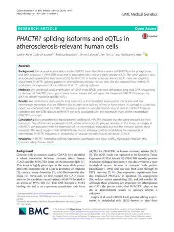
Amaral et al. BMC Cancer (2016) 16:602Fig. 4 S6K isoforms increase tumor growth in Nude mice. a Tumorgrowth curves of Nude mice injected subcutaneously with PC3-luccells overexpressing S6K isoforms (p70-S6K1, p85-S6K1 and p54-S6K2)or GFP control. Tumors measurements began 14 days after injectionand proceeded until day 44. b Tumor growth curves of Nude miceinjected subcutaneously with PC3-luc cells with knockdown for S6K1and S6K2 isoforms or shRNA control. Tumors measurements began11 days after injection and proceeded until day 39. * p 0,05,** p 0,01 e *** p 0,001; n 7We also reported that S6K expression is positivelyrelated to an increase in PC3-luc cells migration. Sincetumor cell motility is a fundamental part of the metastaticcascade, our data imply an involvement of all S6K isoforms in raising metastatic capability of PC3-luc cells. Infact, p70-S6K1 has been described as a regulator of cellmotility [32] and several human tumorigenic cell lines,such rhabdomyosarcoma (Rh1 and Rh30), breast cancer(MDA-MB-468) and cervical adenocarcinoma (HeLa) wereshown to alter their motility and invasion capabilities inresponse to rapamycin and these phenomena were regulated by mTOR through S6K1 and 4E-BP1 [33]. p70-S6K1can directly bind to F-actin and is localized at the actinarc of migrating cells [34, 35]. In ovarian cancer cells,p70-S6K1 regulates cytoskeleton organization and cellmigration by activating the GTPases Rac1 and Cdc42,involved, respectively, in dictating forward movement andmigration’s direction [34]. Indeed, p70-S6K1 knockdownovarian cancer cells migrate less and exhibit reduceddirectional persistence [34]. Our results show that S6K1promotes cell migration in PC3-luc cell line and thatS6K2 is also involved in the regulation of this process.Page 6 of 9S6K2 knockdown was also effective in raising docetaxelsensitivity in PC3-luc cells. It has been reported that S6K2is activated by FGF-2 (Fibroblast Growth Factor-2) andphosphorylates PDCD4, a repressor of the translation ofanti-apoptotic proteins such as XIAP and Bcl-xL [36].PDCD4 phosphorylation by S6K2 causes its degradationand leads to survival and chemoresistance in lung cancercells [36, 37]. Although studies relating S6Ks to chemotherapy resistance are scarce, several reports indicate thatmTOR is involved in drug resistance against chemotherapy [38–43]. Niu et al. [44] showed that mTOR inhibitionby rapamycin raises apoptosis and sensitivity to docetaxelinduced cytotoxity in several human metastatic lung cancer cell lines treated with different concentrations of docetaxel. Treatment with NVP-BEZ235-a PI3K/mTOR dualinhibitor–was able to sensitize human castration resistantprostate cancer cell lines C4-2 and C4-2AT6 resistant todocetaxel, indicating that mTOR inhibition can evenovercome chemoresistance in castration resistant prostatecancer [45]. However, the specific role of mTOR in developing chemotherapy resistance is still poorly understood,yet of current clinical importance. Here, we show thatdocetaxel resistance in PC3-luc cell line may be at least inpart mediated by S6K, especially p54-S6K2.The oncogenic role of S6K was also confirmed in vivo.The overexpression of all S6K isoforms was able to enhance tumor formation in Nude mice and the knockdownof S6Ks isoforms was able to reduce tumor growth in vivo.This result corroborates to Du et al. [31], that demonstrated that Nude mice injected with human metastaticprostate cancer cell line C4-2B with mTOR knockdownpresented a greater reduction in tumor volume whencompared to control group. As it was shown that p70S6K1 played a major role in increasing tumor formation invivo, we decided to test the effectiveness of a novel S6K1inhibitor, PF-4708671.We demonstrated that targeting S6K1 with PF-4708671provided reduced cell migration and proliferation in PC3cells, but had no effect on DU145 cell line (Fig. 5). Apossible explanation for this difference is that PC3 cells,but not DU145, are PTEN null. PTEN deletion leads toincreased PI3K activity and therefore hyperactivation ofmTORC1/S6K [46]. Thus, it is possible that the mechanisms inducing proliferation and migration in PC3 cellsare more related to S6K activity than in DU145 cells and,therefore, more susceptible to PF-4708671 effect.PF-4708671 is a cell-permeable piperazinyl-pyrimidinecompound that specifically inhibits p70-S6K1 with a Ki of20 nM and IC50 of 160 nM and exhibits no significantinhibition of S6K2 or other AGC kinases [25], which mayexplain the reduced effect to abolish S6 phosphorylationcomparing to rapamycin (Fig 5a), since S6K2 may still acton S6 protein. Due to its recent development, there areonly a few reports regarding PF-4708671 use in anti-
Amaral et al. BMC Cancer (2016) 16:602Page 7 of 9Fig. 5 S6K1 inhibitor PF47086701 inhibits cell proliferation and migration of prostate cancer cells. a Western blotting analysis of PF47086701efficiency. b Proliferation assay in DU145 and PC3 cell lines treated with PF47086701. c Scratch relative area of DU145 and PC3 cells treatedwith PF47086701. *p 0.05, **p 0.01, ***p 0,001, n 3cancer therapies [3, 25]. S6K1 inhibition by PF-4708671was shown to sensitize resistant colorectal cancer cells toselumetinib [47], to decrease cell migration and invasionof MDA-MB-231 human breast cancer cell line [48] andto inhibit cell invasion and proliferation in human lungcancer cell lines and tumorigenesis in Nude mice [49].Hence, our results ratify that PF-4708671 might be a novelpotential adjuvant in metastatic prostate cancer drugtreatment.ConclusionsIn summary, our study demonstrated that S6K overexpression enhances cell viability, migration and resistanceto docetaxel in PC3-luc prostate cancer cell line andtumor volume in Nude mice. In addition, we showed thatthe often neglected S6K2 is also involved in these processes and might be a potential target to restore docetaxelsensitivity in advanced prostate cancer. The S6K1 inhibitor PF-4708671 was particularly effective in reducing cellmigration of PC3 and DU145 cell lines, suggesting that itcould represent a possible adjuvant to prevent prostatecancer progression to its advanced state.Additional fileAdditional file 1: Figure S1. Transient overexpression of different S6Ksisoforms in DU145 and PC3 cell lines. (A) Western blotting analysis oftransfection efficiency. (B) Proliferation assay in DU145 and PC3 cell linestransfected with p70-S6K1, p85-S6K1 and p54-S6K2. (C) Colony formationassay of DU145 and PC3 cells transfected with p70-S6K1, p85-S6K1 andp54-S6K2. *p 0.05, **p 0.01, ***p 0,001, n 3. (TIF 2571 kb)AbbreviationseEF2K, eukaryotic Elongation Factor 2 Kinase; eIF4B, eukaryotic translationInitiation Factor 4B; FBS, fetal bovine serum; mTOR, mammalian Target OfRapamycin; MTT, 12 mM of 3-(4, 5-methylthiazol-2-yl)-2, 5-diphenyl-tetrazoliumbromide; rpS6, ribosomal protein S6; S6K, S6 Kinase.AcknowledgementsWe thank prof. Bryan Strauss from the Cancer Institute of São Paulo forgently revise the manuscript. We thank prof. Marcio Torsoni, prof. AdrianaTorsoni and prof. Marciane Milanski from Laboratory of Disorders of
Amaral et al. BMC Cancer (2016) 16:602Metabolism (LabDiMe), and all of its members, from University of Campinas,for all experimental support.FundingThis work was supported by the São Paulo Research Foundation (FAPESP),grant number 2012/13558-7 and fellowships from FAPESP and CNPq.Authors’ contributionsCLA participated in the viruses transductions, viability, migration, docetaxelresistance and in vivo experiments and drafted the manuscript. LBFparticipated in the proliferation, migration and S6K1 inhibitor experimentsand performed initial standardization of protocols. RET performed andcoordinated in vivo experiments and participated in design of experiments.MRT participated in initial cloning. ICBP participated in initial cloning. MCBperformed and coordinated viruses production and participated in design ofexperiments. FMS conceived of the study and participated in its design andcoordination. All authors have read and approved this manuscript.Availability of data and materialsDatasets supporting the conclusions of this article are available afterpublishing in the FigShare repository.Competing interestsThe authors declare that they have no competing interests.Consent to publishNot applicable.Ethics approval and consent to participateAll animal experiments were performed in accordance to guidelines of theinternal committee of ethics in animal research of the Faculty of Medicine ofthe University of São Paulo and approved by the same.Author details1Laboratory of Disorders of Metabolism, School of Applied Sciences,University of Campinas, R. Pedro Zaccaria, 1300, sala LA 421, 13484-350Limeira, São Paulo, Brazil. 2Viral Vector Laboratory, Center for TranslationalInvestigation in Oncology/LIM24, Cancer Institute of São Paulo, School ofMedicine, University of São Paulo, São Paulo, Brazil. 3Brazilian BiosciencesNational Laboratory, Brazilian National Center for Research in Energy andMaterials, Campinas, São Paulo, Brazil.Received: 24 March 2016 Accepted: 26 July 2016References1. Torre LA, Bray F, Siegel RL, Ferlay J, Lortet-Tieulent J, Jemal A. Global cancerstatistics, 2012. CA Cancer J Clin. 2015;65(2):87–108.2. DeSantis CE, Lin CC, Mariotto AB, Siegel RL, Stein KD, Kramer JL, et al.Cancer treatment and survivorship statistics, 2014. CA Cancer J Clin.2014;64(4):252–71.3. Tavares MR, Pavan ICB, Amaral CL, Meneguello L, Luchessi AD, SimabucoFM. The S6K protein family in health and disease. Life Sci. 2015;131:1–10.4. Magnuson B, Ekim B, Fingar DC. Regulation and function of ribosomalprotein S6 kinase (S6K) within mTOR signalling networks. Biochem J.2012;441(1):1–21.5. Karni R, De Stanchina E, Lowe SW, Sinha R, Mu D, Krainer AR. The geneencoding the splicing factor SF2/ASF is a proto-oncogene. Nat Struct MolBiol. 2007;14(3):185–93.6. Kremer CL, Klein RR, Mendelson J, Browne W, Samadzedeh LK, Vanpatten K,et al. Expression of mTOR signaling pathway markers in prostate cancerprogression. Pros
using automated cell counter on days 2, 4 and 6 after transfection. Docetaxel resistance assay Docetaxel resistance assay was performed as previously described by Uzoh et al. [24], with slight modifications. PC3-luc cells with stable S6K knockdown or overexpres-sion were seeded at a density of 8 105 cells/well in 24-well plates and incubated .