Transcription
Brief CommunicationCELLULAR REPROGRAMMINGVolume 16, Number 2, 2014ª Mary Ann Liebert, Inc.DOI: 10.1089/cell.2013.0069Improving Viability and Transfection Efficiencywith Human Umbilical Cord Wharton’s Jelly CellsThrough Use of a ROCK InhibitorAdam J. Mellott, BS,1 Megan E. Godsey, BS,2 Heather E. Shinogle, BA,3David S. Moore, PhD,3 M. Laird Forrest, PhD,4 and Michael S. Detamore, PhD1,5AbstractDifferentiating stem cells using gene delivery is a key strategy in tissue engineering and regenerative medicineapplications. Nonviral gene delivery bypasses several safety concerns associated with viral gene delivery;however, leading nonviral techniques, such as electroporation, subject cells to high stress and can result in poorcell viabilities. Inhibition of Rho-associated coiled-coil kinase (ROCK) has been shown to mitigate apoptoticmechanisms associated with detachment and freezing of induced pluripotent stem cells and embryonic stemcells; however, inhibiting ROCK in mesenchymal stromal cells (MSCs) for improving gene delivery applications has not been reported previously. In this study, we hypothesized that ROCK Inhibitor (RI) wouldimprove cell viability and gene expression in primary human umbilical cord mesenchymal stromal cells(hUCMSCs) when transfected via Nucleofection . As hypothesized, the pre-treatment and post-treatment ofhUCMSCs transfected via nucleofection with Y-27632-RI significantly improved survival rates of hUCMSCsand gene expression as measured by green fluorescent protein intensity. This study provides the first comparative look at the effect of Y-27632-RI on hUCMSCs that underwent transfection via nucleofection andshows that using Y-27632-RI in concert with nucleofection could greatly enhance the utility of differentiatingand reprogramming hUCMSCs for tissue engineering applications.but low cell viabilities (Andre and Mir, 2010; Ear et al., 2001;Golzio et al., 2010). Thus, a method is needed that will enableprimary cells and stem cells to be transfected at a high efficiency while maintaining acceptable cell viabilities for tissueengineering applications.Over the past 5 years, several observations have beenmade regarding stem cell apoptosis related to detachmentand freezing protocols as related to RhoA guanosine triphosphate (GTP) signaling pathways (Ohgushi and Sasai2011; Xu et al., 2012; Zhang et al., 2011; Zhang et al.,2013). In particular, several research groups have noted thatinhibition of the Rho-associated coiled-coil kinase (ROCK)appeared to increase cell survival by mitigating negative effects associated with cell dissociation and thawing(Claassen et al., 2009; Emre et al., 2010; Gauthaman et al.,2010a; Gauthaman et al., 2010b; Shi et al., 2013). TheY-27632 ROCK Inhibitor (Y-27632-RI) appeared to be especially useful for improving stem cell viability in humanIntroductionMany tissue engineering strategies employ genedelivery approaches to differentiate stem cells towardterminal lineages ( Jang et al., 2004; Kofron and Laurencin2006; Shin et al., 2010; Zhang and Godbey 2006). While viralgene delivery remains popular for many tissue engineeringapplications due to high efficiency, concerns regarding toxicity, immunogenicity, and oncogenesis from insertionalmutagenesis still remain (Check 2005; Thomas et al., 2003).Nonviral vectors are an alternative to viral vectors and inmany cases are able to circumvent the safety concerns associated with viral vectors. However, in primary cells andstem cells, nonviral vectors usually exhibit low transfection efficiencies compared to their viral vector counterparts(Mellott et al., 2013). Significant technological advancements have been made in the last decade regarding electroporation that have led to increased transfection efficiencies,1Bioengineering Program, University of Kansas, Lawrence, KS, 66045.Department of Biomedical Engineering, Duke University, Durham, NC, 27708.3Microscopy and Analytical Imaging Lab, University of Kansas, Lawrence, KS, 66045.4Department of Pharmaceutical Chemistry, University of Kansas, Lawrence, KS, 66047.5Department of Chemical and Petroleum Engineering, University of Kansas, Lawrence, KS, 66045.291
92induced pluripotent cells (iPSCs) and embryonic stem cells(ESCs) ( Joo et al., 2012; Narumiya et al., 2000). Furthermore, the use of Y-27632-RI was shown not to affect thepluripotency of ESCs (Watanabe et al., 2007). Chatterjeeet al. (2011) were the first group to use RI to aid in thetransfection of human iPSCs via Nucleofection , an electroporative technique developed by Lonza Group Ltd.(Basel, Switzerland).Human umbilical cord mesenchymal stromal cells(hUCMSCs) have a number of advantages over other cellsources and hold great potential for clinical translation, aswe have reviewed extensively (Bailey et al., 2007a; Wanget al., 2009). Unfortunately, hUCMSCs are difficult totransfect, and few studies are available on the transfection ofhUCMSCs. On the basis of its aforementioned success inother applications, we hypothesized that Y-27632-RI wouldimprove cell viability and transfection efficiency forhUCMSCs that are transfected via nucleofection. In thisstudy, transfection efficiency, gene expression, and cell viability were evaluated for hUCMSCs transfected via nucleofection with green fluorescent protein (GFP), with orwithout Y-27632-RI.Materials and MethodsProcurement and expansion of hUCMSCshUCMSCs were isolated from Wharton’s jelly of humanumbilical cords obtained from the Kansas University MedicalCenter Hospital (IRB# 10951), Lawrence Memorial Hospital(IRB# LMH 08-2), and Stormont-Vail Hospital (IRB approved, no reference number) for a total of five cords used inthe study (n 5). Four cords were from males that were bornat full term and one cord was from a female born at 38.3weeks, all under normal delivery conditions. Maternal agewas not available. We isolated hUCMSCs according to ourprevious published protocol (Devarajan et al., 2013).hUCMSCs were cultured in traditional hUCMSC medium[10% fetal bovine serum (FBS-MSC Qualified) and 1%penicillin-streptomycin in low glucose Dulbecco’s ModifiedEagle Medium (DMEM); Life Technologies, Grand Island,NY, USA)]. hUCMSC medium was changed three times perweek, and hUCMSCs were maintained at 37 C with 5% CO2in a cell culture–grade incubator. hUCMSCs were trypsinizedwith 0.05% trypsin-EDTA (1 · ) (Life Technologies) at 80–90% confluency. All hUCMSCs were expanded to passage 2(P2) for the experiments. Five umbilical cords (n 5) in totalwere used for this study. All experiments were performed intriplicate for each cord.Cell characterizationAt P2, a subculture of cells from each cord was characterized through cell-surface marker identification via flowcytometry on a MoFlo XDP fluorescence-activated cell sorter(FACS) (Beckman Coulter, Brea, CA, USA). hUCMSCswere characterized using the following antibodies and secondary antibodies: STRO-1 mouse immunoglobulin M (IgM)(2.5:200; 1 mg/mL; R&D Systems, Minneapolis, MN, USA);Alexa Fluor 568 rabbit anti-mouse IgG (2:200; 2 mg/ mL;Life Technologies); CD105 mouse IgG (2.5:200; 1 mg/mL;R&D Systems); Qdot 525 donkey anti-mouse IgG (2:200;1 lM; Life Technologies); human CD45 preconjugated toQdot 800 (2:200; Life Technologies); human CD73 pre-MELLOTT ET AL.conjugated to fluorescein isothiocyanate (FITC) (5:200; BDBiosciences, San Jose, CA, USA); human CD34 preconjugated to Brilliant Violet (5:200; BD Biosciences); humanCD90 preconjugated to allophycocyanin (APC) (5:200; BDBiosciences). A total of 20,000 events were recorded for eachsample. Positive identification of cell markers was defined asfluorescent emission that exceeded the fluorescent thresholdof cells stained with corresponding isotype (negative) controls. The isotype controls used in these studies were rabbitIgG Alexa Fluor 568, donkey IgG Qdot 525, IgG2 Qdot 800(all from Life Technologies), and IgG1 FITC, IgG1 BrilliantViolet, and IgG1 APC (all from BD Biosciences). The cellcharacterization experiments were repeated three times foreach cord.RI treatments and transfectionOn the day of transfection, medium from all wells wasremoved, and cells were washed twice with phosphatebuffered saline (PBS). Afterward, cells were incubated for 1 hat 37 C in traditional hUCMSC medium (10% FBS-MSCQualified/1% penicillin-streptomycin/low-glucose DMEM)or traditional hUCMSC medium with 10 lM of Y-27632-RI(Reagents Direct, Encinitas, CA, USA). After 1 h, hUCMSCswere washed twice with PBS, trypsinized, and then resuspended in 95 lL of 4D Nucleofector P1 Primary Solution (P1PS) (Lonza) at a density of 500,000 cells per 95 lL ina 50-mL conical tube (Phenix Research Products, Candler,NC, USA). 5 lL of P1PS or 5 lL of pmaxGFP (1 mg/mL;Lonza) was added to each sample, depending on the group, tobring the final cell suspension volume to 100 lL. hUCMSCswere separated into five groups consisting of three replicatesper group. Groups 1 and 3 received no pmaxGFP. Groups 2,4, and 5 each received 5 lL of pmaxGFP (1 mg/mL; Lonza).Groups 1, 2, 3, and 4 were cultured in traditional hUCMSCmedium before and after transfection, while Group 5 wascultured in traditional hUCMSC medium with 10 lM of Y27632-RI (Reagents Direct) before and after transfection. Ourpreliminary experiments revealed that a concentration of10 lM of Y-27632-RI used both before and after transfectionwere preferred for effective transfection. hUCMSC suspensions from Groups 1 and 2 were immediately transferred tosix-well plates (BD Biosciences) containing 1.5 mL of traditional hUCMSC medium and incubated at 37 C. hUCMSCsuspensions from Groups 3, 4, and 5 were transferred to 100-lL4D Nucleofection cuvettes. Each cuvette was gently tappedtwice and then placed in a 4D Nucleofector (Lonza) andnucleofected with the program FF-104. hUCMSCs were incubated at room temperature (*22 C) for 10 min and thentransferred to a six-well plate (BD Biosciences) containing1.5 mL of traditional hUCMSC medium (Groups 3 and 4) ortraditional hUCMSC medium with 10 lM of Y-27632-RI(Reagents Direct) (Group 5) and incubated at 37 C.Fluorescent microscopyAt 24 h and 48 h after transfection, hUCMSCs were collected for analysis. A 0.5-lL amount of Hoechst 33342 dye(Life Technologies) was added to each well, and hUCMSCswere incubated for 10 min at 37 C. Afterward, hUCMSCswere imaged using an Olympus IX81 inverted epifluorescence microscope (Olympus America, Center Valley,PA, USA) with an Olympus LUCPlanFL 20X 0.4 NA
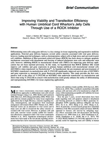
ROCK INHIBITOR STEM CELL GENE DELIVERYobjective (Olympus). Images were captured using the software, SlideBook (Intelligent Imaging Innovations (3i),Denver, CO, USA). A mercury arc lamp was used with thefollowing excitation filters (Excitation/Emission) for imagecollection: Hoechst (387 – 11 nm/447 – 60 nm) and GFP(494 – 20 nm/531 – 22 nm). For each sample that was imaged, a montage was generated from 25 (five by five arrangement) neighboring fields of view that were alignedtogether to generate one comprehensive composite image ofthe sample. All experiments were repeated three times foreach umbilical cord at 24 h and 48 h.FACS analysisImmediately after imaging, cells were washed twice,trypsinized, and transferred into 5-mL polypropylene roundbottomed tubes (BD Biosciences). A 0.5-lL amount ofpropidium iodide (PI) (1 mg/mL; Life Technologies) wasadded to each sample just before analysis. hUCMSCs wereanalyzed via flow cytometry on the Beckman Coulter MoFloXDP FACS. A total of 20,000 events were recorded for eachsample analyzed. Flow cytometry was used to analyze bothcell viability and transfection efficiency. Live hUCMSCswere characterized as hUCMSCs expressing Hoechst at anintensity of 102 relative fluorescence units (RFU) or above,with expression of PI at an intensity below 100 RFU. DeadhUCMSCs were characterized as hUCMSCs that expressedHoechst at an intensity below 102 RFU and expressed PI atan intensity of above 100 RFU. GFP-positive hUCMSCswere characterized as live hUCMSCs that expressed GFP atan intensity of 100 RFU or greater. Transfection efficiency93was determined by dividing the number of live GFP-positivecells in a sample by the total population of the sample. Allexperiments were repeated three times for each cord at 24 hand 48 h post-transfection. An example of how cell populations were gated is provided in Figure 1 and the statisticsfor all samples from an entire umbilical cord are displayedin Table 1.StatisticsAll values are reported as means – standard deviations. Aone-way analysis of variance (ANOVA) was performedwith a Tukey’s post hoc test to assess statistical significancewith n 5 cords. Statistical significance was set at p 0.05.Results and DiscussionAs hypothesized, hUCMSCs treated with RI displayedsignificantly increased survival rates and transfection efficiencies after nucleofection than hUCMSCs that were nottreated with RI. Gene delivery is a powerful tool for reprogramming hUCMSCs as demonstrated by Baksh et al. (2007)and Devarajan et al. (2013). Until now, nonviral deliverymethods have suffered from poor transfection efficiency withhigh cell viability or high transfection efficiency with poorcell viability. RhoA GTP signaling pathways are criticalfor inducing several apoptotic mechanisms in response tounfavorable environmental changes (Ichikawa et al., 2013;Ohgushi et al., 2010). Inhibiting ROCK can reduce apoptosisassociated with detachment and freezing protocols (Harbet al., 2008; Ichikawa et al., 2012; Koyanagi et al., 2008;Kurosawa, 2012; Olson, 2008). One of the key disadvantagesFIG. 1. Flow cytometry gating parameters used to quantify cell numbers. The set of histograms displayed are an arbitraryselection of a single replicate for each treatment from one umbilical cord out of five tested. Nuc ( / - ) designates whethercells were Nucleofected or not. DNA ( / - ) designates whether cells received 5 lg of pmaxGFP or not. RFU, relativefluorescence units; RI, ROCK Inhibitor. Color images available online at www.liebertpub.com/cell
Table 1. Gating StatisticsTreatmentReplicateCell numberfrom gateNuc (-)DNA 19,57119,226 – 38518,80518,93318,98918,909 – 9419,75319,75619,73819,749 – 1019,32319,17819,16119,221 – 8919,03319,29818,87619,069 – 213Nuc (-)DNA ( )Nuc ( )DNA (-)Nuc ( )DNA ( )Nuc ( )DNA ( )10 lM RIGFP ( )GFP %0.0%0.0%0.0%0.0%0.2%1.4%1.2%0.93 – 0.64%16.5%25.8%8.2%16.8 – 0%5.0%4.7%3.8%4.5 – 0.62%36.3%29.8%29.6%31.9 – 3.8%91.2%97.0%96.9%95.0 – 3.3%11.9%12.1%10.1%11.4 – 1.1%93.3%96.9%97.0%95.7 – 2.1%1.9%1.4%1.4%1.6 – 0.32%3.8%7.9%2.6%4.8 – 2.8%8.8%3.0%3.1%5.0 – 3.3%88.1%97.9%89.9%92.0 – 5.2%6.7%3.2%3.0%4.3 – 2.1%92.9%92.6%93.6%93.0 – 0.53%43.4%36.5%59.6%46 – 12%RI, Rock Inhibitor.FIG. 2. (A) Intensity maps of cell density with corresponding flow cytometry histograms of Hoechst signal distribution24 h after transfection. (B) Intensity maps of GFP expression with corresponding flow cytometry histograms of GFPexpression 24 h after transfection. The set of images and corresponding histograms are an arbitrary selection from one cordout of five tested. Nuc ( / - ) designates whether cells were Nucleofected or not. DNA ( / - ) designates whether cellsreceived 5 lg of pmaxGFP or not. Scale bar, 500 lm. RFU, relative fluorescence units; GFP, green fluorescent protein; RI,ROCK Inhibitor. Color images available online at www.liebertpub.com/cell94
ROCK INHIBITOR STEM CELL GENE DELIVERYof using nonviral physical delivery methods such as electroporation is the need to dissociate cells from adherent surfaces.However, by using Y-27632-RI to mitigate some of the apoptotic mechanisms induced by cell detachment and electricshock, it might be possible to rescue positively transfectedcells from cell death, which was the primary goal of this study.Flow cytometry analysis revealed that the cell populationswere mostly nonhematopoietic because the hUCMSCs were98.1 – 0.04% negative for CD34 expression and 90.4 – 1.1%negative for CD45 expression. The expression of CD90, akey mesenchymal stem cell marker, was detected in 98.5 –0.54% of cells. The expression of remaining key mesenchymal stem cell markers, CD73 (12.0 – 7.8%), CD105(10.2 – 0.15%), and STRO-1 (3.4 – 0.33%), were low andrelatively variable, suggesting subpopulations may existwithin each cell population that displayed surface epitopesconsistent with mesenchymal stem cell markers, as reviewedby (Wang et al., 2009; Wang et al., 2011).As was seen in the microscopy images at 24 h (Fig. 2A)and 48 h (Fig. 3A) after transfection, hUCMSCs that werenot subjected to nucleofection displayed high cell densitiescompared to hUCMSCs that were subject to nucleofection.95However, hUCMSCs that were treated with 10 lM ofY-27632-RI before and after transfection displayed a greatercell density than hUCMSCs that were subject to nucleofection and not treated with Y-27632-RI, as shown in microscopy images and corroborated by flow cytometry data.There was a clear increase in both cell viability andtransfection efficiency between the experimental group ofhUCMSCs that was nucleofected and treated with Y-27632RI and the group of hUCMSCs that was nucleofected andnot treated with Y-27632-RI at both 24 h and 48 h aftertransfection (Fig. 4). Cell viability was 3.3 times greater inhUCMSCs treated with Y-27632-RI than hUCMSCs thatwere not treated with Y-27632-RI 24 h after transfection( p 0.05), whereas cell viability was 3.2 times greater inhUCMSCs treated with Y-27632-RI than hUCMSCs nottreated with Y-27632-RI 48 h after transfection ( p 0.01).Transfection efficiency was 4.6 times greater in hUCMSCstreated with Y-27632-RI than hUCMSCs not treated withY-27632-RI 24 h after transfection ( p 0.01). At 48 h aftertransfection, transfection efficiency was 4.8 times greater inhUCMSCs treated with Y-27632-RI than hUCMSCs nottreated with Y-27632-RI ( p 0.05).FIG. 3. (A) Intensity maps of cell density with corresponding flow cytometry histograms of Hoechst signal distribution48 h after transfection. (B) Intensity maps of GFP expression with corresponding flow cytometry histograms of GFPexpression 48 h after transfection. The set of images and corresponding histograms are an arbitrary selection from one cordout of five tested. Nuc ( / - ) designates whether cells were Nucleofected or not. DNA ( / - ) designates whether cellsreceived 5 lg of pmaxGFP or not. Scale bar, 500 lm. RFU, relative fluorescence units; GFP, green fluorescent protein; RI,ROCK Inhibitor. Color images available online at www.liebertpub.com/cell
96MELLOTT ET AL.stromal cells for an electroporative gene delivery strategy.Transfection efficiency significantly increased four-fold andcell viability increased three-fold in hUCMSC populationsthat were treated with 10 lM of Y-27632-RI before and aftertransfection compared to hUCMSC populations not treatedwith Y-27632-RI. Although this study focused onhUCMSCs and provides an example for evaluating the effect of Y-27632-RI, other cell types undergoing electroporation may benefit from Y-27632-RI treatment, althoughdosing levels, application of treatment, and timing oftreatment should be tailored for each cell type and application. The use of Y-27632-RI provides an opportunity tobenefit strategies that combine both stem cell therapy andgene therapy for regenerative medicine applications.AcknowledgmentsFIG. 4. Live/dead and transfection efficiency collected viaflow cytometry 24 h and 48 h after transfection. Groups thatdid not express GFP were not plotted in the bar chart. (*)Statistically significant difference ( p 0.05) fromhUCMSCs that underwent Nucleofection without RIsupplement; (#) statistically significant difference ( p 0.01)from hUCMSCs that underwent Nucleofection without RIsupplement. The results are representative of cells collectedfrom five different umbilical cords (n 5) and are reportedas statistical means. All experiments were repeated threetimes. Error bars represent standard deviations. Nuc ( / - )designates whether cells were Nucleofected or not. DNA( / - ) designates whether cells received 5 lg of pmaxGFPor not. RI, ROCK Inhibitor.The difference in GFP intensity may have been a result ofan increased number of live cells present and able to readhere to a surface when treated with Y-27632-RI as opposedto hUCMSCs that were not treated with Y-27632-RI. Theflow cytometry histograms were consistent with microscopyimages in showing that a greater number of hUCMSCstreated with Y-27632-RI survived and expressed GFP atvarying intensities at both 24 h (Fig. 2B) and 48 h (Fig. 3B)after transfection than hUCMSCs that were not treated withY-27632-RI. Furthermore, the data from the histogramssuggested that there might be a relationship between GFPexpression and cell density. Further formal studies areneeded to verify if an actual relationship exists.hUCMSCs treated with Y-27632-RI at both 24 h and 48 hpost-transfection displayed an increase in cell viability and afar more substantial increase in transfection efficiency compared to hUCMSCs that were not treated with Y-27632-RI.Thus, future studies are needed to explore whether a synergistic phenomenon is occurring in which Y-27632-RI is notonly rescuing dying cells, but also improving cell health tofacilitate expression of GFP in cells that may not have beenpreviously able to express GFP. Further long-term studies areneeded to determine whether Y-27632-RI can prolong andsustain gene expression in hUCMSCs. Additionally, followup studies are needed to determine whether Y-27632-RI cannegatively affect multipotency character and downstreamdifferentiation of hUCMSCs for tissue engineering applications (Watanabe et al., 2007; Pakzad et al., 2010).For the first time, it was demonstrated that Y-27632-RIenhanced survival and gene expression in mesenchymalWe would like to acknowledge the efforts of the nursingstaffs at the University of Kansas Hospital (Kansas City,KS), Lawrence Memorial Hospital (Lawrence, KS), andStormont-Vail (Topeka, KS) for assisting us in obtaininghuman umbilical cords. Furthermore, we would like to acknowledge the efforts of Peggy Keefe and Austin Smith fortheir assistance on this project. This project was funded by theNational Institutes of Health (NIH) and the state of Kansas.Author Disclosure StatementThe authors declare that there are no conflicts of interest.ReferencesAndre, F.M., and Mir, L.M. (2010). Nucleic acids electrotransfer in vivo: Mechanisms and practical aspects. Curr GeneTher 10, 267–280.Bailey, M., Wang, L., Bode, C., Mitchell, K., and Detamore, M.(2007a). A comparison of human umbilical cord matrix stemcells and temporomandibular joint condylar chondrocytes fortissue engineering temporomandibular joint condylar cartilage. Tiss. Eng. 13, 2003–2010.Baksh, D., Yao, R., and Tuan, R.S. (2007). Comparison ofproliferative and multilineage differentiation potential ofhuman mesenchymal stem cells derived from umbilical cordand bone marrow. Stem Cells 25, 1384–1392.Chatterjee, P., Cheung, Y., and Liew, C. (2011). Transfectingand nucleofecting human induced pluripotent stem cells. J.Vis. Exp. Oct 5;(56). pii: 3110. doi: 10.3791/3110.Check, E. (2005). Gene therapy put on hold as third child develops cancer. Nature 433, 561.Claassen, D.A., Desler, M.M., and Rizzino, A. (2009). ROCKinhibition enhances the recovery and growth of cryopreservedhuman embryonic stem cells and human induced pluripotentstem cells. Mol. Reprod. Dev. 76, 722–732.Devarajan, K., Forrest, M.L., Detamore, M.S., and Staecker, H.(2013). Adenovector-mediated gene delivery to humanumbilical cord mesenchymal stromal cells induces inner earcell phenotype. Cell. Reprogram. 15, 43–54.Ear, T., Giguere, P., Fleury, A., Stankova, J., Payet, M.D., andDupuis, G. (2001). High efficiency transient transfection ofgenes in human umbilical vein endothelial cells by electroporation. J. Immunol. Methods 257, 41–49.Emre, N., Vidal, J.G., Elia, J., O’Connor, E.D., Paramban, R.I.,Hefferan, M.P., Navarro, R., Goldberg, D.S., Varki, N.M.,Marsala, M., and Carson, C.T. (2010). The ROCK inhibitor
ROCK INHIBITOR STEM CELL GENE DELIVERYY-27632 improves recovery of human embryonic stem cellsafter fluorescence-activated cell sorting with multiple cellsurface markers. PloS One 5, e12148.Gauthaman, K., Fong, C.Y., and Bongso, A. (2010a). Effect ofROCK inhibitor Y-27632 on normal and variant human embryonic stem cells (hESCs) in vitro: Its benefits in hESCexpansion. Stem Cell Rev. 6, 86–95.Gauthaman, K., Fong, C.Y., Subramanian, A., Biswas, A., andBongso, A. (2010b). ROCK inhibitor Y-27632 increasesthaw-survival rates and preserves stemness and differentiationpotential of human Wharton’s jelly stem cells after cryopreservation. Stem Cell Rev. 6, 665–676.Golzio, M., Escoffre, J.M., Portet, T., Mauroy, C., Teissie, J.,Dean, D.S., and Rols, M.P. (2010). Observations of themechanisms of electromediated DNA uptake—from vesiclesto tissues. Curr. Gene Ther. 10, 256–266.Harb, N., Archer, T., and Sato, N. (2008). The Rho-RockMyosin signaling axis determines cell-cell integrity of selfrenewing pluripotent stem cells. PloS One 3, e3001.Ichikawa, H., Nakata, N., Abo, Y., Shirasawa, S., Yokoyama,T., Yoshie, S., Yue, F., Tomotsune, D., and Sasaki, K. (2012).Gene pathway analysis of the mechanism by which the Rhoassociated kinase inhibitor Y-27632 inhibits apoptosis inisolated thawed human embryonic stem cells. Cryobiology64, 12–22.Ichikawa, H., Kanoh, Y., Shirasawa, S., Yokoyama, T., Yue, F.,Tomotsune, D., and Sasaki, K. (2013). Unique kinetics ofOct3/4 microlocalization following dissociation of humanembryonic stem cell colonies. Ann. Anat. 195, 50–56.Jang, J.H., Houchin, T.L., and Shea, L.D. (2004). Gene deliveryfrom polymer scaffolds for tissue engineering. Expert Rev.Med. Devices 1, 127–138.Joo, H., Choi, D.-K., Lim, J., Park, J.-S., Lee, S.-H., Song, S.,Shin, J., Lim, D.-S., Kim, I., Hwang, K.-C., and Koh, G.Y.(2012). ROCK suppression promotes differentiation and expansion of endothelial cells from embryonic stem cell-derivedFlk1( ) mesodermal precursor cells. Blood 120, 2733–2744.Kofron, M.D., and Laurencin, C.T. (2006). Bone tissue engineering by gene delivery. Adv. Drug Deliv. Rev. 58, 555–576.Koyanagi, M., Takahashi, J., Arakawa, Y., Doi, D., Fukuda, H.,Hayashi, H., Narumiya, S., and Hashimoto, N. (2008). Inhibition of the Rho/ROCK pathway reduces apoptosis duringtransplantation of embryonic stem cell-derived neural precursors. J. Neurosci. Res. 86, 270–280.Kurosawa, H. (2012). Application of Rho-associated proteinkinase (ROCK) inhibitor to human pluripotent stem cells. J.Biosci. Bioeng. 114, 577–581.Mellott, A.J., Forrest, M.L., and Detamore, M.S. (2013). Physical non-viral gene delivery methods for tissue engineering.Ann. Biomed. Eng. 41, 446–468.Narumiya, S., Ishizaki, T., and Uehata, M. (2000). Use andproperties of ROCK-specific inhibitor Y-27632. MethodsEnzymol. 325, 273–284.Ohgushi, M., and Sasai, Y. (2011). Lonely death dance ofhuman pluripotent stem cells: ROCKing between metastablecell states. Trends Cell Biol. 21, 274–282.Ohgushi, M., Matsumura, M., Eiraku, M., Murakami, K., Aramaki, T., Nishiyama, A., Muguruma, K., Nakano, T., Suga,H., Ueno, M., Ishizaki, T., Suemori, H., Narumiya, S., Niwa,H., and Sasai, Y. (2010). Molecular pathway and cell stateresponsible for dissociation-induced apoptosis in humanpluripotent stem cells. Cell Stem Cell 7, 225–239.97Olson, M.F. (2008). Applications for ROCK kinase inhibition.Curr. Opin. Cell Biol. 20, 242–248.Pakzad, M., Totonchi, M., Taei, A., Seifinejad, A., Hassani,S.N., and Baharvand, H. (2010). Presence of a ROCK inhibitor in extracellular matrix supports more undifferentiatedgrowth of feeder-free human embryonic and induced pluripotent stem cells upon passaging. Stem Cell Rev. 6, 96–107.Shi, J., Wu, X., Surma, M., Vemula, S., Zhang, L., Yang, Y.,Kapur, R., and Wei, L. (2013). Distinct roles for ROCK1 andROCK2 in the regulation of cell detachment. Cell Death Dis.4, e483.Shin, S., Salvay, D.M., and Shea, L.D. (2010). Lentivirus delivery by adsorption to tissue engineering scaffolds.J. Biomed. Mater. Res. A 93:1252–1259.Thomas, C.E., Ehrhardt, A., and Kay, M.A. (2003). Progressand problems with the use of viral vectors for gene therapy.Nat. Rev. Genet. 4, 346–358.Wang, L., Tran, I., Seshareddy, K., Weiss, M.L., and Detamore,M.S. (2009). A comparison of human bone marrow-derivedmesenchymal stem cells and human umbilical cord-derivedmesenchymal stromal cells for cartilage tissue engineering.Tiss. Eng. Part A 15, 2259–2266.Wang, L., Ott, L., Seshareddy, K., Weiss, M.L., and Detamore, M.S.(2011). Musculoskeletal tissue engineering with human umbilical cord mesenchymal stromal cells. Regen. Med. 6, 95–109.Watanabe, K., Ueno, M., Kamiya, D., Nishiyama, A., Matsumura, M., Wataya, T., Takahashi, J.B., Nishikawa, S., Nishikawa, S., Muguruma, K., and Sasai, Y. (2007). A ROCKinhibitor permits survival of dissociated human embryonicstem cells. Nat. Biotechnol. 25, 681–686.Weiss, M.L., and Troyer, D. (2008). Stem cells in the umbilicalcord. Stem Cell Reviews 6, 155–162.Xu, B., Song, G., Ju, Y., Li, X., Song, Y., and Watanabe, S.(2012). RhoA/ROCK, cytoskeletal dynamics, and focal adhesion kinase are required for mechanical stretch-inducedtenogenic differentiation of human mesenchymal stem cells.J. Cell. Physiol. 227, 2722–2729.Zhang, K., Zhang, H., Xiang, H., Liu, J., Liu, Y., Zhang, X.,Wang, J., and Tang, Y. (2013). TGF-b1 induces the dissolution of tight junctions in human renal proximal tubularcells: Role of the RhoA/ROCK signaling pathway. Int. J. Mol.Med. 32, 464–468.Zhang, L., Valdez, J.M., Zhang, B., Wei, L., Chang, J., and Xin,L. (2011). ROCK inhibitor Y-27632 suppresses dissociationinduced apoptosis of murine prostate stem/progenitorcells and increases their cloning efficiency. PloS One 6,e18271.Zhang, X., and Godbey, W.T. (2006). Viral vectors for genedelivery in tissue engineering. Adv. Drug
analyzed via flow cytometry on the Beckman Coulter MoFlo XDP FACS. A total of 20,000 events were recorded for each sample analyzed. Flow cytometry was used to analyze both cell viability and transfection efficiency. Live hUCMSCs were characterized as hUCMSCs expressing Hoechst at an intensity of 102 relative fluorescence units (RFU) or above,