Transcription
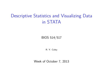
Discovering and Visualizing Patterns in EEG DataErik W. Anderson Catherine Chong†Gilbert A. Preston‡Cláudio T. Silva§University of UtahBarrow Neurological InstituteUniversity of UtahNew York UniversityFigure 1: Our system is composed of many linked views. Structural MRI data is presented in a 3D visualization (top left) whose transfer functionis manipulated using 2D widgets (bottom right). Users may select individual EEG sensors (yellow sphere) in the 3D visualization to explore itsraw signal (bottom left) and time-frequency representation (middle right). The user also interacts with the time-frequency query area (top right)to generate spectral queries. The time-frequency display and query area are colormapped using an interactive widget (middle left). Combinedwith the settings in the parameter dialog, the query is issued and relevant sensors are colored blue according to their correlation with the query.A BSTRACTBrain activity data is often collected through the use of electroencephalography (EEG). In this data acquisition modality, the electricfields generated by neurons are measured at the scalp. Although thistechnology is capable of measuring activity from a group of neurons, recent efforts provide evidence that these small neuronal collections communicate with other, distant assemblies in the brain’scortex. These collaborative neural assemblies are often found byexamining the EEG record to find shared activity patterns.In this paper, we present a system that focuses on extractingand visualizing potential neural activity patterns directly from EEGdata. Using our system, neuroscientists may investigate the spectraldynamics of signals generated by individual electrodes or groupsof sensors. Additionally, users may interactively generate querieswhich are processed to reveal which areas of the brain may exhibitcommon activation patterns across time and frequency. The utilityof this system is highlighted in a case study in which it is used toanalyze EEG data collected during a working memory experiment. g‡ e-mail: gpreston@gmail.com§ e-mail: csilva@nyu.edu† e-mail:Index Terms: Computer Graphics [I.3.4]: Graphics Utilities—Application Packages, Electroencephalography1I NTRODUCTIONElectroencephalography (EEG) is a technology developed to investigate the alert and working brain by reading the electric fields generated by small neuron groups in the cortex [29]. Since its first usein humans in 1924, EEG has had a profound impact on our understanding of the brain [9, 41]. The processing and analysis of EEGdata falls into two general categories: time domain analysis andspectral analysis.Analysis of EEG data in terms of its signal amplitude has leadto many discoveries regarding the functional characteristics of various areas of the brain [6]. However, exploration of EEG data in thefrequency domain has provided additional insight into brain function [2]. Although interpreting EEG data in the time domain remains a powerful tool for neuroscientists, we focus on the benefitsand difficulties of spectral analysis. Our system (Figure 1) focuseson exploring spectral patterns generated by the brain by examiningthe collection of time-frequency decompositions generated by anEEG electrode array.One important utilization of EEG analysis is for the study ofbrain function expressed as coordinated neural activity. Identification of these activity patterns allow scientists to develop newtheories regarding neurological disorders [32], overall brain function [11], as well as identify potential new treatments for devastating diseases [31].
Determining neural activation patterns sub-serving a specificbrain function is a difficult problem. Functional imaging techniques, such as functional magnetic resonance imaging (fMRI),have provided insight into areas of the brain that are connected.fMRI studies remain powerful mechanisms to examine coordination between different areas of the brain. Buccino et al. provide just one example of fMRI used to uncover functional connections within the brain [8]. These functional studies have revealedthat many processes in the brain are spread across cortical regionsthroughout the brain, as shown in Figure 2. Unfortunately, fMRIstudies are limited in temporal resolution and, because it measuresmetabolic rates, cannot provide insights into spectral dynamics.Discovering important activity characteristics has several usesin the field of neuroscience. These relationships may be monitored and manipulated to diagnose and treat psychological disorders, respectively [30]. Additionally, insights generated from exploring the various functions of different spatio-temporal patternsfrom EEG often lead to a better understanding of brain functionthat can be exploited to create new and more effective drug therapyregimens [22]. Finally, understanding the temporal and spectral underpinnings of different brain functions is useful when determiningthe processing and analysis parameters used to properly interpretneurophysiological data.Ilmoniemi et al. have shown that patterns present in EEG datamay be used to identify potential neural circuits [20]. Our system extends this concept by allowing users to query EEG recordsusing time-frequency patterns of their own design. This queryingparadigm enhances the flexibility of the system, allowing scientiststo explore EEG data in a new way.Contributions. In this paper, we make the following contributions: We demonstrate that the ability to query collections of timefrequency planes with user-generated examples provides scientists with an effective new tool for the exploration of EEGdata. We present a novel technique for the exploration of timefrequency patterns through the use of filtered, normalizedcross-correlogram images. We show that our system enhances the analysis of EEG dataduring working memory studies, expediting the validation ofstated hypotheses.This paper is organized as follows. After a brief discussion ofrelated work in Section 2, Section 3 discusses the details of our system. Section 4 presents the process of generating time-frequencyqueries, while Section 5 shows how these queries are used to extractpatterns across EEG data. In Sections 6 and 7 we present featuresin the data elucidated by the use of our technique in the context ofa working memory study. A robust discussion of this technique isfound in Sections 8 and 9.2R ELATED W ORKDue to the maturity of EEG technology, many analysis techniqueshave been developed to process the associated signal data. Somestudies use event-related potential (ERP) analysis [25] in whichthe processing and interpretation of EEG data occurs in the timedomain. However, many studies forgo ERP analysis and use processing techniques in the frequency domain [14]. Insights gainedfrom the spectral dynamics computed from EEG data have lead toa richer understanding of overall brain functions [16].Finding a collection of EEG sensors that record similar activityis useful for neuroscientists. Often, this analysis consists of forming a graph where the vertices represent individual electrodes andthe edges are generated by examining coherence between its twoFigure 2: Brain activity related to working memory is generated inmore than just one part of the brain. Almost every part of the brainexhibits various patterns of activation spanning the temporal cortex(green), the parietal cortex (blue), the frontal cortex (light red) andthe prefrontal cortex (dark red).nodes. However, these methods are often plagued with visualization difficulties, including excessive clutter and computation time.To address these issues, ten Caat et al. introduced coherence analysis using data-driven functional units [42].To begin exploring the rhythms of brain function, raw EEG datamust be processed. Several EEG processing environments havebeen created to support spectral analysis of EEG signals. The Matlab package EEGLab [12] is a well-used EEG processing suite providing powerful tools to neuroscientists. Although EEGLab implements several advanced analysis tools, it is a more general toolwhereas our system focuses on investigating spectral patterns expressed across multiple EEG electrodes.Identifying important signals and sources to explore EEG timeseries remains an important problem. Ilmoniemi et al. use transcranial magnetic stimulation (TMS) as a known signal for the interrogation of EEG data [20]. However, signal source separationusing independent component analysis (ICA) [45] may also be auseful mechanism to determine relevant patterns to search for whenexploring EEG data holistically [39].As recent studies have suggested that the spectral dynamics associated with neural processes are important, we focus our effortsin the frequency domain. With the advent of robust time-frequencydecompositions, investigating the evolution of brain activity in thefrequency domain is possible [26,40]. Evidence suggesting that thespectral dynamics of brain activity is mounting [2, 37]. Blanco etal. introduced pioneering new methods for time-frequency analysisfor EEG data [5]. In their work, they applied the Gabor transformto investigate the spectral dynamics of EEG data using robust visualization to enhance their analyses. In this work, we enhance someof their techniques with interactive visualization and exploration ofboth the spectral dynamics as well as the spatial characteristics associated with EEG. Our system is a step to extend more traditionalspatio-temporal ERP analysis techniques [43] with additional information regarding the frequency content of EEG signals.Aggregating collections of time-frequency planes into a singleconcise visualization or analysis is difficult. Dimensionality reduction techniques such as Locally Linear Embedding [34] andIsoMap [44] are able to organize these data into local neighborhoodrelationships. However, if only a measure of similarity is required,Gonzalez and Woods argue that correlation provides an acceptableestimation of similarity between two different images [17].
Explore EEG DataExtract Time-frequency PatternsFrequencyVisualize Time-frequency DataScalar Value to AddVisualize Raw EEG DataGenerate Time-frequency QueryCompute Cross-CorrelogramFilter Cross-CorrelogramsHighlight Similar Sensor DataFigure 3: Our system supports two primary functions: exploring EEGdata and extracting neural circuits. Users interact with the systemprimarily to extract and visualize neural circuits. To achieve the bestresults, users iteratively refine time-frequency queries and set filterparameters for the results. Here, yellow ovals represent steps thatusers have direct control over.TimeFigure 4: Creating a time-frequency query. The user interacts primarily with a 2D time-frequency plane in which a circular brush (blueoutline) controls the scalar field being painted. When paint strokesoverlap, their respective values are either additave or subtractive, allowing scientists to gradually change the time-frequency query beingconstructed. The time and frequency coordinates along with theirsupports of the kernel are displayed during interaction to ensure theuser knows exactly how the query is being manipulated.between the query and the data from each EEG sensor. However,in order to use these mechanics, the user must first construct theappropriate query to use.4.13S YSTEM OVERVIEWOur system is designed to facilitate the exploration of EEG datawith respect to the identification of functionally important timefrequency patterns. Figure 1 shows the user interface provided byour application. The choices made during the development of oursystem were designed to enhance the application’s intuitive usability while maintaining flexibility and functionality.The general workflow for the use of our system is illustrated inFigure 3. Our system begins with the user loading EEG and MRIdata. Each EEG sensor is displayed as a sphere in a 3D visualization. In this case, each sensor is rigidly registered to the structural MRI data. Next, the user sets the upper and lower frequencies to parametrize time-frequency decompositions. The user thenexplores both the raw time series data as well as its spectral representation. At this time, the user sets a query to issue to the system;this may be created from a time series present in the EEG collectionor through a brushable canvas. Once the query is defined (Sec. 4),cross-correlograms are computed between the query and the timefrequency planes derived from each EEG electrode (Sec. 5). Eachcross-correlogram is an image representing the cross-correlationstatistics of the query and the time-frequency plane being analyzed.The resulting images are then filtered to provide sensitivity to shiftsin both frequency and time. After filtering, the maximum correlations are displayed by coloring each sensor by its similarity withthe query. Higher correlation values are represented in the final visualization by more saturated color than lower values. In this way,coordinated areas of the brain, as seen by the sensor collection, arehighlighted and may influence future processing and analysis parameters.4EEG Q UERY B Y E XAMPLEHighlighting patterns of interest is performed in our system throughthe use of interactive, user-driven queries. The goal of our queryinterface is to allow the user to construct a pattern for which thesystem will search for without the need to manipulate a raw timeseries. The result of this computation is a measure of similarityEEG Spectral DynamicsAs more is learned about brain activity patterns, it is becomingclearer that EEG analysis in the frequency domain is important.To take advantage of a signal’s frequency content, without losingits temporal dynamics, we employ a time-frequency decompositionfor all incoming EEG data. While there exist many applicable timefrequency transforms to choose from, we have chosen to use the STransform [40]. The S-Transform may be thought of as both a generalization of the Short-Time Fourier Transform and an extensionof the Continuous Wavelet Transform. The transform generates acomplex time-frequency plane by integrating a gaussian window ateach time point as follows:(t τ)2 f 2 f h(τ) e 2 e i2π f τ dτ 2πZ ST (t, f ) where h(τ) is the EEG signal, f is frequency, t is a dyadictranslation in time, the first exponential is the gaussian window function, and the second is a harmonic function. Weuse an implementation of the S-Transform provided by the National Institute of Mental Health (NIMH) MEG Core ckwell).4.2Creating Time-Frequency QueriesThe result of the S-Transform is a complex plane composed of phasors at each time-frequency coordinate. By taking the magnitudeof each phasor, we generate an image of spectral power across timeand frequency. Although it is possible to create images of phasefrom S-Transformed planes, we restrict ourselves to the better understood dynamics of power.Creating time-frequency queries in our system is equivalent tocreating univariate images representing power changes where oneaxis is time and the other is frequency. To form a time-frequencyquery, users interact with time-frequency images. Allowing interaction with both images derived from the data of a single EEG sensoras well as images created from a blank canvas affords additionalflexibility to the user. All interactions with queries are performed
QuerySensor DataCross-CorrelogramFilter MaskFilteredCross-CorrelogramFigure 5: Finding the similarity between an input query and a sensor’s time-frequency data begins with computing the correlation image through convolution. A filter mask is then formed using the usercontrolled sensitivity and lag parameters. This mask is then multiplied with the correlation image. The maximum value of this filteredimage represents the filtered similarity between the input query andsensor data.on a brushable canvas, allowing users to directly modify the timefrequency patterns they wish to search for. Figure 4 shows the result of user interaction to add to and subtract from a time-frequencyquery using our painting interface.To create a query, the user begins with a time-frequency planewith the same dimensions as the transformed EEG data. Each valueon this plane is initially set to zero. The user adds values to thisscalar field by applying brush strokes similar to the airbrush tool inpopular image editing software. Our system uses a circular gaussian kernel for the brush by default, but provides various brush sizesand shapes to increase flexibility (Figure 4). The ability to manipulate user specified queries provides our system the flexibility to beused to analyze EEG experiments eliciting a variety of responsesrather than focusing exclusively on the working memory patternsdescribed by Klimesch [21]. Creating a time-frequency query requires the user to have an idea what patterns are interesting to thephenomenon being studied.Allowing users to interact directly with a time-frequency imageenables powerful modification techniques, such as painting withbrushes. This interaction mechanic is ubiquitous across both commercial and open-source image manipulation tools. In addition tothe concepts of brushes and painting common to image editing environments, global image properties are understood and changedmore easily. Like pictures whose lighting and exposure conditionscan drastically change their appearance, the overall power contained in a time-frequency image can alter the similarity measurebetween a query and the EEG input. To account for these differences, we normalize each image by subtracting its mean from eachelement, then dividing that quantity by the image’s standard deviation. This method of normalization prepares the images for similarity computations using normalized cross-correlation.5C ORRELATION S PACE PATTERN M ATCHINGWe take advantage of the work by Gonzalez and Nuñez to usequickly computed cross-correlations as an acceptable measure ofsimilarity [17]. The notion of using normalized cross correlation asa means for determining similarity between two images stems fromits use as a global approach for template based pattern matching [7].Figure 6: Large collections of time-frequency planes may be projected to a plane to reduce the overall dimensionality of the dataset.Here, IsoMap has been used to position individual planes while attempting to conserve the relative differences between the variousdata points.To measure similarity, we first compute cross-correlograms for eachpair of time-frequency query and transformed EEG data. We thenfilter these images to reduce the impact of template matches outsidethe user-specified time and frequency envelopes. Finally, we determine the similarity by inspecting the filtered cross-correlogram toextract the maximum value indicating the degree to which the querymatches the EEG data.5.1 Pairwise Image Cross-CorrelogramsOnce the user has defined a time-frequency query, we computethe cross-correlogram of the query with each EEG sensor’s timefrequency image. The normalized cross-correlogram, C(u, v), maybe computed by introducing normalization terms to the crosscorrelation term discussed by Lewis [23] as follows:C(u, v) 1( f (x, y) f )(q(x u, y v) q̄) n 1 x yσ f σqwhere f (x, y) is an EEG sensor’s time-frequency image, q(x, y)is the time-frequency query, f and q̄ are the means of the EEGand query data respectively, and σ f and σq are the standard deviations of the EEG and query data, respectively. However, by separating the normalization of each image from the convolution, we cantake advantage of Fourier transforms to compute the cross correlations for the entire image domain. Our system uses the FFTW libraries [15] to quickly compute Fourier transforms during this process.5.2 Filtering Cross-Correlogram ImagesOnce a cross-correlogram image is computed for a query and EEGtime-frequency plane, it must be filtered to ensure that the pointsof maximum correlations lie in the appropriate time and frequencywindows. A cross-correlogram is defined on a plane of time andfrequency. However, unlike its inputs, the origin of the crosscorrelogram is at the center of the image. These details gives rise tothe realization that high values of correlation offset from the originin the temporal domain represent strong query matches that lag intime. Likewise, departures from the origin in the other dimensionreflect patterns undergoing frequency drift, that is they appear atslightly higher or lower frequencies than the query.To ensure that patterns exhibited in both the query image as wellas in an EEG sensor’s time-frequency representation occur in appropriate time lags and frequency shifts, we allow the user to control the filtering parameters. The user sets the minimum and maximum time lag as well as the size of the frequency envelope. Theseparameters define a mask used to filter each cross-correlogram. InFigure 5, we see how an input query and time-frequency imagefrom an EEG sensor are combined to first form a cross-correlogram.The user settings define a filter mask that is then multiplied withthe cross-correlogram image to form the filtered cross-correlogram.
Figure 7: Using a data driven query to extract a neural circuit results in an overall high correlation because the query exists exactly in the data.However, carefully constructed user generated queries may yield similar results. Here, the differences between data driven and user generatedqueries are explored. On the left is the result of a user-generated query (top inset), while the rendering on the right is driven by a time-frequencyimage from the EEG collection (bottom inset).Since we multiply the filter mask with the cross-correlogram, weensure the range of the filter is [0,1] by composing it as the maximum of two separable filters. In practice, we have found that linear(tent) filters in both time and frequency provide appropriate results.5.3Computing SimilarityAlthough the visualization and analysis of small collections oftime-frequency planes is straightforward, examining large groupsremains difficult. To address this problem, techniques such asIsoMap [44] may be employed as in Figure 6. While organizingimage collections in this way strives to maintain a notion of similarity between neighboring points, fine control over differences islost.The construction and application of the filter mask acts as auser-specified weighting function specifying acceptable shifts in thequery pattern in both time and frequency. Our solution, using filtered cross-correlogram images, allows us to selectively tune ourfiltering process in both the temporal and spectral domains. Oncethe filter is parametrized by the user and applied, determining thedegree of similarity between the query and the EEG data is straightforward. Because the cross-correlogram is formed by convolution,we know that the point of highest intensity represents the point ofmaximum similarity.6S PECTRAL DYNAMICSINW ORKING M EMORYWorking memory, sometimes referred to as short-term memory, isresponsible for temporarily storing and retrieving information. Ithas been described as the short-term retention of information thatno longer exists in the environment and the manipulation of thisinformation for subsequent use in guiding behavior [3, 13]. Forexample, memorizing a new telephone number while trying to finda pen and paper to write it down exercises working memory.Previous studies have discovered that working memory performance is governed by specific oscillation patterns [36]. Of particular interest are the frequency bands of theta (3–7 Hz) and alpha(8–13 Hz) [21]. Additional studies have shown that working memory also relies on a functional neural circuit [19, 35]. These studieshighlight the advantages of coupling circuit-based approaches withspectral analysis.Although the brain is a highly distributed system, it is organizedinto spatial regions that play roles in various functions. Figure 2shows that almost all areas of the brain are involved with workingmemory processing and performance. Unfortunately, discoveringthe precise functional circuitry of the brain is a difficult task. Investigating cortical areas using more metabolic resources than othersthrough functional magnetic resonance imaging (fMRI) is a common technique [8]. However, Ilmoniemi et al. found that usingEEG to track signals induced by transcranial magnetic stimulation(TMS) uncovered coordinated areas of the brain [20]. This workrelied on using EEG recordings and the locations of the sensors toinvestigate the spatio-temporal relationships of coordinated areas ofthe brain. This type of analysis strengthens the more complicatedprocess of localizing the signal’s source on the cortical surface.7A S TUDYOFW ORKING M EMORYOur system has been used in the analysis of a working memoryexperiment similar to that performed by Ilmoniemi et al. [20]. Inthis experiment, a verbal working memory task was given to eachparticipant while 64 channel EEG was collected at 500 Hz usingan Electrical Geodesics GES 300 System (http://www.egi.com).Repetitive transcranial magnetic stimulation (rTMS) was then applied to the dorsal lateral prefrontal cortex (DLPFC) in the experimental group, while the control group received sham stimulation,the simulated application of rTMS. The goal of this experiment wasto explore the effects of rTMS on working memory.This study of verbal working memory performance was conducted using the Sternberg paradigm as the basic task [38]. TheSternberg task presents a string of consonants, followed by a brief(1.5 sec) maintenance period in which nothing is displayed. Afterthe maintenance period, a single letter is presented. The participantmust determine whether the probe letter was present in the originalstring as quickly and accurately as possible.Measuring Working Memory Performance During the workingmemory task, EEG data is collected. Clark et al. have shownthat working memory performance can be predicted from the spectral evolution of the alpha band of frequencies [10]. Similarly,Klimesch showed that working memory performance may be estimated by monitoring the oscillation of theta and alpha frequencybands [21]. We use the S-Transform [40] to decompose the rawEEG signal into a complex time-frequency plane to investigate itsspectral evolution.The collection of time-frequency planes computed from rawEEG data are further processed to extract energies (phasor magnitude) in the theta and alpha band of frequencies, commonly between3 and 7 Hz and 8 and 13 Hz, respectively. Individual mean alphafrequencies are computed from EEG collected during a rest period.
Figure 8: Our system helps provide evidence supporting the hypothesis that the application of rTMS enhances the performance of workingmemory. Using the query pictured above, we extracted a working memory circuit before stimulation with rTMS (left) and then after (right).Although the circuit extracted is similar in both cases, the overall similarity measure is markedly different. The expected spectral evolution ismore evident after stimulation.These individualized measurements are then compared to the samemeasures taken during the experiment. As Klimesch points out,the shifts in the mean frequency and the energy densities indicateworking memory performance during the task [21].Manipulating Working Memory rTMS has shown the ability tomanipulate the performance of various neural substrates [33]. Infact, previous work indicates that careful application of TMS mayeither enhance or disrupt working memory processes [24, 31]. Ourexperimental approach applied 10Hz rTMS to facilitate workingmemory performance.Although the details through which rTMS improves workingmemory performance are still unknown, previous studies show thatits effects are robust [31]. Here, we use the application of rTMSwithin the study to validate the findings presented by Ilmoniemi etal. [20]. We then show that our system is capable of using carefullycrafted queries to reproduce the same results without the benefit ofa known, induced signal.Visualizing Working Memory Performance Although this taskfocuses on verbal working memory, prior work shows that verbaland spatial memory tasks activate similar cortical regions - the prefrontal, frontal, parietal and temporal cortices [4, 27]. These worksrely primarily on fMRI and positron emission tomography (PET)to reveal functionally connected regions of the brain by examiningblood flow and changes in localized metabolism.To explore EEG data associated with working memory performance, queries expressing power in the theta and alpha bands offrequencies are created. After the user generates and submits atime-frequency query (Section 4), time-frequency images for eachEEG sensor are correlated with it (Section 5). Following this, theuser interacts with sliders representing the filtering parameters usedto determine sensitivity in the frequency and temporal domains(See Figure 5). Finally, the maximum value of the filtered crosscorrelogram image is found and used as the similarity of the timefrequency image to the user’s query (Section 5.3).This filtered cross-correlation measure of similarity is then presented in the three-dimensional representation of the sensor network registered to an MRI. The degree of similarity is indicated byblue spheres with highly saturated color representing high levels ofsimilarity and unsaturated spheres representing low similarity. Toreduce clutter in the final visualization, the user may further filterthe coloring of EEG sensors by thresholding the similarity measuresto display.The results of the user’s queries represent the similarity of eachEEG sensor’s time-frequency transform with the input query. Figure 7 shows that cortical regions activated during a working memory task are extracted using both known patterns found withinthe EEG record (data-driven query) as well as user-supplied patterns (user-driven query). The sensor coll
data falls into two general categories: time domain analysis and spectral analysis. Analysis of EEG data in terms of its signal amplitude has lead to many discoveries regarding the functional characteristics of vari-ous areas of the brain [6]. However, exploration of EEG data in the frequency domain has provided additional insight into brain func-