Transcription
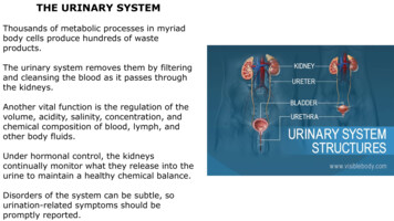
Human Physiology/The Urinary System1Human Physiology/The Urinary System The Immune System — Human Physiology — The respiratory system Homeostasis — Cells — Integumentary — Nervous — Senses — Muscular — Blood — Cardiovascular — Immune — Urinary — Respiratory— Gastrointestinal — Nutrition — Endocrine — Reproduction (male) — Reproduction (female) — Pregnancy — Genetics — Development —AnswersIntroductionThe Urinary System is a group oforgans in the body concerned withfiltering out excess fluid and othersubstances from the bloodstream. Thesubstances are filtered out from thebody in the form of urine. Urine is aliquid produced by the kidneys,collected in the bladder and excretedthrough the urethra. Urine is used toextract excess minerals or vitamins aswell as blood corpuscles from thebody. The Urinary organs include thekidneys, ureters, bladder, and urethra.The Urinary system works with theother systems of the body to helpmaintain homeostasis. The kidneys arethe main organs of homeostasisbecause they maintain the acid base balance and the water salt balance of the bloodFunctions of the Urinary SystemOne of the major functions of the Urinary system is the process of excretion. Excretion is the process of eliminating,from an organism, waste products of metabolism and other materials that are of no use. The urinary systemmaintains an appropriate fluid volume by regulating the amount of water that is excreted in the urine. Other aspectsof its function include regulating the concentrations of various electrolytes in the body fluids and maintaining normalpH of the blood. Several body organs carry out excretion, but the kidneys are the most important excretory organ.The primary function of the kidneys is to maintain a stable internal environment (homeostasis) for optimal cell andtissue metabolism. They do this by separating urea, mineral salts, toxins, and other waste products from the blood.They also do the job of conserving water, salts, and electrolytes. At least one kidney must function properly for lifeto be maintained. Six important roles of the kidneys are:Regulation of plasma ionic composition. Ions such as sodium, potassium, calcium, magnesium, chloride,bicarbonate, and phosphates are regulated by the amount that the kidney excretes.Regulation of plasma osmolarity. The kidneys regulate osmolarity because they have direct control over how manyions and how much water a person excretes.Regulation of plasma volume. Your kidneys are so important they even have an effect on your blood pressure. Thekidneys control plasma volume by controlling how much water a person excretes. The plasma volume has a directeffect on the total blood volume, which has a direct effect on your blood pressure. Salt(NaCl)will cause osmosis to
Human Physiology/The Urinary Systemhappen; the diffusion of water into the blood.Regulation of plasma hydrogen ion concentration (pH). The kidneys partner up with the lungs and they togethercontrol the pH. The kidneys have a major role because they control the amount of bicarbonate excreted or held onto.The kidneys help maintain the blood Ph mainly by excreting hydrogen ions and reabsorbing bicarbonate ions asneeded.Removal of metabolic waste products and foreign substances from the plasma. One of the most important thingsthe kidneys excrete is nitrogenous waste. As the liver breaks down amino acids it also releases ammonia. The liverthen quickly combines that ammonia with carbon dioxide, creating urea which is the primary nitrogenous endproduct of metabolism in humans. The liver turns the ammonia into urea because it is much less toxic. We can alsoexcrete some ammonia, creatinine and uric acid. The creatinine comes from the metabolic breakdown of creatinephospate (a high-energy phosphate in muscles). Uric acid comes from the break down of nucleotides. Uric acid isinsoluble and too much uric acid in the blood will build up and form crystals that can collect in the joints and causegout.Secretion of Hormones The endocrine system has assistance from the kidney's when releasing hormones. Renin isreleased by the kidneys. Renin leads to the secretion of aldosterone which is released from the adrenal cortex.Aldosterone promotes the kidneys to reabsorb the sodium (Na ) ions. The kidneys also secrete erythropoietin whenthe blood doesn't have the capacity to carry oxygen. Erythropoietin stimulates red blood cell production. TheVitamin D from the skin is also activated with help from the kidneys. Calcium (Ca ) absorption from the digestivetract is promoted by vitamin D.CC: Chapter Check: Name the role of the kidneys and how they work?2
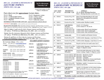
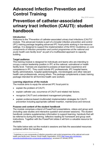
Human Physiology/The Urinary SystemOrgans in the Urinary SystemKidneys And Their StructureThe kidneys are a pair of bean shaped,reddish brown organs about the size of yourfist.It measures 10-12 cm long. They arecovered by the renal capsule, which is atough capsule of fibrous connective tissue.Adhering to the surface of each kidney istwo layers of fat to help cushion them.There is a concaved side of the kidney thathas a depression where a renal artery enters,and a renal vein and a ureter exit the kidney.The kidneys are located at the rear wall ofthe abdominal cavity just above thewaistline, and are protected by the ribcage.They are considered retroperitoneal, whichmeans they lie behind the peritoneum. Thereare three major regions of the kidney, renalcortex, renal medulla and the renal pelvis.The outer, granulated layer is the renalcortex. The cortex stretches down inbetween a radially striated inner layer. Theinner radially striated layer is the renal medulla. This contains pyramid shaped tissue called the renal pyramids,separated by renal columns. The ureters are continuous with the renal pelvis and is the very center of the kidney.1. Renal pyramid2. Interlobar artery3. Renal artery4. Renal vein5. Renal hylum6. Renal pelvis7. Ureter8. Minor calyx9. Renal capsule10. Inferior renal capsule11. Superior renal capsule12. Interlobar vein13. Nephron14. Minor calyx15. Major calyx16. Renal papilla17. Renal column3
Human Physiology/The Urinary SystemRenal VeinThe renal veins are veins that drain the kidney. They connect the kidney to the inferior vena cava. Because theinferior vena cava is on the right half of the body, the left renal vein is generally the longer of the two. Unlike theright renal vein, the left renal vein often receives the left gonadal vein (left testicular vein in males, left ovarian veinin females). It frequently receives the left suprarenal vein as well.Renal ArteryThe renal arteries normally arise off the abdominal aorta and supply the kidneys with blood. The arterial supply ofthe kidneys are variable and there may be one or more renal arteries supplying each kidney. Due to the position ofthe aorta, the inferior vena cava and the kidneys in the body, the right renal artery is normally longer than the leftrenal artery. The right renal artery normally crosses posteriorly to the inferior vena cava. The renal arteries carry alarge portion of the total blood flow to the kidneys. Up to a third of the total cardiac output can pass through therenal arteries to be filtered by the kidneys.UretersThe ureters are two tubes that drain urine from the kidneys to the bladder. Each ureter is a muscular tube about 10inches (25 cm) long. Muscles in the walls of the ureters send the urine in small spurts into the bladder, (a collapsiblesac found on the forward part of the cavity of the bony pelvis that allows temporary storage of urine). After the urineenters the bladder from the ureters, small folds in the bladder mucosa act like valves preventing backward flow ofthe urine. The outlet of the bladder is controlled by a sphincter muscle. A full bladder stimulates sensory nerves inthe bladder wall that relax the sphincter and allow release of the urine. However, relaxation of the sphincter is also inpart a learned response under voluntary control. The released urine enters the urethra.Urinary BladderThe urinary bladder is a hollow, muscular and distendible or elastic organ that sits on the pelvic floor (superior tothe prostate in males). On its anterior border lies the pubic symphysis and, on its posterior border, the vagina (infemales) and rectum (in males). The urinary bladder can hold approximately 17 to 18 ounces (500 to 530 ml) ofurine, however the desire to micturate is usually experienced when it contains about 150 to 200 ml. When thebladder fills with urine (about half full), stretch receptors send nerve impulses to the spinal cord, which then sends areflex nerve impulse back to the sphincter (muscular valve) at the neck of the bladder, causing it to relax and allowthe flow of urine into the urethra. The Internal urethral sphincter is involuntary. The ureters enter the bladderdiagonally from its dorsolateral floor in an area called the trigone. The trigone is a triangular shaped area on thepostero-inferior wall of the bladder. The urethra exits at the lowest point of the triangle of the trigone. The urine inthe bladder also helps regulate body temperature. If the bladder becomes completely void of fluid, it causes thepatient to chill.4
Human Physiology/The Urinary System5UrethraThe urethra is a muscular tube that connects thebladder with the outside of the body. The function ofthe urethra is to remove urine from the body. Itmeasures about 1.5 inches (3.8 cm) in a woman but upto 8 inches (20 cm) in a man. Because the urethra is somuch shorter in a woman it makes it much easier for awoman to get harmful bacteria in her bladder this iscommonly called a bladder infection or a UTI. Themost common bacteria of a UTI is E-coli from the largeintestines that have been excreted in fecal matter.Female urethraFemale urethra (labeled at bottom right.)In the human female, the urethra is about 1-2 incheslong and opens in the vulva between the clitoris and thevaginal opening.Men have a longer urethra than women. This meansthat women tend to be more susceptible to infections ofthe bladder (cystitis) and the urinary tract.Male urethraIn the human male, the urethra is about 8 inches longand opens at the end of the head of the penis.The length of a male's urethra, and the fact it contains anumber of bends, makes catheterisation more difficult.The urethral sphincter is a collective name for themuscles used to control the flow of urine from theurinary bladder. These muscles surround the urethra, sothat when they contract, the urethra is closed. There are two distinct areas of muscle: the internalsphincter, at the bladder neck and the external, or distal, sphincter.Human males have much stronger sphincter musclesthan females, meaning that they can retain a largeamount of urine for twice as long, as much as 800mL,i.e. "hold it".NephronsA nephron is the basic structural and functional unit ofthe kidney. The name nephron comes from the Greekword (nephros) meaning kidney. Its chief function is toregulate water and soluble substances by filtering theblood, reabsorbing what is needed and excreting the rest as urine. Nephrons eliminate wastes from the body, regulateblood volume and pressure, control levels of electrolytes and metabolites, and regulate blood pH. Its functions arevital to life and are regulated by the endocrine system by hormones such as antidiuretic hormone, aldosterone, andparathyroid hormone.Male Sphincter urethrae muscle - The male urethra laidopen on its anterior (upper) surface. (Region visible,but muscle not labeled.)
Human Physiology/The Urinary SystemEach nephron has its own supply of blood from two capillary regions from the renal artery. Each nephron iscomposed of an initial filtering component (the renal corpuscle) and a tubule specialized for reabsorption andsecretion (the renal tubule). The renal corpuscle filters out large solutes from the blood, delivering water and smallsolutes to the renal tubule for modification.GlomerulusThe glomerulus is a capillary tuft that receives its blood supply from an afferent arteriole of the renal circulation. Theglomerular blood pressure provides the driving force for fluid and solutes to be filtered out of the blood and into thespace made by Bowman's capsule. The remainder of the blood not filtered into the glomerulus passes into thenarrower efferent arteriole. It then moves into the vasa recta, which are collecting capillaries intertwined with theconvoluted tubules through the interstitial space, where the reabsorbed substances will also enter. This thencombines with efferent venules from other nephrons into the renal vein, and rejoins with the main bloodstream.Afferent/Efferent ArteriolesThe afferent arteriole supplies blood to the glomerulus. A group of specialized cells known as juxtaglomerular cellsare located around the afferent arteriole where it enters the renal corpuscle. The efferent arteriole drains theglomerulus. Between the two arterioles lies specialized cells called the macula densa. The juxtaglomerular cells andthe macula densa collectively form the juxtaglomerular apparatus. It is in the juxtaglomerular apparatus cells thatthe enzyme renin is formed and stored. Renin is released in response to decreased blood pressure in the afferentarterioles, decreased sodium chloride in the distal convoluted tubule and sympathetic nerve stimulation of receptors(beta-adrenic) on the juxtaglomerular cells. Renin is needed to form Angiotensin I and Angiotensin II whichstimulate the secretion of aldosterone by the adrenal cortex.Glomerular Capsule or Bowman's CapsuleBowman's capsule (also called the glomerular capsule) surrounds the glomerulus and is composed of visceral(simple squamous epithelial cells) (inner) and parietal (simple squamous epithelial cells) (outer) layers. The viscerallayer lies just beneath the thickened glomerular basement membrane and is made of podocytes which send footprocesses over the length of the glomerulus. Foot processes interdigitate with one another forming filtration slits that,in contrast to those in the glomeruluar endothelium, are spanned by diaphragms. The size of the filtration slitsrestricts the passage of large molecules (eg, albumin) and cells (eg, red blood cells and platelets). In addition, footprocesses have a negatively-charged coat (glycocalyx) that limits the filtration of negatively-charged molecules, suchas albumin. This action is called electrostatic repulsion.The parietal layer of Bowman's capsule is lined by a single layer of squamous epithelium. Between the visceral andparietal layers is Bowman's space, into which the filtrate enters after passing through the podocytes' filtration slits. Itis here that smooth muscle cells and macrophages lie between the capillaries and provide support for them. Unlikethe visceral layer, the parietal layer does not function in filtration. Rather, the filtration barrier is formed by threecomponents: the diaphragms of the filtration slits, the thick glomerular basement membrane, and the glycocalyxsecreted by podocytes. 99% of glomerular filtrate will ultimately be reabsorbed.The process of filtration of the blood in the Bowman's capsule is ultrafiltration (or glomerular filtration), and thenormal rate of filtration is 125 ml/min, equivalent to ten times the blood volume daily. Measuring the glomerularfiltration rate (GFR) is a diagnostic test of kidney function. A decreased GFR may be a sign of renal failure.Conditions that can effect GFR include: arterial pressure, afferent arteriole constriction, efferent arterioleconstriction, plasma protein concentration and colloid osmotic pressure.Any proteins that are roughly 30 kilodaltons or under can pass freely through the membrane. Although, there is someextra hindrance for negatively charged molecules due to the negative charge of the basement membrane and thepodocytes. Any small molecules such as water, glucose, salt (NaCl), amino acids, and urea pass freely intoBowman's space, but cells, platelets and large proteins do not. As a result, the filtrate leaving the Bowman's capsule6
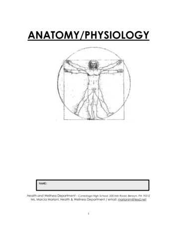
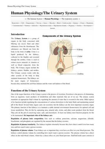
Human Physiology/The Urinary System7is very similar to blood plasma in composition as it passes into the proximal convoluted tubule. Together, theglomerulus and Bowman's capsule are called the renal corpuscle.Proximal Convoluted Tubule (PCT)The proximal tubule can be anatomically divided into two segments: the proximal convoluted tubule and theproximal straight tubule. The proximal convoluted tubule can be divided further into S1 and S2 segments based onthe histological appearance of it's cells. Following this naming convention, the proximal straight tubule is commonlycalled the S3 segment. The proximal convoluted tubule has one layer of cuboidal cells in the lumen. This is the onlyplace in the nephron that contains cuboidal cells. These cells are covered with millions of microvilli. The microvilliserve to increase surface area for reabsorption.Fluid in the filtrate entering the proximal convoluted tubule is reabsorbed into the peritubular capillaries, includingapproximately two-thirds of the filtered salt and water and all filtered organic solutes (primarily glucose and aminoacids). This is driven by sodium transport from the lumen into the blood by the Na /K ATPase in the basolateralmembrane of the epithelial cells. Much of the mass movement of water and solutes occurs in between the cellsthrough the tight junctions, which in this case are not selective.The solutes are absorbed isotonically, in that the osmotic potential of the fluid leaving the proximal tubule is thesame as that of the initial glomerular filtrate. However, glucose, amino acids, inorganic phosphate, and some othersolutes are reabsorbed via secondary active transport through cotransport channels driven by the sodium gradient outof the nephron.Loop of the Nephron or Loop of HenleThe loop of Henle (sometimes knownas the nephron loop) is a U-shapedtube that consists of a descending limband ascending limb. It begins in thecortex, receiving filtrate from theproximal convoluted tubule, extendsinto the medulla, and then returns tothe cortex to empty into the distalconvoluted tubule. Its primary role isto concentrate the salt in theinterstitium, the tissue surrounding theloop.Descending limbIts descending limb is permeabletowaterbutcompletelyimpermeable to salt, and thusonly indirectly contributes to theconcentration of the interstitium.As the filtrate descends deeperinto the hypertonic interstitiumThe Nephron Loop or Loop of Henle.of the renal medulla, water flowsfreely out of the descending limbby osmosis until the tonicity of the filtrate and interstitium equilibrate. Longer descending limbs allow moretime for water to flow out of the filtrate, so longer limbs make the filtrate more hypertonic than shorter limbs.Ascending limb
Human Physiology/The Urinary SystemUnlike the descending limb, the ascending limb of Henle's loop is impermeable to water, a critical feature ofthe countercurrent exchange mechanism employed by the loop. The ascending limb actively pumps sodiumout of the filtrate, generating the hypertonic interstitium that drives countercurrent exchange. In passingthrough the ascending limb, the filtrate grows hypotonic since it has lost much of its sodium content. Thishypotonic filtrate is passed to the distal convoluted tubule in the renal cortex.Distal Convoluted Tubule (DCT)The distal convoluted tubule is similar to the proximal convoluted tubule in structure and function. Cells lining thetubule have numerous mitochondria, enabling active transport to take place by the energy supplied by ATP. Much ofthe ion transport taking place in the distal convoluted tubule is regulated by the endocrine system. In the presence ofparathyroid hormone, the distal convoluted tubule reabsorbs more calcium and excretes more phosphate. Whenaldosterone is present, more sodium is reabsorbed and more potassium excreted. Atrial natriuretic peptide causes thedistal convoluted tubule to excrete more sodium. In addition, the tubule also secretes hydrogen and ammonium toregulate pH. After traveling the length of the distal convoluted tubule, only 3% of water remains, and the remainingsalt content is negligible. 97.9% of the water in the glomerular filtrate enters the convoluted tubules and collectingducts by osmosis.Formation of UrineUrine is formed in three steps: Filtration, Reabsorption, and Secretion.FiltrationBlood enters the afferent arteriole and flows into the glomerulus. Blood in the glomerulus has both filterable bloodcomponents and non-filterable blood components. Filterable blood components move toward the inside of theglomerulus while non-filterable blood components bypass the filtration process by exiting through the efferentarteriole. Filterable Blood components now take on plasma like form called glomerular filtrate. A few of thefilterable blood components are water, nitrogenous waste, nutrients and salts (ions). Nonfilterable blood componentsinclude formed elements such as blood cells and platelets along with plasma proteins. The glomerular filtrate is notthe same consistency as urine, as much of it is reabsorbed into the blood as the filtrate passes through the tubules ofthe nephron.ReabsorptionWithin the peritubular capillary network, molecules and ions are reabsorbed back into the blood. Sodium Chloridereabsorbed into the system increases the osmolarity of blood in comparison to the glomerular filtrate. Thisreabsorption process allows water (H2O) to pass from the glomerular filtrate back into the circulatory system.Glucose and various amino acids also are reabsorbed into the circulatory system. These nutrients have carriermolecules that claim the glomerular molecule and release it back into the circulatory system. If all of the carriermolecules are used up, excess glucose or amino acids are set free into the urine. A complication of diabetes is theinability of the body to reabsorb glucose. If too much glucose appears in the glomerular filtrate it increases theosmolarity of the filtrate, causing water to be released into the urine rather than reabsorbed by the circulatory system.Frequent urination and unexplained thirst are warning signs of diabetes, due to water not being reabsorbed.Glomerular filtrate has now been separated into two forms: Reabsorbed Filtrate and Non-reabsorbed Filtrate.Non-reabsorbed filtrate is now known as tubular fluid as it passes through the collecting duct to be processed intourine.8
Human Physiology/The Urinary SystemSecretionSome substances are removed from blood through the peritubular capillary network into the distal convoluted tubuleor collecting duct. These substances are Hydrogen ions, creatinine, and drugs. Urine is a collection of substances thathave not been reabsorbed during glomerular filtration or tubular reabsorbtion.Maintaining Water-Salt BalanceIt is the job of the kidneys to maintain the water-salt balance of the blood. They also maintain blood volume as wellas blood pressure. Simple examples of ways that this balance can be changed include ingestion of water,dehydration, blood loss and salt ingestion.Reabsorption of waterDirect control of water excretion in the kidneys is exercised by the anti-diuretic hormone (ADH), released by theposterior lobe of the pituitary gland. ADH causes the insertion of water channels into the membranes of cells liningthe collecting ducts, allowing water reabsorption to occur. Without ADH, little water is reabsorbed in the collectingducts and dilute urine is excreted. There are several factors that influence the secretion of ADH. The first of thesehappen when the blood plasma gets too concentrated. When this occurs, special receptors in the hypothalamusrelease ADH. When blood pressure falls, stretch receptors in the aorta and carotid arteries stimulate ADH secretionto increase volume of the blood.Reabsorption of SaltThe Kidneys also regulate the salt balance in the blood by controlling the excretion and the reabsorption of variousions. As noted above, ADH plays a role in increasing water reabsorption in the kidneys, thus helping to dilute bodilyfluids. The kidneys also have a regulated mechanism for reabsorbing sodium in the distal nephron. This mechanismis controlled by aldosterone, a steroid hormone produced by the adrenal cortex. Aldosterone promotes the excretionof potassium ions and the reabsorption of sodium ions. The release of Aldosterone is initiated by the kidneys. Thejuxtaglomerular apparatus is a renal structure consisting of the macula densa, mesangial cells, and juxtaglomerularcells. Juxtaglomerular cells (JG cells, also known as granular cells) are the site of renin secretion. Renin is anenzyme that converts angiotensinogen (a large plasma protein produced by the liver) into Angiotensin I andeventually into Angiotensin II which stimulates the adrenal cortex to produce aldosterone. The reabsorption ofsodium ions is followed by the reapsorption of water. This causes blood pressure as well as blood volume toincrease.Atrial natriuretic hormone (ANH) is released by the atria of the heart when cardiac cells are streatched due toincreased blood volume. ANH inhibits the secretion of renin by the juxtaglomerular apparatus and the secretion ofthe aldosterone by the adrenal cortex. This promotes the excretion of sodium. When sodium is excreted so is water.This causes blood pressure and volume to decrease.HypernatremiaAn increase in plasma sodium levels above normal is hypernatremia. Sodium is the primary solute in theextracellular fluid. Sodium levels have a major role in osmolarity regulation. For excitable cells the electrochemicalgradient for sodium across the plasma membrane is critical for life. Water retention and an increased blood pressureusually are signs of hypernatremia. If the plasma sodium levels are below normal it is called hyponatremia. Signs ofthis are low plasma volume and hypotension.9
Human Physiology/The Urinary SystemDiureticsA diuretic (colloquially called a water pill) is any drug that elevates the rate of bodily urine excretion (diuresis).Diuretics also decrease the extracellular fluid (ECF) volume, and are primarily used to produce a negativeextracellular fluid balance. Caffeine, cranberry juice and alcohol are all weak diuretics. In medicine, diuretics areused to treat heart failure, liver cirrhosis, hypertension and certain kidney diseases. Diuretics alleviate the symptomsof these diseases by causing sodium and water loss through the urine. As urine is produced by the kidney, sodiumand water – which cause edema related to the disease – move into the blood to replace the volume lost as urine,thereby reducing the pathological edema. Some diuretics, such as acetazolamide, help to make the urine morealkaline and are helpful in increasing excretion of substances such as aspirin in cases of overdose or poisoning. Theantihypertensive actions of some diuretics (thiazides and loop diuretics in particular) are independent of their diureticeffect. That is, the reduction in blood pressure is not due to decreased blood volume resulting from increased urineproduction, but occurs through other mechanisms and at lower doses than that required to produce diuresis.Indapamide was specifically designed with this is mind, and has a larger therapeutic window for hypertension(without pronounced diuresis) than most other diuretics. Chemically, diuretics are a diverse group of compounds thateither stimulate or inhibit various hormones that naturally occur in the body to regulate urine production by thekidneys. Alcohol produces diuresis through modulation of the vasopressin system.Diseases of the KidneyDiabetic nephropathy (nephropatia diabetica), also known as Kimmelstiel-Wilson syndrome and intercapillaryglomerulonephritis, is a progressive kidney disease caused by angiopathy of capillaries in the kidney glomeruli. It ischaracterized by nodular glomerulosclerosis. It is due to longstanding diabetes mellitus, and is a prime cause fordialysis in many Western countries.In medicine, hematuria (or "haematuria") is the presence of blood inthe urine. It is a sign of a large number of diseases of the kidneys andthe urinary tract, ranging from trivial to lethal.Kidney stones, also known as nephrolithiases, urolithiases or renalcalculi, are solid accretions (crystals) of dissolved minerals in urinefound inside the kidneys or ureters. They vary in size from as small asa grain of sand to as large as a golf ball. Kidney stones typically leavethe body in the urine stream; if they grow relatively large beforepassing (on the order of millimeters), obstruction of a ureter anddistention with urine can cause severe pain most commonly felt in theflank, lower abdomen and groin. Kidney stones are unrelated togallstones.Case Study I was 34 weeks pregnant when I noticed blood in myurine. I immediately went to my OBGYN where I was told that I had abladder infection and given an antibiotic. The next morning Iexperienced the most intense pain. I was rushed to the ER where I wastold that I had kidney stones. The doctors explained that there wasAn image of a kidney stone.nothing they could do as long as I was pregnant. The next 3 weeks ofmy life were filled with intense pain and multiple painkillers. After I delivered my baby, CAT scans were done and Iwas informed that I had 6 kidney stones. It took three more weeks for me to pass all of the stones the largestmeasuring 5 mm. The stones were tested and I was informed that my body had been building up calcium due to mypregnancy and this was the cause of the kidney stones. I continued to have kidney pain for 6 months after passing thestones. I now live my life on a low calcium diet and the hope that my body will not develop more kidney stones.10
Human Physiology/The Urinary SystemPyelonephritis When an infection of the renal pelvis and calices, called pyelitis, spreads to involve the rest of thekidney as well, the result is pyelonephritis. It usually results from the spread of fecal bacterium Escherichia coli fromthe anal region superiorly
Human Physiology/The Urinary System 4 Renal Vein The renal veins are veins that drain the kidney.They connect the kidney to the inferior vena cava. Because the inferior vena cava is on the right half of the body