Transcription
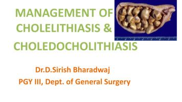
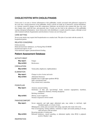
CHOLECYSTITIS WITH CHOLELITHIASISCholecystitis is an acute or chronic inflammation of the gallbladder, usually associated with gallstone(s) impacted inthe cystic duct, causing distension of the gallbladder. Stones (calculi) are made up of cholesterol, calcium bilirubinate,or a mixture caused by changes in the bile composition. Gallstones can develop in the common bile duct, the cysticduct, hepatic duct, small bile duct, and pancreatic duct. Crystals can also form in the submucosa of the gallbladdercausing widespread inflammation. Acute cholecystitis with cholelithiasis is usually treated by surgery, although severalother treatment methods (fragmentation and dissolution of stones) are now being used.CARE SETTINGSevere acute attacks may require brief hospitalization on a medical unit. This plan of care deals with the acutely ill,hospitalized patient.RELATED CONCERNSCholecystectomyFluid and electrolyte imbalances, see Nursing Plan CD-ROMPsychosocial aspects of careTotal nutritional support: parenteral/enteral feedingPatient Assessment DatabaseACTIVITY/RESTMay report:FatigueMay exhibit:RestlessnessCIRCULATIONMay exhibit:Tachycardia, diaphoresis, lightheadednessELIMINATIONMay report:Change in color of urine and stoolsMay exhibit:Abdominal distensionPalpable mass in right upper quadrant (RUQ)Dark, concentrated urineClay-colored stool, steatorrheaFOOD/FLUIDMay report:Anorexia, nausea/vomitingIntolerance of fatty and ―gas-forming‖ foods; recurrent regurgitation, heartburn,indigestion, flatulence, bloating (dyspepsia)Belching (eructation)May exhibit:Obesity; recent weight lossNormal to hypoactive bowel soundsPAIN/DISCOMFORTMay report:May exhibit:RESPIRATIONSevere epigastric and right upper abdominal pain, may radiate to mid-back, rightshoulder/scapula, or to front of chestMidepigastric colicky pain associated with eating, especially after meals rich in fatsPain severe/ongoing, starting suddenly, sometimes at night, and usually peaking in 30 min,often increases with movementRecurring episodes of similar painRebound tenderness, muscle guarding, or abdominal rigidity when RUQ is palpated;positive Murphy’s sign
May exhibit:Increased respiratory rateSplinted respiration marked by short, shallow breathingSAFETYMay exhibit:Low-grade fever; high-grade fever and chills (septic complications)Jaundice, with dry, itching skin (pruritus)Bleeding tendencies (vitamin K deficiency)TEACHING/LEARNINGMay report:Discharge planconsiderations:Familial tendency for gallstonesRecent pregnancy/delivery; history of diabetes mellitus (DM), IBD, blood dyscrasiasDRG projected mean length of inpatient stay: 4.3 daysMay require support with dietary changes/weight reductionRefer to section at end of plan for postdischarge considerations.DIAGNOSTIC STUDIESBiliary ultrasound: Reveals calculi, with gallbladder and/or bile duct distension (frequently the initial diagnosticprocedure).Oral cholecystography (OCG): Preferred method of visualizing general appearance and function of gallbladder,including presence of filling defects, structural defects, and/or stone in ducts/biliary tree. Can be done IV (IVC)when nausea/vomiting prevent oral intake, when the gallbladder cannot be visualized during OCG, or whensymptoms persist following cholecystectomy. IVC may also be done perioperatively to assess structure andfunction of ducts, detect remaining stones after lithotripsy or cholecystectomy, and/or to detect surgicalcomplications. Dye can also be injected via T-tube drain postoperatively.Endoscopic retrograde cholangiopancreatography (ERCP): Visualizes biliary tree by cannulation of the common bileduct through the duodenum.Percutaneous transhepatic cholangiography (PTC): Fluoroscopic imaging distinguishes between gallbladder diseaseand cancer of the pancreas (when jaundice is present); supports the diagnosis of obstructive jaundice and revealscalculi in ducts.Cholecystograms (for chronic cholecystitis): Reveals stones in the biliary system. Note: Contraindicated in acutecholecystitis because patient is too ill to take the dye by mouth.Nonnuclear CT scan: May reveal gallbladder cysts, dilation of bile ducts, and distinguish betweenobstructive/nonobstructive jaundice.Hepatobiliary (HIDA, PIPIDA) scan: May be done to confirm diagnosis of cholecystitis, especially when bariumstudies are contraindicated. Scan may be combined with cholecystokinin injection to demonstrate abnormalgallbladder ejection.Abdominal x-ray films (multipositional): Radiopaque (calcified) gallstones present in 10%–15% of cases;calcification of the wall or enlargement of the gallbladder.Chest x-ray: Rule out respiratory causes of referred pain.CBC: Moderate leukocytosis (acute).Serum bilirubin and amylase: Elevated.Serum liver enzymes—AST; ALT; ALP; LDH: Slight elevation; alkaline phosphatase and 5-nucleotidase aremarkedly elevated in biliary obstruction.Prothrombin levels: Reduced when obstruction to the flow of bile into the intestine decreases absorption of vitamin K.NURSING PRIORITIES1.2.3.4.Relieve pain and promote rest.Maintain fluid and electrolyte balance.Prevent complications.Provide information about disease process, prognosis, and treatment needs.DISCHARGE GOALS1.2.3.4.5.Pain relieved.Homeostasis achieved.Complications prevented/minimized.Disease process, prognosis, and therapeutic regimen understood.Plan in place to meet needs after discharge
NURSING DIAGNOSIS: Pain, acuteMay be related toBiological injuring agents: obstruction/ductal spasm, inflammatory process, tissue ischemia/necrosisPossibly evidenced byReports of pain, biliary colic (waves of pain)Facial mask of pain; guarding behaviorAutonomic responses (changes in BP, pulse)Self-focusing; narrowed focusDESIRED OUTCOMES/EVALUATION CRITERIA—PATIENT WILL:Pain Control (NOC)Report pain is relieved/controlled.Demonstrate use of relaxation skills and diversional activities as indicated for individual situation.ACTIONS/INTERVENTIONSRATIONALEPain Management (NIC)IndependentObserve and document location, severity (0–10 scale),and character of pain (e.g., steady, intermittent, colicky).Assists in differentiating cause of pain, and providesinformation about disease progression/resolution,development of complications, and effectiveness ofinterventions.Note response to medication, and report to physician ifpain is not being relieved.Severe pain not relieved by routine measures may indicatedeveloping complications/need for further intervention.Promote bedrest, allowing patient to assume position ofcomfort.Bedrest in low-Fowler’s position reduces intra-abdominalpressure; however, patient will naturally assume leastpainful position.Use soft/cotton linens; calamine lotion, oil (Alpha Keri)bath; cool/moist compresses as indicated.Reduces irritation/dryness of the skin and itchingsensation.Control environmental temperature.Cool surroundings aid in minimizing dermal discomfort.Encourage use of relaxation techniques, e.g., guidedimagery, visualization, deep-breathing exercises. Providediversional activities.Promotes rest, redirects attention, may enhance coping.Make time to listen to and maintain frequent contact withpatient.Helpful in alleviating anxiety and refocusing attention,which can relieve pain.
ACTIONS/INTERVENTIONSRATIONALEPain Management (NIC)CollaborativeMaintain NPO status, insert/maintain NG suction asindicated.Removes gastric secretions that stimulate release ofcholecystokinin and gallbladder contractions.Administer medications as indicated:Anticholinergics, e.g., atropine,propantheline (Pro-Banthı-ne);Relieves reflex spasm/smooth muscle contraction andassists with pain management.Sedatives, e.g., phenobarbital;Promotes rest and relaxes smooth muscle, relieving pain.Narcotics, e.g., meperidine hydrochloride (Demerol),morphine sulfate;Given to reduce severe pain. Morphine is used withcaution because it may increase spasms of the sphincter ofOddi, although nitroglycerin may be given to reducemorphine-induced spasms if they occur.Monoctanoin (Moctanin);This medication may be used after a cholecystectomy forretained stones or for newly formed large stones in thebile duct. It is a lengthy treatment (1–3 wk) and isadministered via a nasal-biliary tube. A cholangiogram isdone periodically to monitor stone dissolution.Smooth muscle relaxants, e.g., papaverine (Pavabid),nitroglycerin, amyl nitrite;Relieves ductal spasm.Chenodeoxycholic acid (Chenix), ursodeoxycholicacid (Urso, Actigall);These natural bile acids decrease cholesterol synthesis,dissolving gallstones. Success of this treatment dependson the number and size of gallstones (preferably three orfewer stones smaller than 20 min in diameter) floating ina functioning gallbladder.Antibiotics.To treat infectious process, reducing inflammation.Prepare for procedures, e.g.:Endoscopic papillotomy (removal of ductal stone);Extracorporeal shock wave lithotripsy (ESWL);Choice of procedure is dictated by individual situation.Shock wave treatment is indicated when patient has mildor moderate symptoms, cholesterol stones in gallbladderare 0.5 mm or larger, and there is no biliary tractobstruction. Depending on the machine being used, thepatient may sit in a tank of water or lie prone on awater-filled cushion. Treatment takes about 1–2 hr and is75%–95% successful. Note: This procedure iscontraindicated in patients with pacemakers orimplantable defibrillators.Endoscopic sphincterotomy;Procedure done to widen the mouth of the common bileduct where it empties into the duodenum. This proceduremay also include the manual retrieval of stones from theduct by means of a tiny basket or balloon on the end ofthe endoscope. Stones must be smaller than 15 mm.
ACTIONS/INTERVENTIONSRATIONALEPain Management (NIC)CollaborativeSurgical intervention.Cholecystectomy may be indicated because of thesize of stones and degree of tissue involvement/presence of necrosis. Refer to CP: Cholecystectomy.NURSING DIAGNOSIS: Fluid Volume, risk for deficientRisk factors may includeExcessive losses through gastric suction; vomiting, distension, and gastric hypermotilityMedically restricted intakeAltered clotting processPossibly evidenced by[Not applicable; presence of signs and symptoms and establishes an actual diagnosis.]DESIRED OUTCOMES/EVALUATION CRITERIA—PATIENT WILL:Hydration (NOC)Demonstrate adequate fluid balance evidenced by stable vital signs, moist mucous membranes, good skinturgor, capillary refill, individually appropriate urinary output, absence of rolyte Management (NIC)IndependentMaintain accurate record of I&O, noting output less thanintake, increased urine specific gravity. Assessskin/mucous membranes, peripheral pulses, and capillaryrefill.Provides information about fluid status/circulatingvolume and replacement needs.Monitor for signs/symptoms of increased/continuednausea or vomiting, abdominal cramps, weakness,twitching, seizures, irregular heart rate, paresthesia,hypoactive or absent bowel sounds, depressedrespirations.Prolonged vomiting, gastric aspiration, and restricted oralintake can lead to deficits in sodium, potassium, andchloride.Eliminate noxious sights/smells from environment.Reduces stimulation of vomiting center.Perform frequent oral hygiene with alcohol-freemouthwash; apply lubricants.Decreases dryness of oral mucous membranes; reducesrisk of oral bleeding.Use small-gauge needles for injections and apply firmpressure for longer than usual after venipuncture.Reduces trauma, risk of bleeding/hematoma formation.
ACTIONS/INTERVENTIONSRATIONALEFluid/Electrolyte Management (NIC)IndependentAssess for unusual bleeding, e.g., oozing from injectionsites, epistaxis, bleeding gums, ecchymosis, petechiae,hematemesis/melena.Prothrombin is reduced and coagulation time prolongedwhen bile flow is obstructed, increasing risk ofbleeding/hemorrhage.CollaborativeKeep patient NPO as necessary.Decreases GI secretions and motility.Insert NG tube, connect to suction, and maintain patencyas indicated.Provides rest for GI tract.Administer antiemetics, e.g., prochlorperazine(Compazine).Reduces nausea and prevents vomiting.Review laboratory studies, e.g., Hb/Hct, electrolytes,ABGs (pH), clotting times.Aids in evaluating circulating volume, identifies deficits,and influences choice of intervention forreplacement/correction.Administer IV fluids, electrolytes, and vitamin K.Maintains circulating volume and corrects imbalances.NURSING DIAGNOSIS: Nutrition: imbalanced, risk for less than body requirementsRisk factors may includeSelf-imposed or prescribed dietary restrictions, nausea/vomiting, dyspepsia, painLoss of nutrients; impaired fat digestion due to obstruction of bile flowPossibly evidenced by[Not applicable; presence of signs and symptoms establishes an actual diagnosis.]DESIRED OUTCOMES/EVALUATION CRITERIA—PATIENT WILL:Nutritional Status (NOC)Report relief of nausea/vomiting.Demonstrate progression toward desired weight gain or maintain weight as individually n Management (NIC)IndependentEstimate/calculate caloric intake. Keep comments aboutappetite to a minimum.Identifies nutritional deficiencies/needs. Focusing onproblem creates a negative atmosphere and may interferewith intake.
ACTIONS/INTERVENTIONSRATIONALENutrition Management (NIC)IndependentWeigh as indicated.Monitors effectiveness of dietary plan.Consult with patient about likes/dislikes, foods that causedistress, and preferred meal schedule.Involving patient in planning enables patient to have asense of control and encourages eating.Provide a pleasant atmosphere at mealtime; removenoxious stimuli.Useful in promoting appetite/reducing nausea.Provide oral hygiene before meals.A clean mouth enhances appetite.Offer effervescent drinks with meals, if tolerated.May lessen nausea and relieve gas. Note: May becontraindicated if beverage causes gas formation/gastricdiscomfort.Assess for abdominal distension, frequent belching,guarding, reluctance to move.Nonverbal si
Cholecystitis is an acute or chronic inflammation of the gallbladder, usually associated with gallstone(s) impacted in the cystic duct, causing distension of the gallbladder. Stones (calculi) are made up of cholesterol, calcium bilirubinate, or a mixture caused by changes in the bile composition. Gallstones can develop in the common bile duct, the cystic duct, hepatic duct, small bile duct .
