Transcription
Delany et al. Journal of Congenital -3(2021) 5:7Journal ofCongenital CardiologyREVIEWOpen AccessDown syndrome and congenital heartdisease: perioperative planning andmanagementDennis R. Delany1* , Stephanie S. Gaydos2, Deborah A. Romeo3, Heather T. Henderson1, Kristi L. Fogg4,Angela S. McKeta1, Minoo N. Kavarana5 and John M. Costello1AbstractApproximately 50% of newborns with Down syndrome have congenital heart disease. Non-cardiac comorbiditiesmay also be present. Many of the principles and strategies of perioperative evaluation and management forpatients with congenital heart disease apply to those with Down syndrome. Nevertheless, careful planning forcardiac surgery is required, evaluating for both cardiac and noncardiac disease, with careful consideration of the riskfor pulmonary hypertension. In this manuscript, for children with Down syndrome and hemodynamically significantcongenital heart disease, we will summarize the epidemiology of heart defects that warrant intervention. We willreview perioperative planning for this unique population, including anesthetic considerations, commonpostoperative issues, nutritional strategies, and discharge planning. Special considerations for single ventriclepalliation and heart transplantation evaluation will also be discussed. Overall, the risk of mortality with cardiacsurgery in pediatric patients with Down syndrome is no more than the general population, except for those withfunctional single ventricle heart defects. Underlying comorbidities may contribute to postoperative complicationsand increased length of stay. A strong understanding of cardiac and non-cardiac considerations in children withDown syndrome will help clinicians optimize perioperative care and long-term outcomes.Keywords: Down syndrome, Trisomy 21, Congenital heart disease, Congenital heart surgery, PerioperativemanagementBackgroundDown syndrome (DS) is the most common chromosomal abnormality with a prevalence of 11.8 per 10,000live births [1]. Approximately 50% of newborns with DShave congenital heart disease (CHD) [2, 3]. The vast majority of these heart defects require surgical or transcatheter intervention, most commonly in infancy. Althoughmany of the principles and strategies of perioperativeevaluation and management for patients with CHD* Correspondence: delanyd@musc.edu1Department of Pediatrics, Division of Pediatric Cardiology, Medical Universityof South Carolina, 10 McClennan Banks Drive, MSC915, Charleston, SC 29425,USAFull list of author information is available at the end of the articleapply to those with DS, there are a number of specific issues that warrant special attention.In this manuscript, for children with DS andhemodynamically significant CHD, we will summarizethe epidemiology of heart defects that warrant intervention. We will review perioperative planning for thisunique population, including anesthetic concerns, common postoperative issues, nutritional strategies, and discharge planning. Special considerations for singleventricle palliation and heart transplantation evaluationwill also be discussed. The Author(s). 2021 Open Access This article is licensed under a Creative Commons Attribution 4.0 International License,which permits use, sharing, adaptation, distribution and reproduction in any medium or format, as long as you giveappropriate credit to the original author(s) and the source, provide a link to the Creative Commons licence, and indicate ifchanges were made. The images or other third party material in this article are included in the article's Creative Commonslicence, unless indicated otherwise in a credit line to the material. If material is not included in the article's Creative Commonslicence and your intended use is not permitted by statutory regulation or exceeds the permitted use, you will need to obtainpermission directly from the copyright holder. To view a copy of this licence, visit http://creativecommons.org/licenses/by/4.0/.The Creative Commons Public Domain Dedication waiver ) applies to thedata made available in this article, unless otherwise stated in a credit line to the data.
Delany et al. Journal of Congenital Cardiology(2021) 5:7MethodsThe authors searched the published literature using avariety of medical subject terms to identify peerreviewed articles on perioperative care issues relevant tothe management of patients with DS and CHD, rangingin age from neonates to adults. We focused on originalmanuscripts published since 2010 but included a number of earlier relevant publications. Non-full text publications or languages other than English were excluded.History of congenital heart surgery in patientswith Down syndromeAlthough aggressive medical management and surgicalintervention for CHD in patients with DS is routinepractice in the current era, that was not always the case.John Langdon Down first described the features of DS in1866 and Jerome Lejeune linked the syndrome to thechromosomal abnormality of trisomy 21 in 1959 [4].Open heart surgery for most congenital heart defects became widely available in the 1960s and early 1970s. Inthe early years of congenital heart surgery, patients withDS were systematically not offered cardiac surgical intervention [5]. However, as outcomes for noncardiac conditions that afflict infants with DS improved and completecardiac repair in infancy became more common for largeseptal defects, it was found that patients with DS generally tolerated surgery well. Societal attitudes changed,and increasingly more infants with DS and CHD werereferred for surgery. Today, it is the standard of care tooffer cardiac surgery to patients with DS with CHD thatotherwise meet criteria for intervention.Epidemiology of CHD in patients with Downsyndrome undergoing surgeryDS is present in nearly 10% of all children undergoingcardiac surgery, including approximately 2% of neonatalcases [6, 7]. Based upon statistics reported from the Society of Thoracic Surgeons Congenital Heart SurgeryPage 2 of 14(STS-CHS) Database, the most common surgical procedures performed in children with DS are summarized inTable 1 [6].Special considerations for perioperative planningPreoperative evaluation and planning for any patient being considered for a cardiac surgical procedure must bethorough and consider the patient’s medical and surgicalhistory, cardiac anatomy, and non-cardiac comorbidities.Additional details regarding cardiovascular disease andextracardiac comorbidities warrant specific attentionwhen planning surgery for patients with DS and CHD.Simple left to right shuntsVentricular septal defect (VSD), atrial septal defect(ASD), and patent ductus arteriosus (PDA) are commontypes of CHD present in children with DS [6, 8, 9]. Inpatients with ASD and VSD, data from the STS-CHSDatabase indicate that those with DS were younger andweighed less for age at time of surgery compared withpatients without genetic anomalies. Specifically, averageages at time of repair of ASD was 1.2 years versus 4.1years, and average ages at time of VSD repair was 4.8months versus 7.4 months (DS versus non-DS respectively) [6].Atrioventricular septal defectsDue to the increased frequency of endocardial cushiondefects with DS, evaluation of left to right shunts shouldspecifically evaluate for inlet-type VSDs, primum-typeASDs, and partial, transitional, or complete atrioventricular septal defects (AVSD). With these defects, special attention should be given to assess for conoseptalmalalignment in VSDs, additional muscular VSDs, andcrossing atrioventricular valve chords in inlet-type VSDs.Repair of complete AVSD is the most common cardiacprocedure for patients with DS [6]. As with any preoperative cardiac work-up of complete AVSD, detailedTable 1 Most common cardiac surgeries in patients with Down syndrome as summarized in the STS-CHS Database [6]Procedure% of patients with Down syndromeComplete atrioventricular septal defect repair33Ventricular septal defect closure19Mitral valve repair/replacement7Partial atrioventricular septal defect repair6Patent ductus arteriosus ligation4Tetralogy of Fallot repair4Atrial septal defect closure4Coarctation/arch repair2Tricuspid valve repair/replacement2Tetralogy of Fallot- atrioventricular septal defect repair1STS-CHS Society of Thoracic Surgeons Congenital Heart Surgery
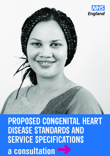
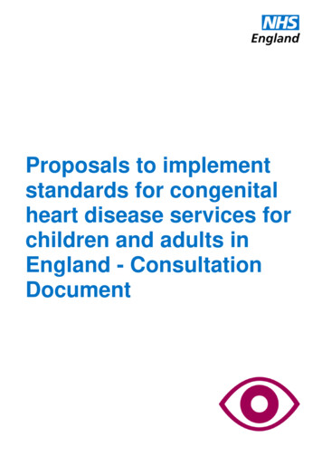
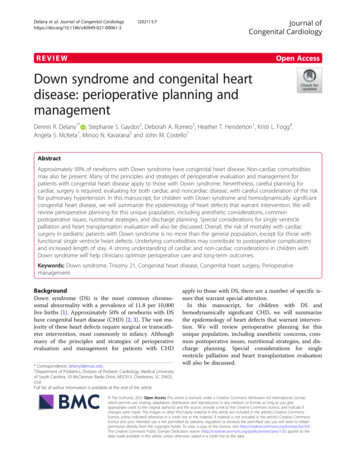
Delany et al. Journal of Congenital Cardiology(2021) 5:7evaluation should be completed of the common atrioventricular valve (AVV), ventricular balance, and ventricular outflow tracts. Structurally, patients with DShave lower rates of left AVV dysplasia (3% with DS vs.24% with non-DS), unbalanced ventricles (3% with DSvs. 25% with non-DS), and left-sided obstructive lesions(1% with DS vs. 30% with non-DS) when compared tothose with complete AVSD without genetic anomalies[10–12]. Additionally, the AVVs in patients with DS aremore likely to be Rastelli Type A, with a divided anteriorbridging leaflet with connection to the crest of the ventricular septum [13]. Excluding a patent foramen ovaleor a small additional secundum ASD, nearly half of patients with DS undergoing AVSD repair have one ormore additional cardiac procedures performed intraoperatively, including PDA ligation, repair of aortic archanomaly, closure of additional VSD, baffle of anomaloussystemic venous drainage, repair of pulmonary venousstenosis, and subaortic stenosis resection [14].Compared to patients without chromosomal abnormality, those with DS tend to present for AVSD repairsurgery at the same age but lower average weight [6].Growth failure at surgery is high at 71% for infants withDS versus 21% of those without chromosomal abnormality [14]. Of note, patients with DS tend to have significant catch up growth after surgery but a largeproportion still have growth failure (27% in DS vs. 7% innon-DS) [14].Tetralogy of FallotIn addition to endocardial cushion defects, the presenceof conoseptal malalignment must also be evaluated. TOFor coexistent TOF and AVSD are more common inchildren with DS than in children without genetic abnormalities [6]. Overall, those with TOF are repairedaround the same age but have a lower weight for age [6].Single ventricle palliationSingle ventricle lesions are occasionally seen in patientswith DS. Structurally, patients with DS are more likelyto have left ventricle dominant lesions, while patientswithout DS are more likely to have right ventricle dominant or complex single ventricle lesions, includingtransposition of the great arteries or left-sided obstructive lesions [15, 16]. Despite generally having more favorable anatomy for single ventricle palliation, the reportedoutcomes in the literature indicate that mortality is relatively high compared to patients without DS. A STSCHS Database study found that there was increased operative mortality in patients with DS at every stage ofsingle ventricle palliation (Stage 1 palliation: 73% in DSvs. 19% in non-DS, Glenn operation: 19% in DS vs. 2%in non-DS, and Fontan operation: 24% in DS vs. 2% innon-DS) [6]. Using Pediatric Cardiac Care ConsortiumPage 3 of 14(PCCC) data, Petersen et al. showed that patients withDS and functional single ventricle heart defects had highin-hospital first-stage mortality at 24%, and for thoseundergoing a Norwood procedure, specifically, hospitalmortality was 68% [15]. These investigators also foundthat only 21% of patients included in the initial cohortcompleted the Fontan procedure, and of those who didnot go to Fontan, 50% were ineligible due tohemodynamic or anatomic concerns [15]. The KaplanMeier Survival following each stage of single ventriclepalliation for DS patients is depicted in Fig. 1.Risk stratification in patients with DS and functionalsingle ventricle heart defects can be challenging due tosmall sample sizes in most studies, but pulmonaryhypertension (PH) is likely one of the main contributingfactors to mortality [6, 16, 17]. Colquitt et al. showedthat patients with DS and single ventricle anatomy haveminimal mortality beyond 2 years of age if their pulmonary vascular resistance (PVR) is less than 3 indexedWoods units in their first year of life [18]. In fact, whenadjusting for PVR, this study found that DS is not associated with increased mortality compared to other nonsyndromic patients undergoing the single ventriclepalliation at their center [18]. These data suggest thatfor patients with DS and functional single ventricle heartdefects, careful patient selection is critical to achievingacceptable outcomes. One must consider the potentialfor decreased responsiveness to PH therapies found inpatients with DS (described below) when planning onstaged procedures [19–21]. In children with DS, performing a pulsatile Glenn operation or fenestrated Fontan operation, if anatomy and hemodynamics allow, hasbeen shown to decrease perioperative and late mortality[22]. If hemodynamics are borderline after Glenn procedure, one should strongly weigh the risks versus benefits of proceeding to Fontan, as one report suggestedthat Fontan completion was not associated with improved survival compared with sustained Glenn physiology [15].Pulmonary hypertensionIn patients with DS who are being evaluated prior to acardiac operation, the work-up should closely evaluatefor signs of PH. More specifically, the concern should befor pulmonary vascular disease (PVD), meaning elevatedpulmonary arterial pressures in the context of changesin the pulmonary vascular bed leading to increased PVR.Children with DS are at more than 20-fold greater riskfor having PH when compared to the general population, and those with CHD are at even greater risk [23].Lung disorders including relative underdevelopment ofthe pulmonary vascular bed (pulmonary hypoplasia) andchronic issues with upper airway obstruction are likelycontributory (World Health Organization Group 3 PH).
Delany et al. Journal of Congenital Cardiology(2021) 5:7Page 4 of 14Fig. 1 Kaplan-Meier transplant-free survival plot conditioned on hospital discharge: a after the Stage 1 procedure in patients with DS withfunctional single ventricle heart defect, b after Glenn procedure in patients with DS with functional single ventricle heart defect, and c afterFontan procedure in patients with DS with functional single ventricle heart defect. DS, Down syndrome; CI, Confidence interval [15]. (Usedwith permission)Left to right intracardiac shunts with longstanding overcirculation create increased pulmonary artery pressure,shear stress, endothelial dysfunction, vascular remodeling, and altered vasoactive mediator expression (WorldHealth Organization Group 1 PH). Additionally, patientswith obstructed pulmonary venous return, mitral stenosis, or left ventricular outflow obstruction (critical aortic valve stenosis or coarctation of the aorta) maydevelop increased PVR (World Health OrganizationGroup 2 PH).Studies from the early 1990s have shown higher PVRthat is more likely to be irreversible in DS patients, andthis phenomenon may develop as early as 6 months ofage [24, 25]. These observations have been correlatedmore recently in a STS-CHS Database report whichfound higher rates of PH present in patients with ASDsor VSDs and DS compared to those without geneticanomalies [6]. Surprisingly, higher rates of PH were notfound in patients with AVSD and DS, likely related tothe fact that, in the current era, most undergo repairwithin the first 6 months of life [6]. In fact, early definitive repair (i.e., within the first 6 months of life) can reduce the incidence of postoperative PH [26]. Acombination of anecdotal experience and data from earlycase series has prompted most centers into performingcomplete AVSD and large VSD repairs at 4–6 months ofage.Clinically, suspicion for PH should be increased inchildren with anatomically large left to right shunts ifthere is a lack of signs and symptoms of pulmonaryovercirculation, lack of failure to thrive, or if increasingcyanosis is present. By echocardiogram, evidence of PHcan be shown using a variety of methods depending onthe type of lesion. In the presence of a restrictive VSDor intact ventricular septum without right ventricularoutflow tract obstruction (RVOTO), signs of PH includea tricuspid regurgitation jet estimating right ventricularpressure greater than half systemic or by intraventricularseptal flattening. If there is a nonrestrictive VSD or largePDA, PH may be suggested by right to left shunting atventricular or great vessel level (in the absence ofRVOTO) or an elevated early diastolic pulmonary regurgitation gradient. Finally, in the absence of left sided obstructive lesions, right to left shunting at the level of thePDA may also provide evidence of PH. As shown by thenumber of caveats based on type of CHD noted above,these echocardiographic findings only suggest that PVRis elevated. If there is clinical or echocardiographic
Delany et al. Journal of Congenital Cardiology(2021) 5:7evidence of PH or the patient is older with a longstanding left to right shunt lesion, then cardiac catheterizationshould be considered to quantify PVR, and, if it is elevated, pulmonary vaso-reactivity testing with oxygen andnitric oxide is typically performed. Alternatively, giventhe risks associated with cardiac catheterization, an infant with initial predominant right to left shunting canbe placed on oxygen while in clinic, observed to see ifthe oxygen saturation increases, and the direction of theshunt on echo can be reassessed. If the direction of theshunt reverses becoming left to right, a cardiaccatheterization can likely be avoided. Of note, there issome evidence that patients with DS may have less response to pulmonary vasodilators, including nitric oxide[20]. In the STARTS trial, sildenafil was tolerated well inpatients with DS, but led to little improvement of PVR[19]. Similarly, in a study by Duffles et al., bosentan didnot have an effect on exercise capacity or quality of lifein adults with pulmonary arterial hypertension related toCHD and DS [21].Following cardiopulmonary bypass (CPB), PH may becaused by a combination of preoperative, intraoperative,and postoperative factors. DS is a known risk factor forsevere postoperative PH [27]. Yet, even in the absence ofDS, CPB is associated with increased PVR in infants andchildren [28–30]. The presence of significant PH soonafter weaning from CPB is predictive of subsequent PHin the ICU and the need for prolonged ventilatory support. Residual lesions, including large left to right shunts,obstruction to pulmonary venous or distal pulmonaryarterial blood flow, or left-sided obstructive lesions maycause PH. Noxious stimuli, particularly suctioning of theendotracheal tube, may trigger a pulmonary hypertensivecrisis. PH may manifest as low cardiac output followinga biventricular repair, particularly when both septa arecompletely intact, or as excessive cyanosis in patientswith palliated single ventricle physiology. In general,children with PH are at increased risk of perioperativecomplications [31, 32]. A combination of relatively simple postoperative strategies should be sufficient both toprevent and to treat pulmonary hypertensive crises inmany at-risk patients (Table 2) [33].Non-cardiac comorbiditiesFeatures of DS may manifest in nearly every organ system, and thus a comprehensive assessment is requiredprior to taking a patient with DS to the OR [34]. Specifically, one should consider the potential for problemswithin the gastrointestinal, respiratory, immunologic,hematologic, endocrine, and neurologic systems. Generally, clinicians involved in preoperative evaluation shouldrefer to the American Academy of Pediatrics guidelinesPage 5 of 14Table 2 Critical care strategies for treatment of PH [33]EncourageAvoid1. Anatomic investigation1. Residual anatomic disease2. Right to left atrial pop off2. Intact atrial septum3. Sedation/analgesia3. Agitation/pain4. Moderate hyperventilation4. Respiratory acidosis5. Moderate alkalosis5. Metabolic acidosis6. Adequate inspired oxygen6. Alveolar hypoxia7. Normal lung volumes7. Atelectasis or overdistension8. Optimal hematocrit8. Excessive hematocrit9. Inotropic support9. Low output and coronary perfusion10. Pulmonary vasodilators10. Pulmonary vasoconstrictorsfor “Health supervision for children with Down syndrome” for age-appropriate screening [35].GastrointestinalAfter CHD, abnormalities of the gastrointestinal tractare the most common anomaly associated with DS, being present in 6% of cases in one large European registrystudy [9]. These include duodenal atresia, duodenal webbing, annular pancreas, anal atresia, Hirschsprung disease, and tracheoesophageal fistula/esophageal atresia. Ifthere are feeding concerns preoperatively or the neonatefails to pass meconium, there should be a low thresholdto further evaluate for these anomalies.RespiratoryThe respiratory manifestations of DS contribute significantly to morbidity and reduced survival in this population [36–38]. The anatomic features are discussed belowin the section on anesthesia management. Functionally,obstructive sleep apnea (OSA) has a high prevalence ofabout 76–97% in children with DS who snored and 55%in those without snoring [39, 40]. The risk of OSA increases with BMI and age [41], although OSA may bemore severe if it presents earlier [40]. If OSA is foundpreoperatively, consideration for adenotonsillectomyshould be taken, although the timing of that procedurerelative to an upcoming cardiac operation will requireclinical judgment. Children with DS and OSA whounderwent adenotonsillectomy showed significant improvement of obstructive and central apneic indices[42–45]. Additionally, OSA likely plays a long-term rolein the development of PH, and adenotonsillectomy mayhelp halt or reverse the progression of PH and rightheart disease [46, 47].Immunologic/hematologicChildren with DS are also more likely to develop infections and to take longer to recover from them [48, 49].Multiple factors are contributory, as children with DS
Delany et al. Journal of Congenital Cardiology(2021) 5:7may have abnormalities of the immune system with Tand B cell lymphopenia, secondary immunodeficiencydue to nutritional and metabolic factors, and anatomicabnormalities with small ear canals, tracheomalacia, tracheal bronchus, and gastroesophageal reflux [48]. Overall, the most frequent type of infections involve therespiratory tract, especially the upper respiratory tract[49, 50]. Additionally, patients with DS and acute respiratory tract infection are more likely to develop acutelung injury or acute respiratory distress syndrome [51].Careful history for recent infections or history of infections requiring hospitalization should be obtainedpreoperatively.Neonates with DS can have neutrophilia,thrombocytopenia/thrombocytosis, polycythemia transient myeloproliferative disorder, and congenital leukemia[52]. Children with DS have a 20-fold increased risk ofleukemia, with acute myeloblastic leukemia as the mostcommon type, and the highest incidence occurs in children younger than 5 years [53]. Interestingly, althoughthe rates of blood cancers are higher in children withDS, most other cancers tend to be lower than non-DSpopulation [53, 54]. For these reasons, a complete bloodcount should be obtained before surgery, and abnormalblood counts should be discussed with hematologist.EndocrineThyroid disorders are common in DS. Hypothyroidismhas a prevalence of 4–18% [55, 56]. If not assessed onthe state newborn metabolic screen, TSH should beobtained in the neonatal period. The risk forhypothyroidism increases with age, and TSH should befollowed annually [35, 57]. Dry skin, thin hair, poor appetite, constipation, poor energy, hypotonia, bradycardia,and behavior changes may be signs of hypothyroidism,but are not always present [58]. Thyroid hormones areimportant for cardiovascular effects of decreasing afterload and increasing heart rate, preload, and contractility.If there are any signs or symptoms of hypothyroidism ora level has not been recently checked, consider checkinga TSH with preoperative labs.Though not generally a problem in the operativeroom, hypothyroidism can contribute to hypotensionpost-operatively and is easily treated with triiodothyronine therapy when identified. Cardiopulmonary bypasshas been shown to decrease thyroid hormone levels [59].A triiodothyronine level less than 0.6 has been associatedwith longer mechanical ventilation and ICU LOS, withhigher vasoactive drip and furosemide cumulative doses[59]. A separate study however, showed no significantcardiac impairment in patients with DS and subclinicalhypothyroid [60]. Additionally, replacement of triiodothyronine post-bypass has shown little benefit [61].Overall, the literature does not support routine use ofPage 6 of 14intraoperative or early postoperative triiodothyroninesupplementation, but larger studies are needed.NeurologicThe degree of cognitive impairment can range from mild(intelligence quotient 50–70) to severe (intelligence quotient 20–35) [35]. Clinicians should also be aware of the1–13% prevalence of seizures and frequent hearing andvision impairment with a prevalence of 60–75% [35].Preoperatively, it is useful to understand the child’s cognitive function, likes and dislikes, and any physical handicaps, and to deliver this information to the anesthesiaand postoperative care teams.Adults with Down syndrome and congenital heart diseaseSpecial attention should be allocated to the surgical andanesthetic preoperative assessment for adult patientswith DS undergoing congenital cardiac surgery. As described above, comorbidities such as obstructive sleepapnea, hypothyroidism, and epilepsy occur with high frequency and may significantly impact a patient’s perioperative course [62]. Hypothyroidism can developslowly over time, and can be difficult to detect clinicallyin adults with DS given overlapping symptomatologywith other aging processes [63, 64].Despite possessing increased rates of many traditionalrisk factors for acquired cardiovascular disease such asobesity, diabetes, sedentary lifestyle, and evidence ofearly aging within other organ systems, atherosclerosis issurprisingly uncommon in adults with DS [65–68]. Incontrast to the general population of adults with similarrisk profiles undergoing cardiac surgery, there is no recommendation for specific preoperative screening forcoronary artery disease in adults with DS. As mentioned,diabetes does occur more commonly in patients with DSand would require additional perioperative monitoringand treatment to maintain goal glucose levels [69]. Theteam should note patients’ fasting serum glucose levelson preoperative labs and, if elevated, consider additionallab screening with a hemoglobin A1C.There should be a high suspicion for dementia in patients with DS over 40 years of age. Alzheimer’s diseaseoccurs in nearly 25% of adults with DS ages 40 to 49years and in up to 55% of those ages 50 to 59 years [70].Preexisting dementia is a significant risk factor for delirium in subacute care settings, and delirium confersbroadly worse clinical and functional patient outcomes[71]. Identification of baseline dementia is additionallyrelevant when considering expected patient behavior andcommunication strategies postoperatively. Similarly, preoperative inquiry of visual and hearing abilities is important given prevalence of sensorineural hearing lossand early cataract formation in adults with DS [62].Lastly, there should be close inspection of dental health
Delany et al. Journal of Congenital Cardiology(2021) 5:7on a preoperative assessment given the higher frequencyof gingivitis and periodontal disease in adults with DS[72]. Such dental issues may raise a patient’s risk ofspontaneous bacterial endocarditis postoperatively and,if possible, should be treated prior to certain cardiothoracic interventions.Cardiac transplantation in children with Down syndromeSometimes surgical repair or palliation is not a good option for patients with DS, and heart transplant may beconsidered. There is wide variation among transplantcenters of how intellectual and developmental disabilitiesare factored into transplant evaluations and determination of candidacy. However, denying a patient with DSthat would otherwise benefit from transplant the possibility of the same evaluation as a non-DS patient is unjust and prohibited by Americans with Disabilities Act(ADA) [73]. In 1995, Sandra Jensen, a 34 year-old withDS and CHD was denied heart-lung transplant solelybased on her underlying chromosomal abnormality.After a national campaign protesting this discrimination,she underwent successful transplant in 1996, becomingthe first patient in the world with DS to receive a lifesaving organ transplant [73, 74].Since this time, several small studies report patientswith DS receiving successful solid organ transplants withequivalent outcomes to non-DS patients [73, 75–78]. Intellectual disability alone does not affect short- andmedium-term outcome in children after heart transplant, supporting the concept that intellectual delay thatoccurs in DS should not alone impact eligibility fortransplant [78–81]. Overall, little is known specificallyabout heart transplant outcomes in patients with DS. Astudy by Broda, et al. evaluated cardiac transplant outcomes in children with various chromosomal abnormalities and found only 64 of 3080 (2%) pediatric hearttransplants were performed in children with variouschromosomal abnormalities, and only five of these patients had DS, one of whom died prior to hospital discharge. Given the small numbers it was hard to makeconclusions about prognosis [76].Although intellectual disability should not precludepatients with DS from a thorough transplant evaluationand possible listing, many other factors need to be considered. Given the scarcity of donor organs, potentialtransplant recipients undergo a rigorous evaluation todetermine their ca
Keywords: Down syndrome, Trisomy 21, Congenital heart disease, Congenital heart surgery, Perioperative management Background Down syndrome (DS) is the most common chromo-live births [1]. Approximately 50% of newborns with DS have congenital heart disease (CHD) [2, 3]. The vast ma-jority of these heart defects require surgical or transcath-