Transcription
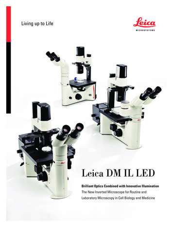
Leica DM IL LEDBrilliant Optics Combined with Innovative IlluminationThe New Inverted Microscope for Routine andLaboratory Microscopy in Cell Biology and Medicine
2Inverted RoutineMicroscopy in a New LightCompact and stable Lean and sturdy design Plenty of space for operation Low stage height Large dimensions and lowcenter of gravity of microscope Large working distancesWide variety of possible applications Cell biology and medicine Micromanipulation Medicine Biotechnology Developmental biology Transgenics Molecular biology Fluorescence applicationsOptical performance and illumination are key elements in microscopy. Both characteristics are unified in the new design of theLeica DM IL LED. As the first inverted routine microscope, theLeica DM IL LED is not only equipped with outstanding Leica HCoptics, but also features innovative LED illumination. The transmitted-light illuminator including optimized condensers and improvedcontrast methods are adapted specifically for cell biology applications. High stability, plenty of space for operation, large workingdistances, illumination without heat development and the separately accommodated electronics provide optimum conditions formicroscopy. The Leica DM IL LED is exceptionally well-suited foruses ranging from various cell and tissue culture examinations inlife sciences, developmental biology studies or micromanipulationin cell biology to living cell examinations in transgenics or electrophysiology.
LEICA DM IL LED – BRILLIANT OPTICS COMBINED WITH INNOVATIVE ILLUMINATIONThe fluorescence version, the Leica DM IL LED Fluo, also offers avariety of possible applications. Optionally, it is also available withthe new LED illumination.Heatable microscope stages and 3-plate cross-stages providegreat flexibility for experiments on living cells under physiologicalconditions.The Leica DM IL LED has a further advantage that distinguishesit from other microscopes in its class: The stand is highly compatible with components of the Leica research microscopes.Objectives, eyepieces, tubes, camera ports, contrast methods.Additionally, special tubes and condensers have been developedfor the Leica DM IL LED.3Integrated fluorescence Manual fluorescence with three filter cubes Integrated shutter Optionally LED, classic mercuryillumination or fiber optic couplingFlexible and modular A full range of optical components Compatible with research stands Unheated and heated stages Large selection of tubes Comprehensive range of accessoriesfor special applications
4The Most Comprehensive Arrayof Contrast MethodsAll available contrast methods can be adapted to individualapplications easily and quickly. Two condensers have been developed specifically for the Leica DM IL LED, which can be used forthe entire magnification range of the respective contrast method.The high-resolution S40/0.45 condenser makes even tiny details ofa specimen visible. Both condensers, the S40/0.45 and S80/0.30,allow for use of phase contrast up to the 63x objective as well asIntegrated Modulation Contrast (IMC) up to the 40x objective.Fatigue-free operationThe ergonomic arrangement of all controls such as the focus dial,brightness controller, condenser height adjustment, objectivenosepiece and XY stage adjustment allow users to be relaxedwhile working with the microscope – even for hours. The heightadjustable stages, Ergo tubes with variable tube height, flexibleviewing height, and the interpupillary distance and diopter settingenable each user to configure his or her personal Leica DM IL LED.The large working distances provide sufficient room for large culture flasks, and the unobstructed view of the specimen area facilitates handling more difficult specimens.
LEICA DM IL LED – BRILLIANT OPTICS COMBINED WITH INNOVATIVE ILLUMINATIONBrightfieldAll Leica brightfield and phase objectives from 2.5x to 100x canbe used for stained specimens. The bright field method can beused even for low magnification levels without condenser. Aworking distance of 200 mm is realized simply by unscrewing thecondenser head.5PAP smear, BrightfieldPhase contrastPhase contrast is used primarily in live cell microscopy to makestructures in unstained specimens visible. Three preadjusted lightrings on a slider allow phase contrast for all objectives from 5x to63x. No readjustment is necessary when changing the objectives.The intelligent LED illumination adjusts the brightness automatically when switching between the phase contrast and brightfieldmethod.Integrated Modulation Contrast (IMC)IMC creates relief-type images and has proven to be an alternative to Differential Interference Contrast (DIC), particularly inmicromanipulation. The IMC developed by Leica Microsystemsdoes not require special objectives because the IMC modulator isnot integrated into the objective, but operated via a separate slider.The IMC illumination slider is encoded and controls the LED illumination. IMC is available for both condensers and for standard 10x,20x, 32x and 40x objectives.FluorescenceIncident-light fluorescence is an integral part of the Leica DM ILLED Fluo microscope variant. The fluorescence slider holds threefilter blocks. The transmitted light method and fluorescence canbe used simultaneously. This way, object structures can be clearlyassigned. Each filter block comprises an optimally matched combination of excitation, reflection and barrier filters. Illumination canbe generated via the Leica SFL100 LED illumination, the classicmercury illumination or the Leica EL6000 fiber optic coupling.Section taste buds rabbit, Phase ContrastC. elegans, Integrated Modulation Contrast (IMC)Convallaria lilly of the valley,10x, Fluorescence
6Perfectly IlluminatedHigh-intensity and high-contrast 5 watt LED illumination Constant color temperature Automatic brightness adjustment to the contrast method Phase contrast from 5x to 63x Modulation contrast for10x, 20x, 32x and 40x Integrated modulation contrast withoutspecial objectives; for all condensersCost-effective and efficient Low energy consumption No heat buildup LED with a service life of 50,000 hours "Auto-off" function for illuminationRat testes, Integrated Modulation Contrast (IMC)The Leica DM IL LED is an inverted routine microscope withLED illumination for the transmitted-light method. The compactillumination unit includes a precentered light emitting diode thathas a service life of 50,000 hours.The LED, with a service life at least 250 times longer than that ofconventional halogen lamps, is easy to maintain and very costeffective. The 5 watt power of the LED is completely convertedinto light while maintaining uniform color temperature. Nearly nounwanted heat is generated. Optionally, users can activate theintegrated automatic shut off – an additional contribution to energysavings.In particular the phase contrast and IMC are optimized by thewarm hue of the LED. With the help of the attachable filter, theillumination impression can be individually adapted in both directions of the color spectrum.The integrated collector attains optimum light utilization and theintegrated aperture diaphragm creates optimum contrast andresolution for every specimen and every objective.
LEICA DM IL LED – BRILLIANT OPTICS COMBINED WITH INNOVATIVE ILLUMINATION7Anything GoesFor the first time, a condenser concept has been realized in theLeica DM IL LED that allows all contrast methods with all condensers. With a working distance of at least 40 mm and a numerical aperture of 0.45, the S40 condenser is the perfect tool forapplications for which optimum resolution is the most importantparameter. Phase contrast and IMC ensure optimum contrasting.The working distance of at least 80 mm and 0.30 aperture of theS80 condenser are the ideal prerequisites for achieving maximumpossible free room around the specimen and optimum contrastingat the same time. The continuous adjustment of the condenserheight depending on specimen vessel and liquid layer is a one-ofa-kind feature. It ensures maximum flexibility when using peripheral microtools.Perfect for your applications –the S40 and S80 condensersWhether you work with thin sections or thick specimens, phaseand modulation contrast produce a brilliant microscopic image forall specimens and applications.High-intensity and high-contrast –the 5 W LED illumination
8Flexible FluorescenceInnovative LED illumination for fluorescence applicationswith the Leica SFL100.Fluorescence applications, in particular GFP labeling, play an evermore important role in clinical diagnostics and routine microscopy.The Leica DM IL LED Fluo has been designed in consideration ofthis trend. The microscope has been equipped with a fluorescenceaxis and a 3-position slider to ensure fast and easy switching todifferent fluorochromes. The slider glides smoothly in an elaborately constructed dovetail guide. An extensive, constantly growing range of filters allows a wide variety of fluorescence examinations. The filter blocks are optimized for minimizing stray light.Excitation, reflection and barrier filters adapt to your application.Transmitted-light methods can be used simultaneously or as analternative so that fluorescent and nonfluorescent structures canbe clearly assigned. An integrated shutter protects the specimenagainst bleaching.The Leica DM IL LED Fluo is a routine fluorescence microscopethat allows users to choose between classic illumination (halogen,mercury or high-pressure xenon lamps), the ”cold” light guide coupling of the Leica EL6000 and the new LED illumination, Leica SFL100.This gives users the ability to excite and examine fluorochromesto see more detail under the microscope. A dark background andthe bright emitted fluorescence produce brilliant color images.The field of applications ranges from DAPI (UV) for nuclear staining to CY5 (IR) for immunohistochemical arrays. TheLeica DM IL LED Fluo thus is ahigh-performance instrument foruse in immunology, cytology, virology – and in any field that requires fluorescence techniques for living specimens.Leica DM IL LED with fluorescence axis and 3-position slider
LEICA DM IL LED – BRILLIANT OPTICS COMBINED WITH INNOVATIVE ILLUMINATION9Capturing Every DetailA large selection of tubes is available for the Leica DM IL LED. Alltubes can be rotated individually by 360 and are equipped with a1x tube lens and an eyepiece holder for HC optics.In addition, two special tubes have been developed for theLeica DM IL LED:– ILB binocular tube with a viewing angle of 45 – ILT trinocular tube with a viewing angle of 45 and vertical camera port with selectable light path (100% photo or 100% visual).The port is positioned 88 mm to the side and allows an unobstructed view of the specimen at all times. It is also possible tocenter the camera port.Nine other tubes from the range of accessories for upright Leicamicroscopes are also available. These include different tubeswith fixed viewing angles and Ergo tubes with variable viewingangles, Ergo tubes with camera port and different splittings of thelight path.Leica DM IL LED with trinocular tubefor transmitted-light applicationsApart from the Ergo Modules for variable height adjustment, LeicaMicrosystems offers a drawing attachment for special examinations and a discussion attachment for two observers.A large selection of TV adapters is available for a wide variety ofcamera types. Leica digital cameras offer many advantages forlive cell microscopy. The product range includes everything fromcolor cameras for various applications to monochrome camerasystems for fluorescence applications. Leica digital cameras offervariable resolutions for live imaging; Resolutions range between1.3 and 12 megapixels at a color depth of up to 14 bits per colorchannel.Discussion attachment for two observerson the Leica DM IL LED
10Properly CulturedIn live cell microscopy, the right microscope stage and the corresponding accessories are important prerequisites for the bestresults. In addition to the fixed stages with or without mechanicalstage, Leica Microsystems offers 3-plate cross-stages with different inserts for a wide variety of culture flasks. All microscopes arealso available with heating stages or heating inserts. The sturdymechanical system and the compact stand ensure best possiblestability.Leica DM IL LEDHeating insert for Petri dishesOpticsInfinity corrected (HCS), tube factor 1xField of view20 mmLamp power supplyPower supplyAC input: 100-240 V 0.33-0.19 ADC output: 5 V2AIllumination5 watt LEDFocusingCoarse and fine adjustment, nosepiecefocusing, vertical travel 7 mmObjective Nosepiece4-position, M25x0.75 objective threadStageFixed work stage with 3-point support248 x 212 x 20 mmorHeating stage 248 x 212 x 20 mmincl. TempControl 2000or3-plate stage, 150 x 150 mm insert plate,adjustment range 60 x 40 mmTransmitted-lightilluminator armwith illumination unit, with precentered LEDillumination incl. collector, diffusion filter, irisaperture diaphragm, condenser holderAdditionally for fluorescence versions:Lamp housingInterchangeable lamp housings forfluorescenceFluorescenceIntegrated lamp mount in a massive, stableback panel, integrated fluorescence axis,3-position fluorescence slide for threedifferent filter blocks, dark stop
LEICA DM IL LED – BRILLIANT OPTICS COMBINED WITH INNOVATIVE ILLUMINATION11Overview of the Leica DM IL LEDDM IL LEDFluoDM IL LED OpticsLeica HC optics (infinity corrected)HC objectives: 2.5x–100xObjective NosepieceFour positionsFocusCoaxial coarse and fine adjustment, travel path 7 mm, nosepiece focusingTransmitted-lightilluminator5 watt LED, power supply (in 100-240, out 5 V/2 A)Filter holder for TL filter Ø 32 mm, collector, scattering filter CondenserInterchangeable condenser heads: S40/0.45: available working distance 40 mm,aperture 0.45 S80/0.30: available working distance 80 mm, aperture 0.30 ContrastingPrecentered insert with four positionsInsert for IMC illuminationContrast methodsBrightfield, Phase Contrast, Integrated Modulation ContrastFluorescenceFluorescence slider with three positions for filter cubesManual light stop –FluorescenceilluminationFluorescence LED Leica SFL100,50 W Hg, 100 W Hg, Leica EL6000 fiber optic coupling –StagesFixed stage, fixed heating stage, 3-plate stage, attachable mechanical stage for bothfixed stagesDocumentationCamera port for all Leica digital cameras and common camera models Tubes– Binocular tube 45 , interpupillary distance 55–75 mm, field of view 20 mm– Trinocular phototube 45 , interpupillary distance 55–75 mm, field of view 20 mm,with camera port positioned 88 mm to the side, selectable 100% photo or 100% visualAdditional options in the Leica DM product line:– Standard binocular tube 30 , Ergo binocular tube 15 – Ergo Vario binocular tube 7.5–15 , Ergo Vario binocular tube 0–55 – Ergo Vario binocular tube 5 –32 and eyepiece extension 0–30 mm– Standard trinocular phototube 30 , Ergo Vario trinocular phototube 0 –35 370 mm530 mm187 mm187 mm210 mm415 mm430 mm(Brightfield, 5x–63 x Phase Contrast)(Brightfield, 10x, 20x, 32x, 60x IMC)190 mm210 mm
www.leica-microsystems.comThe statement by Ernst Leitz in 1907, “With the User, For the User,” describes the fruitful collaboration with end users and driving force ofinnovation at Leica Microsystems. We have developed five brand values to live up to this tradition: Pioneering, High-end Quality, Team Spirit,Dedication to Science, and Continuous Improvement. For us, living up to these values means: Living up to Life.Leica Microsystems operates globally in three divisions, where we rankwith the market leaders.LIFE SCIENCE DIVISIONThe Leica Microsystems Life Science Division supports the imagingneeds of the scientific community with advanced innovation andtechnical expertise for the visualization, measurement, and analysisof microstructures. Our strong focus on understanding scientificapplications puts Leica Microsystems’ customers at the leading edgeof science.INDUSTRY DIVISIONThe Leica Microsystems Industry Division’s focus is to supportcustomers’ pursuit of the highest quality end result. Leica Microsystemsprovide the best and most innovative imaging systems to see, measure,and analyze the microstructures in routine and research industrialapplications, materials science, quality control, forensic scienceinvestigation, and educational applications.MEDICAL DIVISIONThe Leica Microsystems Medical Division’s focus is to partner withand support surgeons and their care of patients with the highest-quality,most innovative surgical microscope technology today and into thefuture.Leica Microsystems – an international company with a strong networkof worldwide customer services:Active worldwideTel.Fax2 8870 35002 9878 1055Australia North Ryde 61Austria Vienna 431 486 80 50 01 486 80 50 30Belgium Diegem 322 790 98 502 790 98 68800 248 0123847 405 0164Canada Concord/Ontario 1Denmark Ballerup 454454 01014454 0111France Nanterre Cedex 33811 000 6641 56 05 23 23Germany Wetzlar 4964 41 29 40 0064 41 29 41 55Italy Milan 3902 574 86102 574 03392Japan Tokyo 813 5421 28003 5421 2896Korea Seoul 822 514 65 432 514 65 48 3170 4132 10070 4132 109 8522564 66992564 4163Netherlands RijswijkPeople’s Rep. of China Hong Kong ShanghaiPortugal Lisbon 86 35121 6387 660621 6387 669821 388 911221 385 4668Singapore 656779 78236773 0628Spain Barcelona 3493 494 95 3093 494 95 32Sweden Kista 468 625 45 458 625 45 10Switzerland Heerbrugg 4171 726 34 3471 726 34 44United Kingdom Milton Keynes 44800 298 23441908 246312 1800 248 0123847 405 0164USA Buffalo Grove/lllinoisOrder no.: English 914 675 III/14/xxxxxx. Copyright by Leica Microsystems CMS GmbH, Wetzlar, Germany, 2014.Subject to modifications. LEICA and the Leica Logo are registered trademarks of Leica Microsystems IR GmbH.
rately accommodated electronics provide optimum conditions for microscopy. The Leica DM IL LED is exceptionally well-suited for uses ranging from various cell and tissue culture examinations in life sciences, developmental biology studies or micromanipulation in cell biology to living cell examinations in transgenics or electro-physiology.