Transcription
(2021) 21:277Zhou et al. BMC Plant Biolhttps://doi.org/10.1186/s12870-021-03069-4Open AccessRESEARCHMulti‑omics analysis of the bioactiveconstituents biosynthesis of glandular trichomein Perilla frutescensPeina Zhou1, Mengjiao Yin1, Shilin Dai1, Ke Bao1, Chenglin Song1, Chanchan Liu1* and Qinan Wu1,2,3*AbstractBackground: Perilla frutescens (L.) Britt is a medicinal and edible plant widely cultivated in Asia. Terpenoids, flavonoidsand phenolic acids are the primary source of medicinal ingredients. Glandular trichomes with multicellular structuresare known as biochemical cell factories which synthesized specialized metabolites. However, there is currently limitedinformation regarding the site and mechanism of biosynthesis of these constituents in P. frutescens. Herein, we studiedmorphological features of glandular trichomes, metabolic profiling and transcriptomes through different tissues.Results: Observation of light microscopy and scanning electron microscopy indicated the presence of three distinctglandular trichome types based on their morphological features: peltate, capitate, and digitiform glandular trichomes.The oil of peltate glandular trichomes, collected by custom-made micropipettes and analyzed by LC–MS and GC–MS,contained perillaketone, isoegomaketone, and egomaketone as the major constituents which are consistent with thecomponents of leaves. Metabolomics and transcriptomics were applied to explore the bioactive constituent biosynthesis in the leaves, stem, and root of P. frutescens. Transcriptome sequencing profiles revealed differential regulation ofgenes related to terpenoids, flavonoids, and phenylpropanoid biosynthesis, respectively with most genes expressedhighly in leaves. The genes affecting the development of trichomes were preliminarily predicted and discussed.Conclusions: The current study established the morphological and chemical characteristics of glandular trichometypes of P. frutescens implying the bioactive constituents were mainly synthesized in peltate glandular trichomes. Thegenes related to bioactive constituents biosynthesis were explored via transcriptomes, which provided the basis forunraveling the biosynthesis of bioactive constituents in this popular medicinal plant.Keywords: Perilla frutescens (L.) Britt, Glandular trichomes, Metabolite biosynthesis, TranscriptomeBackgroundIn the plant kingdom, trichomes are specialized tissuesfrom epidermal cells with diverse structures and functions that play essential roles in resistance to stress, UVradiation, secretion, and accumulation of special compounds [1, 2]. One type of trichomes, glandular trichomes (GTs), can synthesize, store, or secrete various*Correspondence: liuchanchan0317@outlook.com; qnwyjs@126.com1College of Pharmacy, Nanjing University of Chinese Medicine,Nanjing 210023, ChinaFull list of author information is available at the end of the articlebioactive metabolites, such as terpenoids, phenylpropanoids, flavonoids, alkaloids, and acyl sugars, and aretherefore known as biochemical cell factories [3]. As aresult, GTs are the main target for studying the synthesis and regulation of secondary metabolites in plants. Thestudy of GTs in medicinal plants has attracted the interest of scientists, such as the GTs of Schizonepeta tenuifolia, Mentha x piperita, and Artemisia annua. Their GTssynthesize and store essential oils, which are widely usedin fragrance industries, pharmaceuticals, and cosmeticproducts [4–6]. The Author(s) 2021. Open Access This article is licensed under a Creative Commons Attribution 4.0 International License, whichpermits use, sharing, adaptation, distribution and reproduction in any medium or format, as long as you give appropriate credit to theoriginal author(s) and the source, provide a link to the Creative Commons licence, and indicate if changes were made. The images orother third party material in this article are included in the article’s Creative Commons licence, unless indicated otherwise in a credit lineto the material. If material is not included in the article’s Creative Commons licence and your intended use is not permitted by statutoryregulation or exceeds the permitted use, you will need to obtain permission directly from the copyright holder. To view a copy of thislicence, visit http:// creat iveco mmons. org/ licen ses/ by/4. 0/. The Creative Commons Public Domain Dedication waiver (http:// creat iveco mmons. org/ publi cdoma in/ zero/1. 0/) applies to the data made available in this article, unless otherwise stated in a credit line to the data.
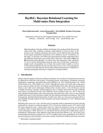
Zhou et al. BMC Plant Biol(2021) 21:277Perilla frutescens (L.) Britt is an edible vegetable andmedicinal plant processed in cosmetics such as skincreams, soaps, and dermatological medicinal preparations [7]. Two kinds of trichomes are present on P.frutescens leaves, flowers, and stems with multicellularstructures: GTs (peltate glandular trichomes PGTs andcapitate glandular trichomes CGTs) and non-glandulartrichomes (NGTs) [8]. Previous reports have noted thatthe GTs in P. frutescens were considered the primary tissue for essential oil synthesis and accumulation, especially PGTs. The number of essential oil componentssynthesized in leaves was positively correlated with thenumber of PGTs [9, 10]. The [14C]-sucrose tracer experiments showed that the monoterpenoid and phenylpropanoid components of the essential oil were synthesizedby PGTs [9]. Histochemical localization further indicatedthat terpenoids, the main ingredients of essential oil,were present in PGTs and not CGTs [10]. Moreover, themetabolite profiles of PGTs in P. frutescens have not beenreported. In addition to essential oil, there are a variety ofother pharmaceutically bioactive secondary metabolitesthat are synthesized and accumulated in P. frutescens,such as phenolic acids (caffeic acid and rosmarinic acid),flavonoids, and volatiles (monoterpenes and sesquiterpenes), which give the plant pleasant green and purplecolors, unique aromas, and medicinal uses [11]. Severalstudies have focused on secondary metabolite profilingof different tissues of P. frutescens, but only a few studies have examined their biosynthetic pathways, especiallymonoterpenes [12].In the present study, we established the morphological and chemical characteristics of PGTs on the leavesPage 2 of 15of P. frutescens. Metabolomics and transcriptome databases from different tissues were used to identify candidate genes involved in the biosynthesis of bioactiveconstituents, and the genes affecting the developmentof trichomes were preliminarily predicted. These resultsenhanced our understanding of the mechanisms underlying the biosynthesis of bioactive constituents in P.frutescens and laid a solid foundation for the future investigation of trichomes, improvement of its health functions, and better-quality medicial plants.ResultsThree types of glandular trichomes are present on aerialsurfaces on leavesThe whole plant of P. frutescens is densely pubescent.Its leaves are green and soft purple on the adaxial andabaxial surfaces, respectively, which is called the “doublecolor P. frutescens.” The primary roots of P. frutescens areshorter than those of the lateral roots (SupplementaryFig. 1). The plants in our study were similar to the 465Pand green/red Perilla in previous reports [11, 13]. Thepubescence of P. frutescens was composed of multicellular GTs and NGTs [8]. The GTs were including PGTs,CGTs and another morphology GTs, with finger-likeshape, named digitiform glandular trichomes (DGTs).These three type GTs all contained three parts includingbasal cells, stalk cells, and head cells, like GTs in otherLamiaceae plants [4, 5]. These three types of GTs locatedon leaves and stems (Fig. 1, Supplementary Fig. 1),which were captured using an SEM and a stereomicroscope. PGTs consisted of multicellular heads, one stalkcell, and one basal cell, which assumed a globular domeFig. 1 The morphology of glandular trichomes in P. frutescens; stereomicroscope and SEM for PGT (a, b), CGT (c, d for one head cell; g, h for twohead cells) to DGT (e, f) (bar 0.05 mm and 0.02 μm for d, h)
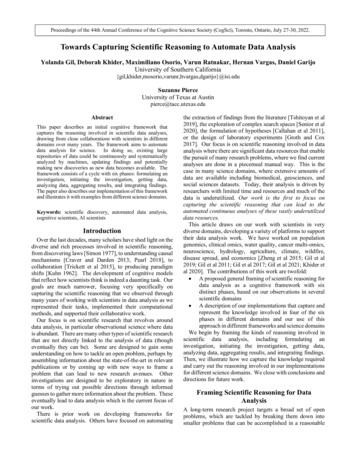
Zhou et al. BMC Plant Biol(2021) 21:277Page 3 of 15shape at maturity (Fig. 1 a, b). It should be noted thatthe secretions of PGTs are yellow when mature. PGTswere approximately 42–47 μm in height and 56–60 μmin diameter (with four and eight secretory cells) whenmature. CGTs were smaller than PGTs, with a 23–28 μmdiameter spherical head and 31–34 μm in height, characterized by a short stalk, one or two-celled head, and onebasal cell (Fig. 1 c, d, g, h). DGTs consisted of one single head cell, a longer stalk cell, and one basal cell (Fig. 1e, f ). They were approximately 61–69 μm in height and10–13 μm in diameter when mature (SupplementaryFig. 2 and 3).Metabolites of PGTs and leaves, stems, and roots of P.frutescensThe components of volatile oil contributed the most tothe aroma of P. frutescens and were the active ingredientsin the drugs. Volatile oil was the primary evaluation criterion for the quality of medicinal materials in the Chinese Pharmacopeia (2015 edition). We measured leaves,stems, roots, and PGT volatiles in plants by GC–MS toinvestigate the volatile constituents. Essential oil was collected by SDE from leaves (enriched in PGTs), stems, androots. Secretions from PGTs were collected using micropipettes diluted with solvent, and then analyzed by GC–MS. There were 16 peaks in PGTs, 19 peaks in leaves, 7peaks in stems, and 7 peaks in roots. 13 peaks have beenpreviously identified, and eight peaks were unknown.The results of the identified peaks are listed in Table 1,and most of the peaks appeared 10 min later. GC–MSpeak mass spectra for the compounds are shown in Supplementary Fig. 4. Based on the GC–MS results, the oilcomposition of PGTs was similar to that of distillatesfrom leaves (Fig. 2a), implying that these PGTs were thedominant source of commercial essential oils. Amongthese compounds, terpenoids were shown as the leading group. Perillaketone, the main compound, accountsfor approximately 50% of the volatile oil of leaves, stems,and PGTs, and the isoegomaketone content in leaves wasclose to 20%, which implied that the chemical type of P.frutescens is PK-II [14]. Moreover, the volatile composition of PGTs, which are known to be the primary tissuesinvolved in the biosynthesis or accumulation of volatileoil, was strikingly similar to that of the leaves, whichshared 11 common identified compounds. The massspectra of the primary compounds in GC–MS are shownin Fig. 2b, containing perillaketone (4), egomaketone (5),isoegomaketone (6), β-caryophyllene (8), and (Z,E)-αfarnesene (11).To comprehensively identify the metabolite profiles ofP. frutescens, the compounds were authenticated usingLC–MS, which was dedicated to the P. frutescens colorand medicinal usage. Therefore, metabolite profiles inthe four organs were tentatively investigated by UPLCESI-Q-TOF–MS/MS through the full scan negative andpositive ESI modes. In our study, the compositions ofdifferent organs showed significant differences. Supplementary Table S1 shows the retention times, molecularformula, and mass spectral data (molecular and fragment ions) of the identified metabolites. LC–MS peaksand mass spectra of the compounds are shown in Supplementary Fig. 5. We identified 71 compounds from P.frutescens through negative and positive modes, of whichflavonoids (16), phenylpropanoids (15), and terpenoidsTable 1 The compounds identified in the essential oils of P. frutescensNoCompund nameRetentiontime (min)FormulaMolecular Mass Type(g/mol)Relative content erpene0.560.3NN10Germacrene terpene0.120.14NNN represented not detected
Zhou et al. BMC Plant Biol(2021) 21:277Page 4 of 15Fig. 2 GC–MS peaks of leaves and PGTs (a), and the mass spectra of major compounds in GC–MS (b); the mass spectra of Caffeic acid (c) andRosmarinic acid (d) in LC–MS(13) accounted for a large proportion. There were 36,63, 46, and 24 compounds identified in the PGTs, leaves,stems, and roots, respectively. Except for roots, the topthree compounds in the other parts were flavonoids,phenylpropanoids, and terpenoids. Caffeic acid and rosmarinic acid were detected as the predominant metabolites [15]. Interestingly, similar to the results of GC–MS,the compounds accumulated in PGTs were strikinglysimilar to leaves, which shared 30 common compounds.The fragmentation behaviors of the above two compounds are shown as examples. The product ion massspectrum of compound 10 (tR 3.2 min) showed a deprotonated ion [M-H] at m/z 179.0320. In the MS/MSspectrum, the distinctive product ion at m/z 135.0446([M-H] -44 amu) corresponded to the loss of a -COOHmoiety, which was confirmed to be caffeic acid (10), withthe elemental formula C9H8O4 (Fig. 2c) [16]. Compound27 (tR 4.85 min) exhibited a deprotonated molecularion at m/z 359.0772. The MS/MS fragmentation patternspossessed three fragment ions at m/z 197.0455, 179.0360,and 161.0243. The fragment ion at m/z 197.0455 wasformed by the loss of m/z 162 fragment (m/z 359–m/z197; [M-H] -162 amu), which was determined to be thecharacteristic of a caffeoyl moiety [17]. The fragmention at m/z 161.0243 corresponded to the deprotonatedcaffeoyl residue. Thus, compound 27 was tentativelyassigned to rosmarinic acid (27) ( C18H16O8) (Fig. 2d).The identified caffeic acid and rosmarinic acid were confirmed using the retention time of the standard mixture(Supplementary Fig. 6 A, B). Perillaketone and isoegomaketone were also identified by LC–MS using a standardmixture (Supplementary Fig. 6 C, D).RNA‑seq and de novo assembly and annotations resultThrough Illumina high-throughput sequencing of different tissues of P. frutescens, 52.8, 41.0, and 42.2 billionclean reads were obtained from leaves, stems, and roots,respectively (Supplementary Table S2 and Table S3). Theclean reads were assembled into contigs and de novoassembled into transcripts using Trinity, yielding a total
Zhou et al. BMC Plant Biol(2021) 21:277of 315,005 transcripts and 117,939 unigenes (Supplementary Table S4). A total of 117,939 unigenes were annotated using common databases including the CDD, KOG,NR, NT, PFAM, SwissProt, TrEMBL, GO, and KEGG,to which approximately 34.43%, 28.8%, 43.94%, 31.45%,25.02%, 42.73%, 43.23%, 46.03%, and 4.74% of unigeneswere mapped, respectively (Supplementary Table S5).Through homologous species comparison in the NRdatabase, 36.32% of the unigenes had the highest homology with Sesamum indicum (Pedaliaceae), followed byErythranthe guttata (Scrophulariaceae) (SupplementaryFig. 7). The details of the GO and KEGG analyses areshown in Supplementary Fig. 8 A and B.Identification of DEGs and annotations resultA total of 10,696 DEGs were collected with the filter criteria FoldChange 2 and q-value 0.05, including 4127upregulated genes and 4088 downregulated genes in Lvs. R, 2548 upregulated and 2574 downregulated in L vs.S, and 3203 upregulated and 2898 downregulated in Svs. R, respectively (Supplementary Fig. 9 A). There were1126 common unigenes in three comparisons (Supplementary Fig. 9 B). DEGs of S vs. R, L vs. S, and L vs. Rcomparisons were assigned to GO classifications shownin Supplementary Fig. 10 A-C. The responses to stimuli,metabolic processes, and cellular processes included inthe biological process were enriched in three comparisons; catalytic activity in molecular function was alsoenriched in three comparisons. To explore the molecularbasis of trichome development among leaves, stems, androots, a large number of unigenes were assigned to GOannotation (128, GO:0,010,026 trichome differentiation;57, GO:0,010,091 trichome branching; 100, GO:0,010,090trichome morphogenesis) (Supplementary Table S6). Inthe above unigenes, most were highly expressed in leavesor stems (Supplementary Fig. 11), which was consistentwith the phenomenon that GTs accumulated at theseorgans. In the KEGG pathway analysis, pathways suchas plant hormone signal transduction and biosynthesisof phenylpropanoids, flavonoids, monoterpenoids, terpenoid backbones, sesquiterpenoids, and triterpenoidswere enriched in S vs. R and L vs. R. As for L vs. S, onlyphenylpropanoid, flavonoid, sesquiterpenoid, and triterpenoid biosynthesis were enriched, revealing the sametrends as the distribution of secondary metabolites (Supplementary Fig. 10 D-F).DEGs related to secondary metabolitesThe pathways annotated in the KEGG database wereinvolved in the biosynthesis of secondary metabolites.We combined the analysis of transcriptomic and metabolomic data to explore key genes involved in terpenoid,flavonoid, and phenylpropanoid biosynthesis in threePage 5 of 15tissues. The metabolism of the terpenoids and polyketides subcategory contained eight pathways, and thelargest number of DEGs (23 unigenes) were mappedto terpenoid backbone biosynthesis (SupplementaryFig. 12). Corresponding to the metabolic profiles andterpenoid biosynthesis, we aimed at the biosynthesis ofterpenoid backbones, monoterpenoids, diterpenoids, sesquiterpenoids, and triterpenoids. Among these unigenes,38 DEGs were identified as encoding 24 key enzymes thatcontrolled terpenoid biosynthesis, and the details arelisted in Supplementary Table S7. These unigenes weremainly mapped in the 2-C-methyl-D-erythritol 4-phosphate and mevalonate pathways (Fig. 3). Of the fourDXS genes, DXS1, DXS2, and DXS4 showed the highestexpression in leaves, and they were located upstream ofterpenoid biosynthesis. These results suggest that DXS isimportant for the generation of 2-C-methyl-D-erythritol4-phosphate pathways and deserves further functionalcharacterization. The genes related to monoterpene biosynthesis also presented the same trends, which may leadto the accumulation of monoterpenes [9].The leaves and stems of P. frutescens showed pleasantpurple and green colors, which were genetically controlled to accumulate a significant amount of anthocyanin (especially malonylshisonin) [18]. In our study,10 DEGs were related to flavonoid biosynthesis andupstream of anthocyanin biosynthesis, which were allhighly expressed in leaves (Fig. 4a). Combined withphenylpropanoid biosynthesis and tyrosine-derivedpathways, 21 unigenes encoding eight key genes wereassigned to rosmarinic acid and caffeic acid biosynthesis(Fig. 4b) [19]. DEGs in the above two pathways showedsimilar expression patterns, except that RASs werehighly upregulated in roots. Metabolites and other DEGsinvolved in phenylpropanoid biosynthesis are listed inSupplementary Fig. 13.DEG related to trichome developmentTrichomes are classified as non-glandular or glandular. Arabidopsis thaliana has single cell non-trichomes,whereas Solanum lycopersicum and Artemisia annuahave multicellular glandular trichomes. The genes relatedto trichome initiation and development have been widelyreported, including TTG1, GL1, and GL3 of A. thaliana;MYC1, MX1, and CD2 of S. lycopersicum; and MIXTA,MYB1, and HD1 of A. annua. According to these reports,46 unigenes, encoding 21 key genes, were screened inour transcriptomes (Fig. 5). Except for the 21 homologous unigenes of A. thaliana, the expression patterns ofhomologous genes of A. annua (13 unigenes) and tomato(12 unigenes) were similar. The homologous geneslike SlMYC1, SlMX1, SlCD2, SlWOOLLY, AaMIXTA,AaMYB1, AaHD1, AaHD8, and AaGWS2, which have
Zhou et al. BMC Plant Biol(2021) 21:277Page 6 of 15Fig. 3 The biosynthesis of terpenoids (a) and heatmap (b) of DEGs involved in monoterpene, diterpenoid and sesquiterpene biosynthesis; AACT,acetyl-CoA acetyltransferase; HMGS, hydroxymethylglutaryl-CoA; HMGCR , hydroxymethylglutaryl-CoA reductase synthase; MVK, mevalonatekinase; PMK, phosphomevalonate kinase; MVD, mevalonate diphosphate decarboxylase; DXS, 1-deoxy-D-xylulose-5-phosphate synthase; DXR,1-deoxy-D-xylulose-5-phosphate reductoisomerase; CMS, 2-C-methyl-D-erythritol 4-phosphate cytidylyltransferase; CMK, 4-diphosphocytidyl-2-C-methyl-D-erythritol kinase; MCS, 2-C-methyl-D-erythritol 2,4-cyclodiphosphate synthase; HDS, 4-hydroxy-3-methylbut-2-enyl diphosphatesynthase; HDR, 4-hydroxy-3-methylbut-2-enyl diphosphate reductase; IDI, isopentenyl pyrophosphate isomerase; GPPS, geranyl diphosphatesynthase; FPPS, farnesyl diphosphate synthase; GGPPS, geranylgeranyl diphosphate synthase; CYP71D55, premnaspirodiene oxygenase; FDFT1,farnesyl-diphosphate farnesyltransferase; GERD, (-)-germacrene D synthase; LUP4, beta-amyrin synthase; LUS, lupeol synthase; NES1, (3S,6E)-nerolidolsynthase; SQLE, squalene monooxygenase. Unigenes levels data represent by TPM. The red letters represent DEGs. Red and green represent highand low expression levels, respectivelybeen reported to promote trichomes development, allexpressed highly in leaves. Among the 46 trichome development-related unigenes, most were TFs belonging toR2R3 MYB, WD40, bHLH, and HD-ZIP IV family.Plant hormones have also been reported to regulatetrichome initiation and development, such as gibberellin (GA) and jasmonic acid (JA) [6, 20]. Our transcriptome data analysis revealed that multiple genes weremapped to activated signaling pathways. In the GA andJA pathways, we identified 24 and 37 unigenes, respectively (Supplementary Fig. 14). Additionally, 16 DELLAand 18 JAZ were screened among these unigenes, whichnegatively regulated the growth and development of trichomes [21, 22]. As expected, most DELLA and JAZ weredownregulated in leaves enriched with trichomes.Gene expression validated by qRT‑PCRFifteen DEGs were selected to verify the accuracy of thetranscriptome data using qRT-PCR. These key structuralgenes are involved in the biosynthetic pathways of terpenoids, flavonoids, and phenolic acids, and trichomeformation. Specific primers were designed using PrimerPremier 5.0 (Supplementary Table S8). In general, theexpression patterns determined by qRT-PCR showedsimilar trends to RNA-Seq (Fig. 6 and SupplementaryTable S9), which confirmed the accuracy of the RNA-Seqresults reported in this study.DiscussionP.frutescens is a well-known medicinal and edible plantthat has been widely cultivated in Asia more than2000 years [23]. The leaves and stems often present greenor purple colors. Volatile oil is one of the main components of P. frutescens, and different chemotypes havebeen described based on the main components of essential oils [24, 25]. The chemotypes are listed in Supplementary Table S10, and their speculated synthesis pathwaysare shown in Supplementary Fig. 15 [14]. P. frutescens inour study was identified as a PK-II type cultivated widelyin China. To further mine nutraceutical and medicinalpotential, we integrated metabolite profiling and transcriptome analysis of different tissues. Additionally, wedisplayed the metabolic profiles of PGT secretion for thefirst time.
Zhou et al. BMC Plant Biol(2021) 21:277Page 7 of 15Fig. 4 The biosynthesis of flavonoids (a) and phenylpropanoids (b) according KEGG. PAL, phenylalanine ammonia-lyase; C4H, trans-cinnamate4-monooxygenase; 4CL, 4-coumarate–CoA ligase; COMT, caffeic acid 3-O-methyltransferase; TAT , tyrosine aminotransferase; HPPR, hydroxypyruvatereductase; RAS, rosmarinate synthase.Unigenes levels data represent by TPM. The red letters represent DEGs and detected compounds. Red andblue represent high and low expression levels, respectivelyMorphological and distribution of GTs in P. frutescensIn previous reports, it has been elaborated that P. frutescens has two types of glandular trichomes (PGTs andCGTs), and PGTs were the primary tissues of biosynthesis and accumulation of essential oils with a predominantnumber and size [9, 10]. We certified another fingerlike shaped GT, named DGT (Fig. 1 and SupplementaryFig. 2 I-L), similar to the type II CGTs with a unicellular base, two elongated stalk cells, and one head cell [10].The number of secretory cells was consistent with published papers (PGT usually with 8 cells, CGTs usuallywith one or two cells) [8–10]. Under a stereomicroscope,the PGTs were filled with yellow essential oil in a largesub-cuticular storage space formed by the separation ofthe cuticle from secretory cells, such as PGTs in peppermint [26]. Compared with PGTs, the storage capacity ofCGTs was limited to smaller sizes and fewer secretorycells. As shown in Supplementary Fig. 2E-G, there weretwo types of CGTs containing colored and transparentglandular heads. Whether there are two types of CGTs,or their developmental sequence, needs to be furtherstudied. As for DGTs, there was only one transparenthead cell that was unknown to have a secretory capacity.Its base cell may enlarge during development, as shownin Supplementary Fig. 2 I-K. PGTs, CGTs and DGTs weredistributed on both the adaxial and abaxial leaf (mostly)
Zhou et al. BMC Plant Biol(2021) 21:277Fig. 5 A map of genes regulation for the growth (a) and development of trichomes. Heatmap of genes related to trichomes growth anddevelopment (b). Unigenes levels data represent by TPM. Red and green represent high and low expression levels, respectivelyPage 8 of 15
Zhou et al. BMC Plant Biol(2021) 21:277Page 9 of 15Fig. 6 Validation of expression patterns of 15 DEGs by qRT-PCR. The relative expression levels were calculated according to the 2 -ΔΔCT methodusing β-actin as internal reference gene. Error bars represented standard error of mean. * indicated significant difference of the expression means atp 0.05 between L and S or L and R (n 4). The relative expression of qRT-PCR was indicated on the left y-axis and the TPM normalized expressionlevel (log2TPM) of RNA sequencing was indicated on the right y-axis
Zhou et al. BMC Plant Biol(2021) 21:277surface and stems and were especially abundant in thecentral vein and the area near the leaf base. In summary,the morphological features and occurrence of GTs inour data were similar to previous reports of P. frutescens,which is also similar to GTs in S. tenuifolia [5] and Mentha x piperita [27], belonging to Labiaceae.Metabolites variation of PGTs, leaves, stems, and rootsMetabolite profiling of PGTs, leaves, stems, and rootsshowed that the most enriched compounds were terpenoids, flavonoids, phenolic acids, and fatty acids (Table 1,Supplementary Table S1), and the confirmed compoundsin PGTs were strikingly similar in leaves. Theoretically,the compounds contained in PGTs should be detectedin leaves, whereas the leaf extracts are more abundantthan PGT secretions. In addition to PGTs, the leaves contained other types of cells and GTs, and the energy andheat generated in the process of ultrasonic extractionresulted in changes or loss of compounds. The method,which used micropipettes to collect secretions fromPGTs, resulted in metabolic profiles closer to those ofcompounds in vivo. Egomaketone was the precursorof perillaketone and isoegomaketone (SupplementaryFig. 15). The contents of perillaketone and isoegomaketone were lower in PGTs than in leaves. In contrast,the egomaketone content was the opposite because theleaves in node 1 (from the tip of the stem to the root) forPGT analysis were younger than leaf extraction (nodes1 and 2), which may reveal the developmental dynamicsof leaf essential oil composition. The relative contents ofmonoterpenoids and sesquiterpenoids in GC–MS werevery low or absent in the roots. In the LC–MS analysisof P. frutescens, phenolic acid and flavonoids were generated from flavonoid and phenylpropanoid biosynthesis,respectively [15]. The purple leaves enriched anthocyanin pigments, such as malonylshisonin [18, 28]. It wasdetected in PGTs and leaves with the highest intensity inPGTs compared with other tissues. Interestingly, PGTswith higher intensity were gold, while the abaxial surfaceof the leaves was purple. Anthocyanin is sensitive to light,temperature, and pH [29]. These analyses confirmed thatPGTs contained the bulk of the bioactive constituentsaccumulated by P. frutescens.Molecular mechanism of metabolites biosynthesisMonoterpenes and sesquiterpenes are the main components of the volatile oil in P. frutescens. Crossing experiments between the principal types of Perilla suggestedthat the different chemotypes were under strict geneticcontrol [30]. The main components of volatile oil inthese chemotypes were generated from the mevalonicand shikimic acid pathways (Supplementary Fig. 15).The presence of the G gene is essential for the initiationPage 10 of 15of monoterpenoid biosynthesis via the mevalonic acidpathway. When the initiation was controlled by the recessive gene h, tans-citral was catalyzed by geranyl pyrophosphate, and under the regulation of multiple genes,perillaketone was finally generated [30]. Although thebiosynthetic pathways for essential oils in Perilla havebeen described, the key genes controlling these pathways are still unknown. However, the lack of the Perillagenome made it a great challenge to unravel the completebiosynthesis pathway of monoterpenes in P. frutescens. Alarge number of unannotated unigenes (44.03%, Supplementary Table S5) in our transcriptome needed furtherexploration, which may be annotated as the key genes forthe synthesis of perillaketone in the future.The key gene for anthocyanidin, ANS, is located in theepidermal cells of the stems, where anthocyanins accumulate [28]. The stem transverse section in Supplementary Fig. 16 shows similar result
sue for essential oil synthesis and accumulation, espe-cially PGTs. e number of essential oil components synthesized in leaves was positively correlated with the number of PGTs [910, ]. e [14C]-sucrose tracer experi-ments showed that the monoterpenoid and phenylpro-panoid components of the essential oil were synthesized by PGTs [9].