Transcription
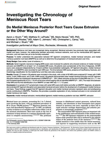
Critical Reviews in Biomedical Engineering, 41(3):183–204 (2013)Compressed Sensing MRI: A ReviewSairam Geethanath,1,* Rashmi Reddy,1 Amaresha Shridhar Konar,1 Shaikh Imam,1 RajagopalanSundaresan,2 Ramesh Babu D.R.,1 & Ramesh Venkatesan2Medical Imaging Research Centre, DSCE, Bangalore, India; 2Wipro-GE Healthcare, India1 * Address all correspondence to: Sairam Geethanath, Ph.D., Director, Medical Imaging Research Centre, Dayananda Sagar College of Engineering,Bangalore, India; Tel.: 91-80-42161733; Sairam.Geethanath@dayanandasagar.eduABSTRACT: Compressed sensing (CS) is a mathematical framework that reconstructs data from highly undersampled measurements. To gain acceleration in acquisition time, CS has been applied to MRI and has been demonstratedon diverse MRI methods. This review discusses the important requirements to qualify MRI to become an optimalapplication of CS, namely, sparsity, pseudo-random undersampling, and nonlinear reconstruction. By utilizing concepts of transform sparsity and compression, CS allows acquisition of only the important coefficients of the signalduring the acquisition. A priori knowledge of MR images specifically related to transform sparsity is required for theapplication of CS. In this paper, Section I introduces the fundamentals of CS and the idea of CS as applied to MRI.The requirements for application of CS to MRI is discussed in Section II, while the various acquisition techniques,reconstruction techniques, the advantages of combining CS and parallel imaging, and sampling mask design problemsare discussed in Section III. Numerous applications of CS in MRI due to its ability to improve imaging speed are reviewed in section IV. Clinical evaluations of some of the CS applications recently published are discussed in SectionV. Section VI provides information on available open source software that could be used for CS implementations.I.INTRODUCTIONMagnetic resonance imaging (MRI), as a biomedical imaging modality, provides images with excellent soft tissue contrast. MRI has been extensivelyused to image detailed structure, function, and metabolism of the organ of interest. The reduction ofMRI acquisition time is an ongoing challenge toenable imaging of certain biological processes thatdemand high spatial and/or spectral resolutions. Toachieve short acquisition times, techniques suchas PI (Ref. 1) and other undersampling strategiessuch as keyhole imaging have been accomplished.These techniques are typically governed by theNyquist sampling rate and hence cannot yieldacceleration beyond the Nyquist limit due to theresulting aliasing artifacts and their ability to resolve them in cases of reduced signal-to-noise ratio(SNR). A mathematical framework called CS thathas been proposed,2 provides for reconstruction ofdata from highly undersampled measurements.2,3CS has been applied to MRI to gain accelerationin acquisition time and it has been demonstratedon diverse MRI methods. Recently, it is also beenused in clinical applications for pediatric imagingwhere reduction in acquisition time is critical for1040-8401/13/ 35.00 2013 by Begell House, Inc.diagnosis.4 The following subsections discuss thefundamentals of CS.A. SparsityA vector is said to be “sparse” if the majority of itscoefficients are equal to zero and very few coefficients will contain all the information. If significantnumbers of these coefficients are exactly zeroes,then the sparsity is said to be “strong.” Generally,there exists a transition band from the few highvalued coefficients to the many zero-valued coefficients, which is called “weak sparsity.” From a signal processing standpoint, a sparse signal has mostof its energy contained in a few measurements whilethe rest of the measurements are zero or negligible.1.Transform Sparsity and CompressionTransform sparsity results in a vector, which becomes sparse on using a mathematical transform.For example, a voltage depicting a sinusoidal characteristic of a particular frequency when recordedover time does not exhibit sparsity. However, on theuse of Fourier transform, the transformed vector hasall of its information in two peaks (ω ωo). Now,183
184Geethanath et al.the sine wave is said to be sparse in the Fourier domain. Most MR images are sparse in some transform domain. For instance, angiography images aresparse in the identity and finite differences domainswhereas cardiac cine images are sparse in the spatiotemporal Fourier domain.Sparsity can also be extended to compressibility, wherein the important coefficients that couldbe used for reconstruction of the original signalcould be retained. Threshold is applied in the regionwhere there is a sharp decrease in the amplitude ofthe coefficients when they are sorted in a descending fashion. This threshold decides the number ofcoefficients to be used to reconstruct. It indicates thecut-off for the important coefficients from the lessimportant ones (analogous to the cut-off frequencyfor impulse response filters in signal processing).Generally, the wavelet transform is most ubiquitously used to compress the images. The reconstructionof the images is done from the sparse coefficients.If this reconstruction is exact, the image is said tobe compressed with no losses, i.e., the differencebetween the original and reconstructed images fromcompressed data is zero. If this is not the case, thecompression is said to be “lossy.” Lossy compression results in loss of some information that is typically not perceivable to the human eye. This information corresponds to the coefficients closer to thezeroes and is not used for reconstruction. This is applicable to MR images as well. However, it shouldbe noted that lossy compression in certain medicalimaging applications involving detection problemsmight not be tolerable and have to be evaluated byradiologists.2.CS: The IdeaThe conventional method of image compressionis performed after the acquisition of the entire image, which typically involves wavelet transformingof the image. Compression is done to accomplishsaving in data storage and to facilitate the transferof such data. Hence, the compressed data consistsof only the most important coefficients that enablenear-perfect reconstruction of the original data.The philosophy of CS is to acquire only theimportant coefficients of the signal during theacquisition by compression and by using the concept of transform sparsity rather than compressionpostacquisition. The implementation of an ideal CSsolution is given by the answer to a simple question:What is the minimum number of coefficients in atransform domain required to acquire data with acceptable SNR and high data fidelity to represent theobject of interest?3.The L0, L1, and L2 NormsThe above question raised in Section I.A.2 can bemathematically cast as an optimization problem.The first half of the question would be to solve forthe minimum number of coefficients in the transform domain required to achieve exact reconstruction. The “norm” function in linear algebra can beused to evaluate this criterion. The Lp norm of a vector x of length n is expressed as follows:(1)In the case of p 0, called the L0 norm, (1) simplifies to the number of nonzero elements in thevector (n in the case of a vector consisting of ones),which is the required solution. The L0 norm is notconvex in nature, and computationally intractable,which results in difficulty in its usage in optimizationproblems.5 However, L0-based CS reconstructionhas been reported recently that utilizes nonconvexoptimization schemes.6 Mathematically, solution toa convex problem or an approximately convex problem is more stable than a nonconvex optimizationscheme due to reliability in convergence. Hence, thenext integer (p 1) norm L1 is generally used, whichis a convex approximation for L0 norm and providesthe absolute sum of the elements of the vector. Itmust be also noted that reconstructions using L1/2and other non-integer norms utilized in a nonconvex framework have also been explored.7 The L1norm penalizes the presence of a large number ofcomponents, which ensures sparsity of the solutionvector. In other words, L1 norm prefers the presence of a few coefficients, which contain the totalenergy of the signal, while the rest of coefficientsare predominantly zeros. The second part of the CSCritical Reviews in Biomedical Engineering
Compressed Sensing MRI: A Reviewproblem is to ensure data fidelity. The L2 norm is anideal choice for ensuring consistency since the normpenalizes small errors relatively lesser and large errors more significantly due to the quadratic nature ofthe norm. Hence, the solution to the CS optimizationconsists of two terms: an L1 norm of the transformcoefficients to ensure sparsity, and an L2 norm of themeasured data and its iterative approximation to account for data consistency. This paper is organizedas follows: Section II reviews the requirements forthe application of CS to MRI; Section III discussesvarious CS techniques such as acquisition and reconstruction techniques such as k-t FOCUSS, k-tBLAST, Bayesian experimental design, k-t groupsparse, modified CS (MCS), motioncompensatedMCS (MC-MCS), and combination of CS and PImethods such as SparseSENSE, phase-constrainedCS (PC-CS) reconstruction, L1-SPIRiT reconstruction techniques, and the mask design problems;Section IV explores various applications of CS, i.e.,cardiac MR, dynamic contrast enhanced MRI (DCEMRI), angiography, functional MRI (fMRI), MRparameter mapping, diffusion-weighted imaging(DWI), arterial spin labeled (ASL), musculoskeletal system, and magnetic resonance spectroscopicimaging (MRSI); Section V discusses the clinicalevaluation of some of the CS applications; and Section VI provides a few resources for open sourcesoftware that could be used for CS implementations.II. REQUIREMENTS FOR APPLICATION OFCS TO MRIThe implementation of CS to MRI is relativelystraightforward. Here, one has to have a priorknowledge of the type of MR images he/she is goingto acquire so that certain unique properties of thatparticular class of images can be utilized optimally,which is not a challenge since this information istypically known. For instance, the person obtainingan MR angiography image will know that the images resulting from MR angiography (MRA) willalso be sparse in its native representation. This priorknowledge provides an idea to the CS implementerabout the utility of application of CS to that of MRmethod. As already mentioned, MRA is sparser inthe finite difference domain or the total variationVolume 41, Number 3, 2013185(TV) norm. There are three fundamental requirements for the application of CS to MRI, detailedbelow.A. Transform SparsityThe fundamentals of transform sparsity have beendescribed in section I.A.1. It is one of the three mostimportant requirements for the implementation ofCS. However, choosing the right transform to exploit sparsity existing in a particular class of MRimages is a challenging task and is a field of ongoing research. Hence, to achieve the purpose, 2D or3D wavelets are generally used, since wavelets result in good compression of medical images. Also,it is required to find an optimal solution to explorethe best transform sparsity for each particular case.Dynamic contrast enhancement MRI (DCE-MRI) isone of the examples wherein spatiotemporal correlations can be exploited to increase transform sparsityalong with the use of wavelets. Thus, the transformsparsity is one of the requirements that allow the CSimplementer to evaluate the sparsity of the imagesand it also provides an insight into the number ofsamples he/she needs to acquire to achieve exact reconstruction, which is completely dependent on thenumber of sparse coefficients that are enough to result in an exact reconstruction (Fig. 1).B.Pseudo-Random UndersamplingAn MRI scanner collects the spatial frequency information, which is called k-space. To apply CSto MRI, the collected k-space has to be randomlyundersampled so that aliasing artifacts are not produced due to the nonprime nature of the vector size(Theorem 1.1, in Ref. 2) as noticed in the case ofsub-Nyquist sampling. A randomly undersampledk‑space, as shown in Fig. 2, results in artifacts thatare incoherent with the image and can be denoisedto obtain the original image. The samples required tobe acquired are typically five to eight times the number of sparse coefficients of the data set.2 Hence, thesparser the transformed data, the fewer the samplesrequired to reconstruct. Random undersampling ofk-space has two specific practical limitations and/or considerations. One of the limitations is that it is
186Geethanath et al.(a)(b)(c)FIGURE 1. Transform sparsity: (a) a 2D MR image of the human brain; (b) representation of the object in2D Daubechies wavelet transform domain; (c) a magnified view of the wavelet coefficients shown in the redsquare in (b)(a)(b)(c)FIGURE 2. Random sampling: (a) k-space of the brain image shown in Fig. 1(a); (b) a sampling mask showingthe sampling locations of the k-space as white pixels while the black pixels represent unsampled locations; (c) theresulting minimum energy reconstruction demonstrates the resulting incoherent artifactsdifficult to achieve pure random k-space samplingsince it requires rapid switching of gradients, whichis impractical due to the resulting eddy currents andrelated artifacts.The second consideration arises from the factthat most of the energy of the data acquired is locatedin the low-frequency components. Hence, a uniformweighting of the samples would result in reducedSNR. To overcome these limitations, variable densitypseudo-random sampling schemes have been proposed and successfully implemented.8 The variabledensity pseudorandom sampling mask consists ofmore samples in the center, with fewer samples in theedges. This prevents the loss of SNR but it requiresoptimal gradient design and new k‑space trajectoriesto sample the chosen points in k‑space. Another approach to retain the SNR is to start from conventionalk‑space trajectories and then introduce incoherentsampling in the same framework by skipping phaseencode values, as demonstrated in Ref. 8.A major factor in the implementation of CSwith regard to undersampling is the dimensionalityof the data. MR images with higher dimensionality(3D or higher) provide better sparsity (similar toCritical Reviews in Biomedical Engineering
Compressed Sensing MRI: A Review187image compression) and hence these images yieldbetter CS performance, i.e., CS technology is better suited to accelerate MR acquisitions of higherdimensional data. This is ideal since MR scansinvolving higher-dimensional data are the ones thatare the most time consuming.C. Nonlinear ReconstructionThe constrained optimization problem put togetherin Sections II.A and II.B can be described by the following equation (reproduced from Ref. 3):(2)where m is the desired image, y is the measuredk‑space data, Ψ is the sparsifying transform, andε is the parameter for error tolerance. This equation can be recast as an unconstrained optimizationproblem using the Lagrange method as shown inEq. (3). Here, λ is the regularization parameter thatneeds to be chosen to balance the data consistencyand sparsity terms. Equation (3) can be solved usingan iterative nonlinear reconstruction method (seeFig. 3). Along with various available methods suchas subspace pursuit, steepest descent method, etc.,the nonlinear conjugate gradient has been well studied and applied.(a)(b)Briefly, the nonlinear conjugate gradient (NCG)calculates the direction of the gradient, which isgiven by the differentiation of the above equation.At each step, the length of the step to be taken inthe gradient direction is given by a line-search parameter. The stopping criteria for the iterations aretwofold, as follows:1. The difference of values of the tolerance parameter between successiveiterations is negligible2. The value of the tolerance parameteris smaller than the chosen ε valueThe precise number of iterations is problem dependent, but the limiting number of iterations for anNCG solution is defined. The reconstructed imagem obtained at the end of this process is the desiredsolution to the following.(3)III. COMPRESSED SENSING TECHNIQUESA. Acquisition and ReconstructionCS has made a significant impact in the field of MRIto minimize the image acquisition time in the lastfew years.3 CS in MRI uses the Fourier coefficients(k‑space samples) to make accurate reconstructions(c)FIGURE 3. Nonlinear reconstruction: (a) k-space of the brain image shown in Fig. 1(a); (b) a variable densitysampling mask showing the sampling locations of the k-space with 33% sampling; (c) the resulting imagereconstructed iteratively using Eq. (3) implemented via NCGVolume 41, Number 3, 2013
188Geethanath et al.from a small subset of k‑space rather than an entire k‑space data. Some MR images are sparse in thepixel representation and other images could havesparse representations in the wavelet transform domain. According to CS theory, images with a sparserepresentation can be recovered from randomlyundersampled k‑space data. MR images need to betransformed to a sparse domain to enable CS reconstruction. We will discuss different CS techniques inthe following paragraphs.1.k-t FOCUSSk-t space FOCal underdetermined system solver (FOCUSS)9 starts by finding a low-resolution estimate ofa sparse signal, and then this solution is pruned to asparse signal representation. This technique uses theprevious iteration solution to implement the pruningprocess by scaling the current solution. Once someentries of the previous solution become zero, thenthese entries will be fixed to zero values. This method obtains a sparser solution with more iterations.These entries corresponding to the zero values on theoriginal spectral support converge to zero during thispruning process. Hence, one of the important requirements of FOCUSS is a reasonable lowresolution estimate that provides the necessary extra constraint toresolve the nonuniqueness of the problem.2.k-t BLASTThe only prerequisites for k-t broad-use linear acquisition speed-up technique (k-t BLAST) reconstruction are spatiotemporal correlations to be presentin the data, and these correlations to be accuratelycaptured by the training scan. The training scan isthe low-resolution scan acquired at the full temporal bandwidth, which is performed either before(for periodic motion) or interleaved with the actualdata acquisition. Training data have been used toresolve aliasing from undersampling along the spatial and temporal dimensions very efficiently. In thistechnique, the information about the object is actually measured rather than simply assumed, as in theview-sharing approach; this method can also be successfully applied to dynamic MRI such as cardiacimaging or inflow imaging.10In dynamic MRI, data sparsity was introduced byapplying the Fourier transformation along the temporal dimension assuming that only parts of the fieldof view (FOV) change at a high temporal rate whileother parts change slowly or remain stationary.10 CSwas achieved by randomly skipping phase-encodinglines in each dynamic frame. Cardiac cine data andFourierencoded velocity data of the carotid arterywere used to test the reconstruction performance.The k-t BLAST results in inhomogeneous distribution of error with largest errors occurring at locationscorresponding to dynamic object edges. In contrast,the error in CS is more homogeneously distributed andsmaller for highly dynamic object features.Spatiotemporal resolution is one of the qualitymeasuring metrics in dynamic MRI such as cardiaccine imaging or functional MRI. It was very difficultto acquire a whole volume image within a singlebreath hold. Various techniques were developed using parallel coil and temporal filtering to overcomethese issues. Simultaneous acquisition of spatialharmonics (SMASH), generalized autocalibratingpartially parallel acquisitions (GRAPPA), partiallyparallel imaging with localized sensitivities (PILS),and sensitivity encoding (SENSE) belong to PItechniques and unaliasing by Fourier-encoding theoverlaps using the temporal dimension (UNFOLD)to remove aliasing using temporal filtering. But stillthese techniques have low SNR, and aliasing artifacts at high reduction factor, these drawbacks areovercome by using the model-based methods suchas k-t BLAST/SENSE and k-t FOCUSS.3.k-t FOCUSS for Dynamic MRIThe k-t FOCUSS technique has also been appliedto dynamic MRI, and it has been shown to performbetter than the k-t BLAST with the following advantages:1. k-t FOCUSS minimizes the L1 norm,which is optimal from the CS perspective, but k-t BLAST does not minimizethe L1.2. Image estimation depends on the parameter p and value of which is 1 ink-t BLAST but this restriction is reCritical Reviews in Biomedical Engineering
Compressed Sensing MRI: A Reviewlaxed in k-t FOCUSS, and any valuebetween 0 to1 can be used. p 0.5 ink-t FOCUSS performs better in stability and reconstruction quality than thep 1 in k-t BLAST.In this technique, temporal average contributions arefirst subtracted from k-t samples and which are converted to the x-f domain using the Fourier transform.The weighting function calculated from the equation15 in Ref. 11 was used first time. A power factor between 0.5 p 1 has been used for the reconstruction.Solution for p 1 was too sparse. p 0.5 was not ableto remove aliasing artifacts and choice of value p 0.5seems optimal in many applications once the weighting matrix is constructed. The k-t FOCUSS algorithmwas used for high-resolution dynamic MRI becauseit showed that k-t FOCUSS was optimal from a CSperspective.12 A method was proposed based on the extension of k-t FOCUSS to a more general framework,in which the prediction provides initial encoding andresidual encoding takes care of the remaining residualsignals. This technique uses a motion estimation andcompensation scheme for sparsifying the signals andalso provides the best undersampling pattern for CS.The proposed method uses the idea from video compression and the MPEG standard.Non-Cartesian k-space trajectories were usedfor the undersampling of k-space to accelerate acquisition. Optimization of the k-space trajectoryfor sparse nonlinear reconstruction was one of thechallenges in CS and the mask designing problem isdiscussed in further detail in Section III.C. This hasresulted in the Bayesian approach for mask design.4.Bayesian Experimental DesignThe objective discussed in the previous section hasled to the Bayesian experimental design. Bayesian experimental design was used for optimizationof k-space trajectories for CS.13 The technique wasproposed for the reconstruction of sparse MRI signal using standard signal processing primitives forthe efficient use of k-space trajectories, which wasdemonstrated for Cartesian and spiral but also extendable. Further, the mask design problems are discussed in Section III.C in detail.Volume 41, Number 3, 2013189Multiple acquisitions of the same region of interest under several different contrast preparationsare done in clinical imaging with structural MRI.14Reconstruction algorithm based on Bayesian CS tojointly reconstruct a set of images from undersampledk-space data with higher fidelity than when the imagesare reconstructed either individually or jointly. Imagegradient coefficients are calculated for each image.All of the images from the same anatomical region,but with different contrast properties, contribute tothe estimation of the hyperparameters, and varianceof image gradients across contrasts for a single volumetric spatial position is single hyper-parameter. Thek-space data belonging to each image are used independently to infer the image gradients. Thus, commonality of image spatial structure across contrastsis exploited without the problematic assumption ofcorrelation across contrasts.CS reconstruction for dynamic cardiac MRIhas been improved by incorporating additional information on the support of the dynamic image inx-f space based on the theory of CS with partiallyknown support by using technique k-t iterative support detection (k-t ISD).15 This technique uses aniterative procedure for alternating between imagereconstruction and support detection in x-f space.Support information from the previous iteration wasused to apply a truncated L1 minimization to obtainthe reconstructed image in x-f space. The methodalternates between CS reconstruction with partiallyknown support (PKS) and adaptive learning of support knowledge used in the next iteration by thresholding the reconstruction.Another CS technique proposed to acceleratethe dynamic MRI, which makes use of the structure within the sparse representation of a signal byenforcing the support component in the form ofgroups, is called k-t group sparse.16 Support estimation, group assignment, and signal recovery arethe three key steps in this technique. In this work, atraining scan was used to identify the support regionin the x-f space. A threshold is set above the noiselevel in the x-f space and elements with intensitiesabove the threshold constitute the support region,then support in x-f space is estimated from the previous step to assign elements in x-f space to distinctgroups. Once the groups are assigned to all elements
190Geethanath et al.in the x-f space, the signal recovery is done by thegroup sparse formulation. However, the use of thistechnique in dynamic MR applications has been limited in terms of the maximum achievable reductionfactor. In general, noiselike artifacts and bad temporal fidelity are visible in standard CS MRI reconstructions when high reduction factors are used. Toincrease the maximum achievable reduction factor,additional or prior information can be incorporatedin the CS reconstruction. This technique exploits thestructure within the sparse representation of a signalby enforcing the support components to be in theform of groups. These groups act like a constraint inthe reconstruction. Figure 4 depicts an illustration ofthe group assignment step of the proposed method(a: 8 8 sparse image, b: group assignment matrixshowing the group number to which element/pixelat the respective location is assigned).The iterative soft thresholding (IST) frameworkhas been used for accelerated MRI using CS.17 Thistechnique is data driven and no tuning of free parameters is required. A Nesterov-type optimal gradient scheme for iterative update along with standardwavelet-based adaptive denoising methods werecombined, which results in a leaner implementation compared with the nonlinear conjugate gradient method. The technique was evaluated on T2weighted brain data, and vascular 3D phase contrastdata show that the image quality of reconstructionsis comparable with those from an empirically tunednonlinear conjugate gradient (NLCG) approach.5.Modified CSIn modified CS, it is assumed that the support (location of nonzero coefficients) of the signal in thesparse domain is partially known and can be usedin reconstructing the sparse coefficients with greateraccuracy.18 Partial knowledge of the signal supportcan be found a priori in many signal processing applications. For example, it is common knowledgethat the locations of the significant Fourier coefficients of the signal are present at the lower frequencies. Such partial knowledge, when introduced intoan L1 minimization objective, results in a sparser solution that is better than conventional CS solutionsat high acceleration factors. Thus, the modified CSproblem introduces the partial support information and aims to find the sparsest signal outside theknown support while satisfying the data constraint.A time series extension of this framework isnamed dynamic modified CS, which assumes thatsupport changes occur slowly in the time series.Thus, the known support for reconstructing thecurrent frame is assumed to come from the supportestimate of the previous frame. This knowledge isthen used in a modified CS framework to find thesignal that satisfies the data constraint and is sparsest outside this known support. The dynamic modified CS algorithm is stated in Ref. 18. The dynamicmodified CS works better than conventional CS athigh acceleration factors. One such technique calledmotion-compensated modified CS combines motionestimation/compensation and modified CS to createFIGURE 4. Formation of groups using threshold valuesCritical Reviews in Biomedical Engineering
Compressed Sensing MRI: A Review191an algorithm that aims to improve the modified CSreconstruction.6. Motion-Compensated(MC-MCS)ModifiedCSThe dynamic modified CS algorithm heavily relies on the assumption that signal support changesslowly over time, i.e., dynamic MCS works well.This assumption is violated in image sequences aspresence of motion causes significant changes inthe support set of adjacent image frames. If thesemotion differences can be corrected, the modifiedCS framework can be exploited to provide more accurate support information. In Ref. 19, motion estimation and compensation methods frequently usedin video processing were used to correct for motionand provide a better estimate of the support.For MC-MCS, two inputs are required, i.e., anoisy measurement and the desired number of motion compensation iterations. The reconstructedis the output. The dynamic MCS is run tosignalcalculate estimates of the sparse representationfor all t. Initially, the estimate of the image frame iscalculated and corresponding vectors are computed.Then, the calculated values are updated.B.Combination of CS and PIAs already mentioned, CS can improve the spatialand/or temporal resolution of the acquired imagesby reducing the number of necessary samples forimage reconstruction. Using CS, a sparse signal canbe recovered from very few samples and thus reduces the scan time without the requirement of improved gradient performance.20Parallel MRI (pMRI) is a method that is used toreduc
Compressed Sensing MRI: A Review Sairam Geethanath,1,* Rashmi Reddy,1 Amaresha Shridhar Konar, 1 Shaikh Imam, Rajagopalan Sundaresan, 2 Ramesh Babu D.R.,1 & Ramesh Venkatesan 1Medical Imaging Research Centre, DSCE, Bangalore, India;