Transcription
Original ResearchInvestigating the Chronology ofMeniscus Root TearsDo Medial Meniscus Posterior Root Tears Cause Extrusionor the Other Way Around?Aaron J. Krych,*† MD, Matthew D. LaPrade,† BS, Mario Hevesi,† MD, PhD,Nicholas G. Rhodes,‡ MD, Adam C. Johnson,‡ MD, Christopher L. Camp,† MD,and Michael J. Stuart,† MDInvestigation performed at Mayo Clinic, Rochester, Minnesota, USABackground: Meniscus root tears are increasingly being recognized. Meniscal extrusion has previously been associated withmedial root tears; however, the relationship between secondary meniscal restraints, such as the meniscotibial (MT) ligament,extrusion, and root tears has yet to be formally evaluated.Purpose: To better understand the association between MT ligament competence, medial meniscal extrusion, and medialmeniscus posterior root tears (MMPRTs) as well as to determine the progression of meniscal extrusion over time.Study Design: Case series; Level of evidence, 4.Methods: Serial magnetic resonance imaging (MRI) scans were reviewed for patients who showed evidence of medial meniscalextrusion and MMPRTs on at least 1 of 2 available MRI scans. All patients were symptomatic at the time of diagnosis. All MRIscans were analyzed independently by 2 board-certified musculoskeletal radiologists. MT ligament disruption, medial meniscalextrusion, and MMPRTs were recorded for each MRI scan. The time between MRI scans, presence of insufficiency fractures, andOuterbridge classification for the medial femur and tibia were also evaluated.Results: Overall, 27 knees in 26 patients were included in this study, with a total of 63 MRI scans analyzed (21 knees with 2 MRIscans, 3 with 3 MRI scans, and 3 with 4 MRI scans). All patients demonstrated clear medial meniscal extrusion and MT ligamentdisruption before the subsequent development of MMPRTs (P .001). Mean extrusion at the time of initial MRI was 3.3 1.1 mmand increased significantly to 5.5 1.8 mm at the time of first imaging with an identified MMPRT (P .001). The mean time betweeninitial MRI and the first identification of an MMPRT on later MRI was 1.7 1.6 years.Conclusion: In a sample of 27 symptomatic knees with serial MRI scans both before and after an MMPRT diagnosis, all patientsdemonstrated MT ligament disruption and associated meniscal extrusion before the development of subsequent medial meniscusroot tears. These findings suggest that MT ligament disruption and medial meniscal extrusion represent early and predisposingevents contributing to MMPRTs. Therefore, this provides a possible explanation of why meniscal extrusion is not corrected withmedial meniscus root repair.Keywords: medial meniscal extrusion; meniscotibial ligament; medial meniscus posterior root tear; MMPRT; extrusionMeniscal tears are common, accounting for 1 in 7 clinicalcases involving the knee, with an estimated prevalence of60 to 70 per 100,000 persons.24-26 Meniscus posterior roottears, first described by Pagnani et al31 in 1991, are definedas bony avulsions or as complete radial tears within 1 cm ofthe meniscal attachments. Root tears result in the loss ofresistance to hoop stresses in a manner functionally equivalent to complete meniscectomy.1,15,19,34 The profoundincrease in tibiofemoral contact pressures, reduced kneestability, and alterations in knee kinematics have piquedinterest in meniscus root tears, especially as their role inrapid articular cartilage degeneration and arthritis progression is increasingly recognized.8,15,34The meniscotibial (MT) ligament plays an important rolein the stabilization and centralization of the medial meniscus.14,32 The MT ligament originates on the posteromedialaspect of the proximal tibia and is anchored to the inferioraspect of the posterior horn of the meniscus.6,20,21,32 Themedial meniscus is less mobile than the lateral meniscus,and injuries to supporting structures such as the MTThe Orthopaedic Journal of Sports Medicine, 8(11), 2325967120961368DOI: 10.1177/2325967120961368ª The Author(s) 2020This open-access article is published and distributed under the Creative Commons Attribution - NonCommercial - No Derivatives License /), which permits the noncommercial use, distribution, and reproduction of the article in any medium, provided the original author and source arecredited. You may not alter, transform, or build upon this article without the permission of the Author(s). For article reuse guidelines, please visit SAGE’s website athttp://www.sagepub.com/journals-permissions.1
2Krych et alligament can worsen meniscal instability and incompetencein the setting of an already destabilized posterior medialmeniscal horn.6,7,32 Meniscal extrusion, defined as meniscaldisplacement beyond the tibial plateau margins on magneticresonance imaging (MRI), is widely accepted as an indicatorfor a meniscal injury.4,14,16 Extrusion is associated with posterior meniscus root tears and the subsequent developmentof osteoarthritis.3,10,14,16 Previous publications have hypothesized that medial meniscus posterior root tears (MMPRTs)may be the underlying cause for extrusion3,9; however, thereis increased recognition that meniscal extrusion can occur inthe absence of a meniscal tear.14 In addition, there is increasing recognition that meniscus root repair alone is not alwaysable to completely correct extrusion.3,5 These findings suggest that extrusion may be caused by additional factors otherthan the meniscus root attachment.To date, there remains a paucity of data evaluatingwhether meniscal extrusion precedes and contributes to thedevelopment of root tears or whether root tears are thereason for subsequent meniscal extrusion. The purpose ofthis study was to better understand the associationbetween MT ligament competence, medial meniscal extrusion, and MMPRTs as well as to determine the progressionof meniscal extrusion over time. We hypothesized that damage to the MT ligament results in progressive meniscalextrusion and eventual MMPRTs.METHODSThis study was conducted after institutional reviewboard approval. A comprehensive institutional radiologydatabase was queried to search finalized radiologyreports for “extrusion,” “root,” and “root tear” between2010 and 2020 using an established technique, asdescribed previously by Krych et al.16 Patients wereincluded if they had at least 2 MRI scans of the sameknee, with at least 1 MRI scan showing evidence of anMMPRT and medial meniscal extrusion. It is importantto note that the timing of extrusion and its relationshipto the presence of root tears upon database query werenot evaluated for inclusion. Namely, patients wereincluded if they had these findings present at any timeduring the imaging evaluation, regardless of chronologicalorder. Patients were excluded if they had a concomitantThe Orthopaedic Journal of Sports Medicinetibial plateau fracture on initial MRI, prior ligamentinjury or meniscal surgery, or surgical interventionbetween the evaluated imaging time points. Of note, allmedial menisci included were otherwise intact withoutevidence of a prior significant injury at the time of inclusion, and no patient developed new ligament tears during the course of follow-up. All patients were clinicallysymptomatic, consistent with medial meniscal injuries,and this was confirmed by a review of their medicalchart.Overall, 27 knees in 26 patients were included in thisstudy, with a total of 63 MRI scans analyzed (21 knees had2 MRI scans, 3 knees had 3 MRI scans, and 3 knees had 4MRI scans). All MRI scans were obtained using our institution’s standard knee protocol on 1.5- or 3-T scanners, withprotocols and sequencing that have varied little over thecourse of the past 10 years. Demographic information wascollected for each patient (Table 1). Each MRI scan wasanalyzed independently by 2 board-certified musculoskeletal radiologists (N.G.R. and A.C.J.). The time between MRIscans was calculated, and the presence and respectiveamount of medial meniscal extrusion were recorded.Previous studies have defined a threshold of meniscalextrusion as a 3-mm displacement of the medial meniscusbeyond the medial tibial plateau, not including osteophytes.11,16,34 Extrusion was measured at the level of themidbody of the meniscus and was classified using the minor( 3 mm) and major ( 3 mm) extrusion classifications asproposed by Costa et al.4 MMPRTs were categorized according to the LaPrade classification.19 The status of the coronary/MT ligament15 and the modified articular cartilageOuterbridge grades were recorded: grade 0 ¼ normal; grade1 ¼ cartilage softening or swelling; grade 2 ¼ cartilagefragmentation, fissuring, or fibrillation extending 50%of the articular thickness; grade 3 ¼ cartilage fragmentation, fissuring, or fibrillation extending 50% of the articular thickness with no subchondral bone exposure; andgrade 4 ¼ complete articular cartilage degeneration withexposure of subchondral bone.16,22The MT ligament was considered to be abnormal on MRIwhen there was a consensus that the ligament was poorlydefined, attenuated, indistinct, or absent.14 The MT ligament was evaluated at 2 points, the posteromedial cornerand anteriorly, with the posteromedial corner being the site*Address correspondence to Aaron J. Krych, MD, Mayo Clinic, 200 First Street SW, Rochester, MN 55905, USA (email: krych.aaron@mayo.edu).†Department of Orthopedic Surgery, Mayo Clinic, Rochester, Minnesota, USA.‡Department of Radiology, Mayo Clinic, Rochester, Minnesota, USA.Final revision submitted May 4, 2020; accepted May 20, 2020.This article and its contents are solely the responsibility of the authors and do not necessarily represent the official views of the National Institutesof Health.One or more of the authors has declared the following potential conflict of interest or source of funding: This study was partially funded by a grant from theNational Institute of Arthritis and Musculoskeletal and Skin Diseases for the Musculoskeletal Research Training Program (T32AR56950). A.J.K. has receivedresearch support from Aesculap/B. Braun, Ceterix Orthopaedics, Exactech, Gemini Medical, and Histogenics; consulting fees from Arthrex, DePuy, JRFOrtho, and Vericel; speaking fees from Arthrex; and royalties from Arthrex; is a board or committee member for the Musculoskeletal Transplant Foundation;and has stock/stock options in Responsive Arthroscopy. M.D.L. has a family member who has received research support from Smith & Nephew; consultingfees from Arthrex, Linvatec, Ossur, and Smith & Nephew; and royalties from Arthrex, Ossur, Smith & Nephew, and Thieme. M.H. has received hospitalitypayments from DePuy Synthes. C.L.C. has received educational support from Arthrex and hospitality payments from Arthrex and Zimmer Biomet. M.J.S. hasreceived research support from Arthrex and Stryker, consulting fees from Arthrex, and royalties from Arthrex. AOSSM checks author disclosures against theOpen Payments Database (OPD). AOSSM has not conducted an independent investigation on the OPD and disclaims any liability or responsibility relatingthereto.Ethical approval for this study was obtained from the Mayo Clinic (study No. PR15-000601-05).
The Orthopaedic Journal of Sports MedicineCause of Medial Meniscal ExtrusionTABLE 1Patient Demographics and Medial MeniscusPosterior Root Tear Types (n ¼ 26 Patients, 27 Knees)aValueSex, n (%)FemaleMaleLaterality, n (%)RightLeftAge at initial MRI, yOuterbridge grade on initial MRIMedial tibial plateauMedial femoral condyleBody mass index, kg/m2Time between first and last MRI, yNo. of MRI scans, n234LaPrade classification, nType 1Type 2Type 2AType 2BType 2CType 3Type 4Type 53 .05 were considered statistically significant. Analyseswere conducted in R 3.4.3 (R Core Team).RESULTSof the most frequent abnormalities. Because of the diminutive size of the MT ligament and its obliquity with respect toour prescribed imaging planes, a single image with anabnormal appearance was considered abnormal, providedthat the adjacent images were not completely normal.The presence of subchondral cysts, edema, collapse, orinsufficiency fractures was also recorded for both themedial femoral condyle (MFC) and medial tibial plateau.Subchondral cysts were described as having sharplydemarcated margins with an increased signal in subchondral bone on T2-weighted imaging.16,33,35 Subchondraledema was identified as a poorly defined area of a decreasedsignal in the underlying bone marrow on T1-weightedimaging and an increased signal on T2-weighted imaging.2,16 Subchondral insufficiency fractures were definedby a low-intensity signal on T1-weighted imaging beneaththe articular weightbearing surfaces of the tibiofemoralcompartment and a subchondral fracture line.33Initially, 40 knees and 79 MRI scans were identified withserial MRI of the same knee. Of these knees, 13 wereexcluded, as initial MRI demonstrated an MMPRT. Therefore, a total of 27 medial meniscus posterior root tears in 26patients (20 female, 6 male) met the study inclusion criteria. All 26 patients were found to have a preceding MRIscan that showed an intact MMPRT. The mean age at thetime of initial MRI before the MMPRT was 55.7 9.9 years(median, 54 years [range, 38-78 years]). Patients were evaluated for a mean of 2.0 2.0 years (median, 1.2 years[range, 0.2-6.4 years]) after their initial date of preMMPRT imaging, with patients having a median of 2 MRIscans (range, 2-4) over the course of follow-up. All patientsprogressed to an MMPRT on MRI during the course of thestudy.Patients underwent their first MRI demonstrating adefinitive MMPRT at a mean of 1.7 1.6 years (median,1.1 years [range, 0.1-6.4 years]) after their initial imagingwithout a root tear. Meniscal extrusion increased significantly from a mean of 3.3 1.1 mm (median, 3.0 mm [range,1.0-5.0 mm]) before the root tear diagnosis to 5.5 1.8 mm(median, 5.0 mm [range, 2.0-9.0 mm]) (P .001) at the timeof the root tear diagnosis. All patients demonstrated MRIevidence of MT ligament disruption (P .001) and visiblemeniscal extrusion (P .001) on MRI before the first notedpresence of an MMPRT.Mean meniscal extrusion measured 5.8 1.9 mm(median, 6.0 mm [range, 2.0-10.0 mm]) at the time of finalimaging. Meniscal extrusion progressed in 26 of the 27knees (96.3%). The mean modified Outerbridge grade progressed from 2.4 1.2 (median, 3 [range, 0-4]) to 3.5 0.8(median, 4 [range, 2-4]) (P .01) in the MFC and from 1.6 1.2 (median, 2 [range, 0-4]) to 3.0 1.2 (median, 3 [range,0-4]) (P ¼ .02) in the medial tibial plateau. Extrusion wasfound to be mildly correlated with the MFC Outerbridgegrade (Spearman r ¼ 0.23; P ¼ .07) and moderately correlated with the tibial Outerbridge grade (r ¼ 0.62; P .001).Similarly, there was a moderate correlation with the presence of an MMPRT and associated MFC (r ¼ 0.45; P .001)and tibial (r ¼ 0.50; P .001) Outerbridge grades. Therewas 1 patient who developed a subchondral insufficiencyfracture. The clinical follow-up identified 9 knees (33.3%)that went on to arthroplasty (2 unicompartmental kneearthroplasty, 7 total knee arthroplasty) at a mean of 2.6 2.5 years after initial MRI.Statistical AnalysisDISCUSSIONDemographics were presented using means and standarddeviations as well as percentages, as appropriate. Proportions were evaluated using the Fisher exact test, and theprogression of tears over time was evaluated using a pairedStudent t test. All statistical tests were 2-sided. P valuesThere is a paucity of literature examining the temporalrelationship between MT ligament disruption, meniscalextrusion, and meniscus root tears. It has yet to be determined if extrusion occurs as a result of meniscus root tearsor if root tears are the final result of damage to secondary20 (77)6 (23)18 (67)9 (33)55.7 9.91.6 2.4 33.0 2.0 1.21.28.22.02133316727080Data are shown as mean SD unless otherwise indicated.MRI, magnetic resonance imaging.a
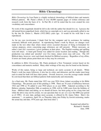
4Krych et alThe Orthopaedic Journal of Sports MedicineFigure 1. Illustration of the proposed progression of meniscal extrusion to a medial meniscus posterior root tear (MMPRT). (A)Meniscotibial (MT) ligament stress and attenuation. (B) Progressive loss of MT ligament function, increased root stress, and earlymeniscal extrusion. (C) MMPRT and associated increasing extrusion. (D) Rapid articular degeneration in the setting of an incompetent extruded meniscus.meniscal restraints and associated extrusion. The purposeof this study was to better understand the associationbetween MT ligament competence, medial meniscal extrusion, and MMPRTs, as well as to determine the progressionof meniscal extrusion over time. The main finding of thisstudy was that MT ligament disruption and associatedmeniscal extrusion predated MMPRTs in all 27 kneesinvestigated, supporting the hypothesis that extrusion andMT ligament abnormalities may increase stresses at themedial meniscus posterior horn root attachment, leadingto MMPRTs.The current literature suggests that medial meniscalextrusion may occur as a result of an MMPRT.3,9 However,the exact chronological and temporal relationship betweenextrusion, secondary restraints, and root tears has yet to beestablished. The current study suggests that medial meniscal extrusion consistently precedes and may potentiallypredispose to a subsequent MMPRT during a series of progressive pathological steps. Given the consistent chronological order on MRI, we propose that MT ligament stress andattenuation (Figure 1A) lead to a progressive loss of MTligament competence, subsequently causing early meniscalextrusion (Figure 1B). As secondary restraints such as theMT ligament are lost, forces are increasingly concentratedat the meniscus root, leading to MMPRTs and furtherextrusion (Figure 1C). Rapid progressive articular cartilagedegeneration ensues as a result of the posterior horn roottear and loss of meniscal function (Figure 1D). Of note, ourstudy demonstrated a temporal correlation but did notprove cause and effect. Additionally, meniscus root tearshave been noted to be associated with ligament (ie, anteriorcruciate ligament) tears in young patients; however, therole of MT ligament injuries in this acute traumatic population has yet to be determined.27A recent study by Krych et al14 investigated the presenceof isolated meniscal extrusion and documented high ratesof associated MT ligament abnormalities (65%), supportingthe above proposed mechanism outlining that MT ligamentstress predisposes to early meniscal extrusion and subsequent root tears. Medial meniscal extrusion is complex, butthere is a relative consensus in the literature that oncepresent, extrusion tends to progress rapidly. This study
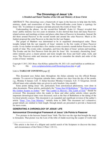
The Orthopaedic Journal of Sports MedicineCause of Medial Meniscal Extrusion5Figure 2. Serial magnetic resonance imaging showing the progression of a meniscotibial (MT) ligament tear, medial meniscalextrusion, and eventual medial meniscus posterior root tear. (A) Initial T2-weighted coronal imaging demonstrates bright MTligament edema (arrow) while the root remains intact (arrowhead). There is a degenerative signal in the meniscus with irregularityand trace extrusion. (B1) Repeat imaging at 4 months demonstrates a further increase in the signal and MT ligament attenuation(arrow), increased extrusion, and (B2) new partial tearing and an increased signal in the meniscus root (arrowhead). (C1) Imaging at6 months from baseline demonstrates marked attenuation of the MT ligament (arrow), substantial extrusion, the progression offemoral chondromalacia, and (C2) a concurrent full-thickness radial root tear (arrowhead). (D) At 8 months, there is radiographicloss of the MT ligament (arrow) and clearly visible complete tearing and displacement of the meniscus root (arrowhead).found that extrusion progressed in 96.3% of the knees analyzed and that mean extrusion increased significantly from3.3 1.1 to 5.5 1.8 mm over a mean follow-up period of 1.7 1.6 years. A previous study of 33 patients with MMPRTsand medial meniscal extrusion demonstrated increasedextrusion from 3.4 to 4.5 mm during a mean interval of only48 days. 28 Another study analyzed the MRI scans ofpatients with confirmed MMPRTs and medial meniscalextrusion and found a positive correlation between theamount of medial meniscal extrusion and the time frominitial injury, further highlighting the progressive natureof extrusion and associated abnormalities; however, thisstudy was limited by a lack of serial MRI scans.9 An additional note of interest is the relative preponderance offemale patients (76.9%) in our study. This is consistent withprevious literature on MMPRTs and is likely multifactorialin cause.17,23 Previously, it has been suggested that once anMMPRT occurs, female patients may be particularly susceptible to increased symptomatic subchondral loading,given their lower bone mineral density at the proximaltibia.17 Further studies using a prospective design will beneeded; however, this study is the first, to our knowledge,that analyzes a longer term progression of serial MRI by 2board-certified musculoskeletal radiologists.All 27 knees in our cohort demonstrated early signs of MTligament disruption during the temporal course, leading toMMPRTs. This is relevant, given that recent studies haveshown that nonanatomic meniscus root repair alone does notfully correct medial meniscal extrusion.3,5 These findingssuggest that other restraints such as the MT ligament mayplay an important role in preventing initial extrusion andmay need to be addressed at the time of meniscus root repair.The relevance of residual extrusion after root repair is wellestablished, with multiple studies demonstrating thatincreased extrusion is associated with inferior outcomesafter repair and an increased risk of arthritis.3,5,10,18 Thebiomechanical ability of the meniscus to function normallyby resisting hoop stresses and protecting the cartilage will
6Krych et alnot be achieved if extrusion is not corrected. Furthermore, ofthe 13 cases excluded because of the presence of an MMPRTon initial MRI, 12 had MT ligament abnormalities with amean associated initial extrusion of 2.4 1.2 mm. Of note,the single patient with an isolated MMPRT and an intactMT ligament demonstrated only 1.0 mm of extrusion, suggesting that MT ligament disruption generally precedesMMPRTs (present in 39 of the 40 knees initially evaluated),but if it remains intact, the MT ligament may keep themeniscus reduced with minimal extrusion, given its role asa secondary restraint.Technical efforts to address residual extrusion after rootrepair are evolving. Koga et al12,13 described a technique inwhich the midbody is centralized and stabilized onto the rimof the tibial plateau to reduce the meniscus and preventextrusion. Results of early reports have been generally positive, suggesting that centralization may better restorenative biomechanics and protect articular cartilage compared with root repair alone.5,12,29,30 Our findings that MTligament disruption occurred before an MMPRT also suggestthat MT ligament repair or functional reconstructionthrough centralization or other procedures may be a usefulaugment to MMPRTs (Figures 1 and 2). Furthermore, MTligament repair may be a viable early intervention before theprogression to an MMPRT, but further investigation withclinical trials is required to document efficacy.This study is not without important limitations. The retrospective design is prone to inherent biases, including relianceon complete and accurate recordkeeping. MRI scans were nottaken at regular intervals but rather were obtained accordingto the discretion of the treating physician. Patients were onlyincluded if they had multiple MRI scans, which may haveselected for patients who were more symptomatic and morelikely to have a quicker progression of extrusion. It isunknown in these knees whether extrusion and MT ligamentabnormalities could have been present sooner, predating clinical symptoms; furthermore, patients who had isolated MTligament disruption or who had MMPRTs but never developed extrusion during the course of 2 to 4 MRI scans wouldnot have been included in this analysis. Given the retrospective and exploratory study design aimed to investigate thetemporal nature of extrusion and root tears, this studystrongly supports, but does not definitively prove, the chronological order of root tears and extrusion. Exceptions mayoccur, and formal prospective series with imaging over multiple and predetermined time points are necessary to furtherinvestigate the temporal phenomena observed. Finally, thesample size of this study was relatively small, given the lownumber of patients with serial MRI scans who did not haveassociated ligamentous injuries. MRI analysis could havealso missed the diagnosis of meniscal extrusion or resultedin unrecognized MMPRTs in some patients, given that thesehave been established to be difficult to consistently visualizeon imaging.18CONCLUSIONIn a sample of 27 symptomatic knees with serial MRI scansboth before and after an MMPRT diagnosis, all patientsThe Orthopaedic Journal of Sports Medicinedemonstrated MT ligament disruption and associatedmeniscal extrusion before the development of subsequentmedial meniscus root tears. These findings suggest that MTligament disruption and medial meniscal extrusion represent early and predisposing events contributing toMMPRTs. Therefore, this provides a possible explanationof why meniscal extrusion is not corrected with medialmeniscus root repair.REFERENCES1. Allaire R, Muriuki M, Gilbertson L, Harner CD. Biomechanical consequences of a tear of the posterior root of the medial meniscus:similar to total meniscectomy. J Bone Joint Surg Am. 2008;90(9):1922-1931.2. Choi JY, Chang EY, Cunha GM, et al. Posterior medial meniscus rootligament lesions: MRI classification and associated findings. AJR AmJ Roentgenol. 2014;203(6):1286-1292.3. Chung KS, Ha JK, Ra HJ, Nam GW, Kim JG. Pullout fixation of posterior medial meniscus root tears: correlation between meniscusextrusion and midterm clinical results. Am J Sports Med. 2017;45(1):42-49.4. Costa CR, Morrison WB, Carrino JA. Medial meniscus extrusion onknee MRI: is extent associated with severity of degeneration or type oftear? AJR Am J Roentgenol. 2004;183:17-23.5. Daney BT, Aman ZS, Krob JJ, et al. Utilization of transtibial centralization suture best minimizes extrusion and restores tibiofemoral contact mechanics for anatomic medial meniscal root repairs in acadaveric model. Am J Sports Med. 2019;47(7):1591-1600.6. DePhillipo NN, Moatshe G, Brady A, et al. Effect of meniscocapsularand meniscotibial lesions in ACL-deficient and ACL-reconstructedknees: a biomechanical study. Am J Sports Med. 2018;46(10):2422-2431.7. El-Khoury GY, Usta HY, Berger RA. Meniscotibial (coronary) liagament tears. Skeletal Radiol. 1984;11(3):191-196.8. Faucett SC, Geisler BP, Chahla J, et al. Meniscus root repair vsmeniscectomy or nonoperative management to prevent knee osteoarthritis after medial meniscus root tears: clinical and economic effectiveness. Am J Sports Med. 2019;47(3):762-769.9. Furumatsu T, Kamatsuki Y, Fujii M, et al. Medial meniscus extrusioncorrelates with disease duration of the sudden symptomatic medialmeniscus posterior root tear. Orthop Traumatol Surg Res. 2017;103(8):1179-1182.10. Kim SJ, Choi CH, Chun YM, et al. Relationship between preoperativeextrusion of the medial meniscus and surgical outcomes after partialmeniscectomy. Am J Sports Med. 2017;45(8):1864-1871.11. Koenig JH, Ranawat AS, Umans HR, Difelice GS. Meniscal root tears:diagnosis and treatment. Arthroscopy. 2009;25(9):1025-1032.12. Koga H, Muneta T, Watanabe T, et al. Two-year outcomes afterarthroscopic lateral meniscus centralization. Arthroscopy. 2016;32(10):2000-2008.13. Koga H, Muneta T, Yagishita K, et al. Arthroscopic centralization of anextruded lateral meniscus. Arthrosc Tech. 2012;1(2):e209-e212.14. Krych AJ, Bernard CD, Leland DP, et al. Isolated meniscus extrusionassociated with meniscotibial ligament abnormality. Knee SurgSports Traumatol Arthrosc. Published online July 22, 2019. doi: 10.1007/s00167-019-05612-115. Krych AJ, Hevesi M, Leland DP, Stuart MJ. Meniscal root injuries.J Am Acad Orthop Surg. 2020;28(12):491-499.16. Krych AJ, Johnson NR, Mohan R, et al. Arthritis progression on serialMRIs following diagnosis of medial meniscal posterior horn root tear.J Knee Surg. 2018;31(7):698-704.17. Krych AJ, Reardon PJ, Johnson NR, et al. Non-operative management of medial meniscus posterior horn root tears is associated withworsening arthritis and poor clinical outcome at 5-year follow-up.Knee Surg Sports Traumatol Arthrosc. 2017;25(2):383-389.
The Orthopaedic Journal of Sports Medicine18. Kwak YH, Lee S, Lee MC, Han HS. Large meniscus extrusion ratio is apoor prognostic factor of conservative treatment for medial meniscusposterior root tear. Knee Surg Sports Traumatol Arthrosc. 2018;26(3):781-786.19. LaPrade CM, James EW, Cram TR, et al. Meniscal root tears: a classification system based on tear morphology. Am J Sports Med. 2015;43(2):363-369.20. LaPrade MD, Kennedy MI, Wijdicks CA, LaPrade RF. Anatomy andbiomechanics of the medial side of the knee and their surgical implications. Sports Med Arthrosc Rev. 2015;23:63-70.21. LaPrade RF, Engebretsen AH, Ly TV, et al. The anatomy of the medialpart of the knee. J Bone Joint Surg Am. 2007;89(9):2000-2010.22. Lee CR, Bin SI, Kim JM, Kim NK. Magnetic resonance imaging findings in symptomatic patients after arthroscopic partial meniscectomyfor torn discoid lateral meniscus. Arthroscopy. 2016;32(11):2366-2372.23. Lee JH, Lim YJ, Kim KB, Kim KH, Song JH. Arthroscopic pulloutsuture repair of posterior root tear of the medial meniscus: radiographic and clinical results with a 2-year follow-up. Arthroscopy.2009;25(9):951-958.24. Logerstedt DS, Snyder-Mackler L, Ritter RC, Axe MJ; OrthopedicSection of the American Physical Therapy Association. Knee
study, with a total of 63 MRI scans analyzed (21 knees had 2 MRI scans, 3 knees had 3 MRI scans, and 3 knees had 4 MRI scans). All MRI scans were obtained using our institu-tion's standard knee protocol on 1.5- or 3-T scanners, with protocols and sequencing that have varied little over the course of the past 10 years. Demographic information was