Transcription
Optical Two-Dimensional Fourier TransformSpectroscopy of SemiconductorQuantum WellsSTEVEN T. CUNDIFF,*,† TIANHAO ZHANG,‡,§ALAN D. BRISTOW,† DENIS KARAISKAJ,† AND XINGCAN DAI††JILA, National Institute of Standards and Technology and University ofColorado, Boulder, Colorado 80309-0440, ‡Department of Physics, Universityof Colorado, Boulder, Colorado 80309-0390RECEIVED ON FEBRUARY 25, 2009CON SPECTUSCoherent light-matter interactions ofdirect-gap semiconductor nanostructuresprovide a great test system for fundamentalresearch into quantum electronics and manybody physics. The understanding gained fromstudying these interactions can facilitate thedesign of optoelectronic devices. Recently, wehave used optical two-dimensional Fouriertransform spectroscopy to explore coherentlight-matter interactions in semiconductorquantum wells. Using three laser pulses togenerate a four-wave-mixing signal, weacquire spectra by tracking the phase of thesignal with respect to two time axes and thenFourier transforming them. In this Account, we show several two-dimensional projections and demonstrate techniques toisolate different contributions to the coherent response of semiconductors.The low-temperature spectrum of semiconductor quantum wells is dominated by excitons, which are electron-hole pairsbound through Coulombic interactions. Excitons are sensitive to their electronic and structural environment, which influences their optical resonance energies and line widths. In near perfect quantum wells, a small fluctuation of the quantumwell thickness leads to spatial localization of the center-of-mass wave function of the excitons and inhomogeneous broadening of the optical resonance. The inhomogeneous broadening often masks the homogeneous line widths associated withthe scattering of the excitons. In addition to forming excitons, Coulombic correlations also form excitonic molecules, calledbiexcitons. Therefore, the coherent response of the quantum wells encompasses the intra-action and interaction of both excitons and biexcitons in the presence of inhomogeneous broadening. Transient four-wave-mixing studies combined with microscopic theories have determined that many-body interactions dominate the strong coherent response from quantum wells.Although the numerous competing interactions cannot be easily separated in either the spectral or temporal domains, theycan be separated using two-dimensional Fourier transform spectroscopy.The most common two-dimensional Fourier spectra are SI(ωτ,T,ωt) in which the second time period is held fixed. Theresult is a spectrum that unfolds congested one-dimensional spectra, separates excitonic pathways, and shows which excitons are coherently coupled. This method also separates the biexciton contributions and isolates the homogeneous and inhomogeneous line widths. For semiconductor excitons, the line shape in the real part of the spectrum is sensitive to the manybody interactions, which we can suppress by exploiting polarization selection rules. In an alternative two-dimensionalprojection, SI(τ,ωΤ,ωt), the nonradiative Raman coherent interactions are isolated. Finally, we show SIII(τ,ωΤ,ωt) spectra thatisolate the two-quantum coherences associated with the biexciton. These spectra reveal previously unobserved many-bodycorrelations.Published on the Web 06/25/2009 www.pubs.acs.org/acr10.1021/ar9000636 CCC: 71.50 2009 American Chemical SocietyVol. 42, No. 9September 20091423-1432ACCOUNTS OF CHEMICAL RESEARCH1423
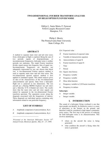
2DFT Spectroscopy of Semiconductors Cundiff et al.IntroductionDirect-gap semiconductors serve an important technologicalfunction in connecting optics and electronics based on theirability to convert light to an electronic excitation and viceversa. The use of semiconductors in this manner motivatesefforts to understand many-body effects, which strongly influence the light-semiconductor interaction. At the same time,semiconductors provide a unique system for studying the fundamental physics of many-body interactions. Thus there areboth applied and fundamental motivations for studying theinteraction of light with semiconductors. The power of multidimensional spectroscopic techniques to disentangle a complex spectral response is providing significant advances in ourunderstanding of how light interacts with semiconductors.The rapid relaxation, on time scales of picoseconds to femtoseconds, of electronic excitations in semiconductors led tothe use of ultrafast optics to probe carrier dynamics. However,it quickly became clear that many-body effects played anessential role in nonlinear optical measurements of semiconductors. The dominance of the many-body effects meant thatthey had to be understood to achieve the goal of understanding the interaction between light and semiconductors. Forexample, theory shows that many-body effects can shift thegain peak of a semiconductor laser.1 In addition, opticallyexcited semiconductors represent an ideal system for studying many-body physics.2 An understanding was developedfrom one-dimensional experiments such as transient fourwave mixing (TFWM). However, many details are obscured bythe presence of multiple competing phenomena. The application of optical two-dimensional Fourier transform (2DFT) spectroscopy to semiconductors has allowed these phenomena tobe separated, thus improving our detailed understanding.1423-1432September 2009hole wave functions. At low temperature, the exciton resonances dominate the absorption and emission spectra ofdirect-gap semiconductors near the band edge.The most common direct-gap semiconductor is GaAs. GaAsgrowth of high-quality heterostructures and because its bandAbsorption of light by a semiconductor creates anelectron-hole pair by promoting an electron from the valenceband to the conduction band. In a direct-gap semiconductorsuch as gallium arsenide (GaAs), the maximum of the valenceband is aligned in momentum space with the minimum of theconduction band. This alignment is important for makingdirect-gap semiconductors good emitters of light, as well asgood absorbers. It also allows excitons to form. Excitons areelectron-hole pairs bound by their Coulomb attraction. Therelative coordinate of an exciton is hydrogenic, while the center-of-mass coordinate is extended. The small masses and thehigh dielectric constant combine to reduce the binding energyto a few millielectronvolts; thus excitons only remain boundat low temperatures. Excitons have a strong dipole momentACCOUNTS OF CHEMICAL RESEARCHbecause of the increased overlap between the electron andis common because of the established technology for epitaxialLinear Optics of Semiconductor Excitons1424FIGURE 1. The top panel shows the band diagram of bulk GaAs(left) and magnetic substates of the three bands (right). The bottompanel shows the linear absorption spectrum of a quantum wellwhere the degeneracy between heavy-hole and light-hole bands islifted by quantum confinement, resulting in two exciton resonances.gap corresponds to near-infrared wavelengths, which are easily accessible with available lasers. A thin ( 10 nm) layer ofGaAs grown between two layers of AlGaAs will display quantum confinement in the growth direction. Near the direct gap,GaAs has two valence bands, known as the heavy-hole andlight-hole bands (see Figure 1, top). In bulk GaAs, the valencebands are degenerate. However, quantum confinement liftsthe degeneracy. Consequently, the low-temperature linearabsorption spectrum of a GaAs quantum well displays twopeaks corresponding to excitons formed between conductionband electrons and heavy or light holes (see Figure 1, bottom). In a quantum well, the heavy-hole exciton has a bindingenergy of approximately 10 meV.Vol. 42, No. 9
2DFT Spectroscopy of Semiconductors Cundiff et al.Coherent Spectroscopy of SemiconductorExcitonsCoherent spectroscopy has been used extensively to studyexcitons in semiconductors during the last 20 years (see ref 3and references therein). Originally the motivation was to measure the dephasing rate of excitons. However, it quicklybecame clear that the signals were dominated by many-bodyeffects. Understanding the many-body effects became the primary goal of much of the work. Structural disorder also playsa critical role in determining the optical properties of nanostructures. Coherent spectroscopy provides an important toolfor separating out the effects of disorder because of its ability to “undo” the effect of inhomogeneous broadening usingphoton-echo techniques.The primary coherent technique used to study excitons hasbeen TFWM using two excitation pulses. Two pulses are incident on the sample with wavevectors ka and kb, with delay τbetween them. The coherent interaction of the pulses generates a signal in direction ks ) 2kb - ka. For a two-level system, the signal is only emitted when ka arrives before kb,4which is defined to be τ 0. TFWM can be physicallydescribed in three steps: (i) The first pulse creates a coherence. (ii) The second pulse converts the coherence to an excited-state or ground-state population depending on the relativephase between the pulses, which varies across the samplebecause of the angle between the pulses. Each population isspatially modulated, creating an effective grating. (iii) The second pulse scatters off of these gratings into the signal direction. The integrated TFWM signal intensity decaysexponentially as a function of τ at twice the dephasing rate,γph.The presence of signals for “negative delay” in TFWMexperiments on semiconductors was evidence for the presence of many-body interactions. These signals were phenomenologically attributed to local fields,5 excitation-induceddephasing,6 biexcitonic effects,7 and excitation-induced shifts.8The relative strengths of these contributions could only beinferred from TFWM experiments.Using TFWM to study disorder in quantum wells was a natural step because the signal from an inhomogeneously broadened system is a photon echo. Thus the decay of theintegrated TFWM signal is 4γph and not the inverse width ofthe inhomogeneous distribution. In quantum wells, the primary form of disorder is fluctuations in the well thickness,which result in fluctuations in the exciton confinement energy.While improvements in epitaxial growth techniques haveimproved sample quality, one monolayer of fluctuations isessentially inevitable. Localization effects of excitons in disordered quantum wells were studied because the lack of Coulomb interactions simplified the situation.9 In quantum wellswhere the monolayer fluctuations were organized into largeislands, the TFWM was observed to oscillate.10 Debate aroseas to whether these oscillations were due to quantum interference and thus were quantum beats or due to simple electromagnetic interference, which was dubbed “polarizationinterference”. While the integrated TFWM signal could notmake this distinction, it was shown that either temporally11 orspectrally12 resolving the signal could. Measurements on disordered quantum wells showed that the beats had signaturesof quantum interference, which was attributed to many-bodycoupling between the localized excitons.13The TFWM signal was observed to depend on the relativepolarizations of the incident beams in surprising ways. Basedon the magnetic substates shown in Figure 1, the signal polarization would depend on the incident polarizations, but otheraspects would not. However, experiment showed that compared with the collinear excitation, the signal for cross-linearpolarization was weaker, decayed faster,14 and changed itstemporal behavior.15 A connection to disorder was establishedby comparing different samples.16 However, it was alsonoticed that the excitation density mattered, suggesting thatmany-body effects played a role.17 Biexcitonic effects alsocontributed to these observations.7 Ultimately, numerical simulations were able to reproduce many of these effects,18although it was difficult to extract significant insight.It is easier to discuss polarization effects on an excitonicbasis rather than the single-particle basis presented in Figure1. The excitonic levels for the heavy-hole exciton are shownin Figure 2. This scheme can be extended to include lighthole states.7 An important aspect of the excitonic basis is thatit naturally includes biexcitonic states. Biexcitons are boundpairs analogous to a hydrogen molecule.The extensive work using 1D techniques, including various forms of TFWM, provided a basic understanding of manybody effects in semiconductors and how they influenced thelight-matter interaction, especially for the exciton resonances.However, many aspects had to be inferred. The spectroscopictechniques were simply inadequate to address the outstanding questions.Two-Dimensional Fourier TransformSpectroscopyThe concept of multidimensional spectroscopy originated innuclear magnetic resonance (NMR).19 In multidimensionalspectroscopy, the nonlinear mixing of multiple electromag-Vol. 42, No. 9September 20091423-1432ACCOUNTS OF CHEMICAL RESEARCH1425
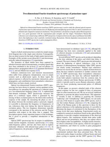
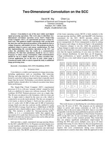
2DFT Spectroscopy of Semiconductors Cundiff et al.useful to take a series of 2DFT spectra as function of the thirdtime. The most common spectra are SI(ωτ,T,ωt), which measures homogeneous widths, SI(τ,ωT,ωt), which isolates Ramancoherences, SII(ωτ,T,ωt), which isolates coupling due to groundstate bleaching, and SIII(τ,ωT,ωt), which isolates two-quantumcoherences.FIGURE 2. Heavy-hole states in the exciton picture. The groundstate corresponds to all electrons in the valence band. For the oneexciton states, one electron has been excited to the conductionband, and it is bound to the hole it left behind to form an exciton.For the two-exciton states, a bound biexciton forms only when theelectrons have opposite spin. While two excitons of the same spindo not bind to form a biexciton, they do interact. Thus the twoexciton states of same spin must be included in the level diagram.Optical transitions between exciton states are denoted by thearrows. The light must be circularly polarized to conserve spin; thehelicity is denoted as σ or σ-.netic fields in a sample produces a signal field. The signal fieldis recorded as a function of either the relative delays, in thecase of pulsed fields, or the relative frequencies, in the case ofmonochromatic fields. For pulsed excitation, a spectrum isgenerated by a multidimensional Fourier transform, whichmeans that the phase of the signal must be recorded and thedelay scanned with steps that are equal and have subwavelength precision.Implementing 2DFT, also known as correlation spectroscopy, with lasers was proposed in 1993.20 The proposal wasbased on Raman excitation of vibrations in molecules. It wasdiscovered that infrared excitation of molecular vibrations wasexperimentally preferable.21,22 2DFT spectroscopy was alsoimplemented in the visible to study electronic transitions indye molecules23 and photosynthesis.24Typically three excitation pulses are used. The incidentpulses have wavevectors ka, kb, and kc. The interaction of thepulses in the sample produces a signal field in the direction ks) -ka kb kc. The first and second pulses have a delay τbetween them, the second and third pulse have a delay T, andthe emission time is t. A three-dimensional spectrum, Si(τ,T,t),can be produced where i denotes the time ordering of theconjugated pulse, ka. For SI(τ,T,t), ka arrives first and dephasing due to inhomogeneous broadening is canceled becausethe phase accumulated during τ is reversed during t. Thisrephasing produces a photon echo in the presence of inhomogeneous broadening; thus it is possible to extract thehomogeneous width from SI(τ,T,t). An SII(τ,T,t) spectrum has kaarriving second. If ka arrives last, an SIII(τ,T,t) spectrum is produced, which is sensitive to two-quantum coherences. Thetime-domain spectra are usually Fourier transformed withrespect to two variables, while the third is held fixed. It is often1426ACCOUNTS OF CHEMICAL RESEARCH1423-1432September 2009Experimental ProceduresA schematic diagram of the experiment is shown in Figure 3.The excitation pulses propagate parallel to one another onthree corners of a square. After passing through a lens, theyfocus to a spot size of 80 µm and overlap. The signal direction, ks, corresponds to the fourth corner of the square. A second lens collimates the beams and makes them parallel. Thewavelength is tuned to the excitonic resonances around 800nm. The average power per beam is 0.1 to 1 mW. Polarization optics are used to produce the desired excitation anddetection conditions.The results presented here are from a multiple quantumwell sample consisting of four periods of 10 nm thick GaAsand 10 nm thick A10.3Ga0.7As barriers. The sample is held at8 K, and the incident pulses produce an excitation densitybetween 2.5 109 and 2.2 1010 cm-2 per layer.To perform a multidimensional Fourier transform on thecomplex signal, its phase must be measured relative to a fixedreference as a function of the delay between the excitationpulses. The delay must be stable and stepped with subwavelength accuracy. To decompose the complex spectrum intoreal and imaginary parts, which can be related to the complex third-order susceptibility, experimental phase shifts mustbe removed by adjusting the “global” phase of the as-measured spectra. The spectra actually correspond to real andimaginary parts of the signal field. The phase shift betweenpolarization in the sample and radiated field needs to be considered to determine the susceptibility.Phase-Locked Excitation. The phase-locked excitationpulses are produced by an actively stabilized apparatus.25The error signal for the servo loop is generated from theinterference of an auxiliary HeNe laser beam that copropagates with the femtosecond pulses. Four phase-lockedpulses are produced by using nested and folded interferometers. The HeNe laser is retroreflected by a dichroicbeam splitter such that it double passes the interferometers.Locking the interferometers using the servo loop providesa fixed delay between the pulses. To change the delay, theservo loops are disabled, and the delay is stepped while theerror signals are monitored. When the delay has beenchanged by the desired amount and the error signal is closeVol. 42, No. 9
2DFT Spectroscopy of Semiconductors Cundiff et al.FIGURE 3. Experimental apparatus.to a zero crossing, the servo loops are re-enabled, whichdrives the error signal to zero. Three of the beams are usedas excitation beams. The fourth is used as a “tracer” beam.Signal Field Measurement. The electric field of the signal is measured using spectral interferometry.26 Spectralinterferometry allows the phase and amplitude of anunknown signal pulse to be extracted by comparing it to areference pulse. In principle, the tracer pulse could serve asthe reference for spectral interferometry because it copropagates with the signal. However, excitation-induced effects inthe sample lead to artifacts if the reference pulse goesthrough the sample. Thus, we derive a reference pulse fromthe tracer and route it around the sample and cryostat.Since the overall (constant) signal phase, not just its spectral variation, is needed, the reference beam must have astable phase throughout the acquisition of a 2DFT spectrum. Unfortunately, the stability of the reference is compromised by routing it around the sample. Thus anadditional feedback loop is needed.Global Phase. A 2DFT spectrum can be decomposed intoreal and imaginary parts related to the third-order complex susceptibility. However the decomposition requires thatthe overall, or “global”, phase be correctly set. The real andimaginary parts are useful because they reflect the underlying physical processes. Additionally, the spectral resolution can be enhanced by suppressing dispersive wings.However, the as-measured 2DFT spectrum has phase offsets that must be corrected. In our apparatus, these phaseoffsets arise from the relative phase of the excitation pulsesat zero delay, from the reference phase relative to the thirdpulse, and from phase shifts between the signal and reference pulses. Previously, these phase offsets were determined by comparing the complex signal spectrum at zerodelay to an auxiliary transient absorption spectrum.25 However, this technique proved to be problematic in semiconductors. Furthermore, the appropriate auxiliary experimentis difficult for some 2DFT spectra such as cross-polarizedexcitation or two-quantum coherences.Recently, we developed an alternative method, based onthe spatial interference pattern at the sample position, fordetermining the global phase.27 A similar method was independently developed for use in infrared 2DFT experiments. 28 The spatial interference pattern between theexcitation beams, including the tracer beam, depends onthe relative phases in the same way as the 2DFT spectrum.By recording the pattern, it is possible to determine thephase differences. The phase shift between the referenceand tracer, as well as propagation-induced phases shifts, aredetermined by recording appropriate spectral interferograms.Co-polarized SIA typical SI(ωτ,T,ωt) of the heavy- and light-hole excitons isshown in Figure 4. This figure shows the real spectrum. Thelaser spectrum is tuned to be centered between the twoexcitonic resonances. The excitation pulses are collinearlypolarized. The 2DFT spectrum displays two diagonal peaksfor the heavy- and light-hole excitons. The heavy-hole peakline shape is “dispersive” in that it is negative above thediagonal and positive below it. For a simple few-level system, the real SI(ωτ,T,ωt) should display an “absorptive” lineshape, that is, one that is purely positive. The dispersive lineshape occurs because the excitonic response is dominatedby many-body effects.29,30 The cross peaks between theexcitons are expected for collinear polarization. For thislaser tuning, their strength lies between those of the diagonal peaks. If the laser is tuned to be centered on the lighthole, the lower left cross peak (labeled “c” in the figure)becomes the strongest feature. For a few-level system, across peak can never be the strongest feature. Many-bodyinteractions between the two excitons can result in a dominant cross peak. 31 Similarly, the many-body interactionbetween the continuum and heavy-hole exciton results ina vertical feature, whereas a diagonal feature is expectedwithout many-body contributions.31Vol. 42, No. 9September 20091423-1432ACCOUNTS OF CHEMICAL RESEARCH1427
2DFT Spectroscopy of Semiconductors Cundiff et al.FIGURE 5. The 2DFT spectrum, SI(ωτ,T,ωt), of the heavy- and lighthole resonances of a GaAs quantum well with cross-linearlypolarized excitation. The numerical values on the color bar arerelative to Figure 4.FIGURE 4. The lower panel shows the real 2DFT spectrum,SI(ωτ,T,ωt), of the heavy- and light-hole resonances for collinearlypolarized excitation. The upper panel shows the linear absorptionspectrum (black line) and the spectrum of the excitation pulses (redline). The photon energies of the exciton resonances are marked bydashed lines. The 2DFT spectrum shows diagonal peaks due theheavy- and light-hole excitons, labeled “a” and “b”, respectively.Cross-peaks between the excitons are marked as “c” and “d”. Thefeature “e” is the biexciton formed from heavy-hole excitons. Thecontinuum contribution is the vertical feature “f”.The elongation of the heavy-hole peak along the diagonal is due to inhomogeneous broadening. In a semiconductor quantum well, inhomogeneous broadening occursbecause of unavoidable fluctuation in the well width. Sincethe quantum confinement energy is inversely proportionalto the width, width fluctuations result in static fluctuationsof the resonance energy. Both the homogeneous and inhomogeneous widths can be extracted from the 2DFT spectrum. The extraction is aided by comparing SI(ωτ,T,ωt) toS II(ω τ,T,ω t). 32 Disorder can localize the electronic states,reducing the strength of many-body interactions.One of the intriguing features is the peak “e”, which isslightly negative due to the formation of biexcitons. For a fewlevel system, the positive diagonal peaks correspond to areduction in the absorption. The generation of biexcitonsresults in an increase in the absorption and thus should havethe opposite sign. Based on the level diagram in Figure 2, thestrength of the biexciton peak should be half that of the exciton peak. It is much weaker than that because many-bodyeffects enhance the exciton peak but not the biexciton peak.Cross-Polarized SIThe SI(ωτ,T,ωt) spectrum is shown in Figure 5 for the sameconditions as Figure 4, except that the first pulse is crosspolarized. The component of the signal that is co-polarizedwith the first pulse is detected. The overall signal is muchweaker. More dramatic changes are the line shape of the1428ACCOUNTS OF CHEMICAL RESEARCH1423-1432September 2009heavy-hole exciton diagonal peak and its strength relativeto the biexciton peak. The line shape is now absorptive,whereas for co-polarized excitation, it was dispersive.These changes occur because cross-polarized excitationsuppresses many-body contributions, which can beexplained in terms of the physical picture for the TFWM signal given above. For cross-polarized excitation, the population grating induced in the 〉 exciton is π out of phasewith that for the -〉 exciton. Consequently, there is no netpopulation grating. Since the many-body interactions arethought to be spin independent, 6 they are spatially uniform and thus do not give a signal. While this understanding had been inferred, the change in the line shape fromdispersive in Figure 4 to absorptive in Figure 5 confirms thisinterpretation.The biexciton peaks in Figures 4 and 5 demonstrate apowerful feature of 2DFT spectroscopy. Biexcitonic featureshave been observed in photoluminescence and TFWMexperiments. In photoluminescence, biexcitons appear as alow-energy shoulder on the exciton line that grows quadratically with excitation intensity. In GaAs quantum wells,the biexciton peak is never resolved from the exciton peak.In the 2DFT spectrum, both peaks show diagonal elongation, resulting in resolved peaks. The diagonal broadeningof the biexciton peak shows that it is inhomogeneouslybroadened by well-width fluctuations. Moreover, it showsthat the two excitons making up the biexciton experiencecorrelated inhomogeneity. Thus they exist on the samelocalization site.The isolation of the biexciton allows its properties to bestudied. For example, the “ridge” of the biexciton line is notexactly diagonal, suggesting that the biexciton bindingenergy varies with the strength of the localization. Thehomogeneous line width of the biexciton can be extractedVol. 42, No. 9
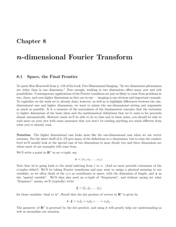
2DFT Spectroscopy of Semiconductors Cundiff et al.as a function of position within the inhomogeneous distribution, which was not possible using 1D spectroscopies.Nonradiative CoherencesAn advantage of 2DFT spectroscopy is its ability to isolatenonradiative coherences, that is, coherences between statesthat are not coupled by a dipole matrix element. Whilethese transitions do not interact directly with light, they doresult in oscillations in some 1D experiments.33-35 However, in 1D experiments, they generally cannot be isolatedfrom other contributions, which means that care is neededin interpreting the results.36In 2DFT experiments, a nonradiative coherence can becreated during one of the time periods, typically the second period, and then converted to a radiative coherence.The phase evolution while the system is in the nonradiative coherence sets the initial phase of the radiative coherence. Thus by recording the signal from the radiativecoherence and taking a Fourier transform with respect tothe time delay corresponding to the nonradiative coherence, it can be isolated.In GaAs quantum wells, there are two examples of nonradiative coherences. The first is a coherence establishedbetween the heavy-hole and light-hole exciton states.34-36Since these states are similar in energy, these coherencesare often known as “Raman” coherences. The other example is a coherence established between the ground stateand a doubly excited state such as a biexciton.33,37 Thesecoherences are known as “two-quantum” coherences.“Raman” Coherences in SI. The Raman coherencebetween the heavy- and light-hole excitons contributes to thespectra in Figures 4 and 5. Specifically, they appear in the offdiagonal peaks labeled “c” and “d” in Figure 4. These peaksalso contain contributions due to coupling between the twoexcitons via bleaching of the ground state. By a scan of thesecond delay, it is possible to produce a different spectrum,SI(τ,ωT,ωt), that separates these contributions and produces apeak due only to the Raman coherences.38Figure 6 shows an experimental SI(τ,ωT,ωt) spectrum. Thepeaks at pωT ) 0 contain all the contributions from pathwaysthat correspond to excited-state emission or ground-statebleaching. Theoretical results agree well with these experimental results.39The widths of the Raman peaks give information aboutscattering processes that affect the relative energy of the twoexciton states. Comparison of the width of the peaks in thecorresponding SI(ωτ,T,ωt) spectrum and their dependences onparameters such as carrier density and temperature yieldFIGURE 6. The magnitude 2DFT spectrum, SI(τ,ωT,ωt), of the heavyand light-hole resonances of a GaAs quantum well for collinearlypolarized excitation. The arrows point to spectral peaks due toRaman coherence between the excitons. The intensity scale hasbeen set so that the central peaks (ωT ) 0) saturate.greater insight into the nature of the scattering processes. Forexample, it is possible to determine whether the fluctuationsin the energy levels that cause dephasing are correlated oranticorrelated.35Two-Quantum Coherences in SIII. A two-quantum coherence occurs between the ground state and a doubly excitedstate. In our case, a two-quantum coherence between theground state
can be separated using two-dimensional Fourier transform spectroscopy. The most common two-dimensional Fourier spectra are S I(ω τ,T,ω t) in which the second time period is held fixed. The result is a spectrum that unfolds congested one-dimensional spectra, separates excitonic pa