Transcription
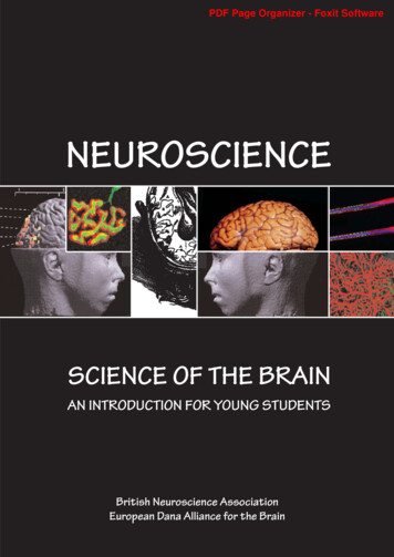
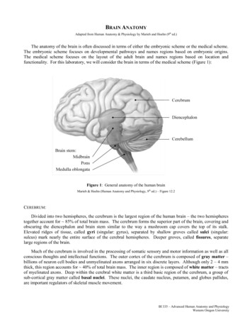
BRAIN ANATOMYAdapted from Human Anatomy & Physiology by Marieb and Hoehn (9th ed.)The anatomy of the brain is often discussed in terms of either the embryonic scheme or the medical scheme.The embryonic scheme focuses on developmental pathways and names regions based on embryonic origins.The medical scheme focuses on the layout of the adult brain and names regions based on location andfunctionality. For this laboratory, we will consider the brain in terms of the medical scheme (Figure 1):Figure 1: General anatomy of the human brainMarieb & Hoehn (Human Anatomy and Physiology, 9th ed.) – Figure 12.2CEREBRUM:Divided into two hemispheres, the cerebrum is the largest region of the human brain – the two hemispherestogether account for 85% of total brain mass. The cerebrum forms the superior part of the brain, covering andobscuring the diencephalon and brain stem similar to the way a mushroom cap covers the top of its stalk.Elevated ridges of tissue, called gyri (singular: gyrus), separated by shallow groves called sulci (singular:sulcus) mark nearly the entire surface of the cerebral hemispheres. Deeper groves, called fissures, separatelarge regions of the brain.Much of the cerebrum is involved in the processing of somatic sensory and motor information as well as allconscious thoughts and intellectual functions. The outer cortex of the cerebrum is composed of gray matter –billions of neuron cell bodies and unmyelinated axons arranged in six discrete layers. Although only 2 – 4 mmthick, this region accounts for 40% of total brain mass. The inner region is composed of white matter – tractsof myelinated axons. Deep within the cerebral white matter is a third basic region of the cerebrum, a group ofsub-cortical gray matter called basal nuclei. These nuclei, the caudate nucleus, putamen, and globus pallidus,are important regulators of skeletal muscle movement.BI 335 – Advanced Human Anatomy and PhysiologyWestern Oregon University
Below are listed the major anatomical regions / landmarks of the cerebrum with their correspondingfunctions (Figures 2 & 3):REGION / LANDMARKFUNCTIONLongitudinal fissureDeep fissure that separates the two hemispheres (right and left) of the cerebrum.Frontal lobeRegion of the cerebrum located under the frontal bone; contains the primarymotor cortex (precentral gyrus) and is involved in complex learning.Parietal lobeRegion of the cerebrum located under parietal bone; contains the primarysensory cortex (postcentral gyrus) and is involved in language acquisition.Central sulcusDeep groove that separates the frontal lobe from the parietal lobe of thecerebrum.Occipital lobeRegion of the cerebrum located under occipital bone; processes visualinformation and is related to our understanding of the written word.Parieto-occipital sulcusGroove on medial surface of hemisphere that separates the parietal lobe from theoccipital lobe of the cerebrum.Temporal lobeRegion of the cerebrum located under temporal bone; processes informationassociated with hearing and equilibrium.Lateral sulcusDeep groove that separates the frontal and parietal lobes from the temporal lobeof the cerebrum.InsulaRegion of the cerebrum deep within the lateral sulcus; processes informationassociated with hearing and equilibrium.Transverse fissureDeep fissure that separates the cerebrum from the cerebellum.Corpus callosumThe major bridge of white fibers that connects the two hemispheres of thecerebrum.FornixBridge of white matter inferior to the corpus callosum; links regions of thelimbic system (‘emotional’ brain) together.Anterior commissureBridge of white fibers found near the anterior tip of the corpus callosum;connects the two hemispheres of the cerebrum.Caudate nucleusBasal nucleus; initiates voluntary movements and coordinates slow skeletalmuscle contractions (e.g., posture and balance)PutamenBasal nucleus; initiates voluntary movements and coordinates slow skeletalmuscle contractions (e.g., posture and balance)Globus pallidusBasal nucleus; initiates voluntary movements and coordinates slow skeletalmuscle contractions (e.g., posture and balance)BI 335 – Advanced Human Anatomy and PhysiologyWestern Oregon University
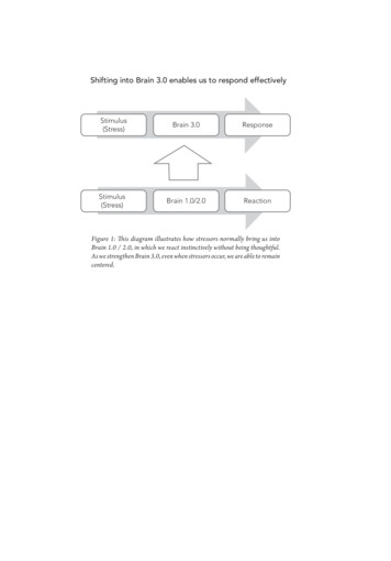
Figure 2: Transverse section of cerebrum showing major regions of cerebral hemispheresMarieb & Hoehn (Human Anatomy and Physiology, 9th ed.) – Figure 12.9Figure 3: Lobes, sulci, and fissures of the cerebral hemispheres (longitudinal fissure not pictured)Marieb & Hoehn (Human Anatomy and Physiology, 9th ed.) – Figure 12.4BI 335 – Advanced Human Anatomy and PhysiologyWestern Oregon University
Exercise 1:Utilize the model of the human brain to locate the following structures / landmarks for thecerebrum: Longitudinal fissureFrontal lobeParietal lobeCentral sulcusPrecentral gyrus Postcentral gyrusOccipital lobeParieto-occipital sulcusTemporal lobeLateral sulcusTransverse fissureCorpus callosumFornixAnterior commissureDIENCEPHALON:Surrounded by the cerebral hemispheres, the diencephalon forms the central core of the brain. Consisting oflargely of three paired structures, the thalamus, hypothalamus, and epithalamus, the diencephalon plays a vitalrole in integrating conscious and unconscious sensory information and motor commands.Below are listed the major anatomical regions / landmarks of the diencephalon with their correspondingfunctions (Figure 4):REGION / LANDMARKFUNCTIONThalamusComposes 80% of diencephalon; major relay point and processing center for allsensory impulses (excluding olfaction).Intermediate massA flattened gray band of tissue connecting the two halves of the thalamus.HypothalamusRegion inferior to thalamus; main regulatory center involved in visceral controlof the body and maintenance of overall homeostasis.Mammillary bodyPea-like structure posterior to hypothalamus; function as relay station inolfactory pathway.InfundibulumNeural stalk originating near mammillary bodies; connects pituitary gland tohypothalamus.Pituitary glandGlandular tissue handing under hypothalamus; important producer and releaserof endocrine hormones.Pineal glandGlandular tissue posterior to the thalamus; important producer and releaser ofendocrine hormones.Posterior commissureBridge of white fibers found inferior to the pineal gland; connects the twohemispheres of the cerebrum.BI 335 – Advanced Human Anatomy and PhysiologyWestern Oregon University
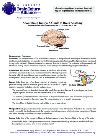
Figure 4: Mid-sagittal section of brain showing diencephalon (includes corpus callosum, fornix, and anterior commissure)Marieb & Hoehn (Human Anatomy and Physiology, 9th ed.) – Figure 12.10Exercise 2:Utilize the model of the human brain to locate the following structures / landmarks for thediencephalon: ThalamusIntermediate massHypothalamus Mammillary bodyInfundibulumPituitary glandPineal glandPosterior commissureBRAIN STEM:The brain stem begins inferior to the thalamus and runs approximately 7 cm before merging into the spinalcord. The brain stem centers produce the rigidly programmed, automatic behaviors necessary for survival.Positioned between the cerebrum and the spinal cord, the brain stem also provides a pathway for fiber tractsrunning between higher and lower brain centers.Below are listed the major anatomical regions / landmarks of the brain stem with their correspondingfunctions (Figure 7):REGION / LANDMARKFUNCTIONMidbrainRegion of brain stem between the diencephalon and pons; contains multiplefiber tracts running between higher and lower neural centers.Cerebral peduncleBulge located on the ventral aspect of the midbrain; contains fiber tracts runningbetween the cerebrum and spinal cord.BI 335 – Advanced Human Anatomy and PhysiologyWestern Oregon University
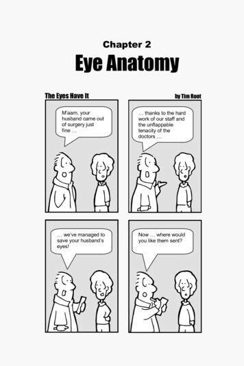
Superior colliculusPart of midbrain (corpora quadrigemina); contains nerve reflex centersinvolved in coordinated eye movements, focusing, and papillary responses.Inferior colliculusPart of the midbrain (corpora quadrigemina); contains nerve reflex centersinvolved in auditory reflexes.PonsRegion of brain stem between the midbrain and medulla oblongata; serves as thebridge (connection) between the two regions, and the cerebellum.Medulla oblongataThe most inferior portion of the brain stem; contains the cardiac, vasomotor, andrespiratory centers.PyramidLongitudinal ridge flanking mid-line of the medulla oblongata; contains fibertracts running between the cerebrum and spinal cord.OliveLocated lateral to the pyramid of the medulla oblongata; regulates impulsepropagation from the cerebrum and midbrain to the cerebellum.Figure 7: Lateral view of the brain stemMarieb & Hoehn (Human Anatomy and Physiology, 9th ed.) – Figure 12.13Exercise 3:Utilize the model of the human brain to locate the following structures / landmarks for thebrain stem: MidbrainCerebral pedunclesSuperior colliculus Inferior colliculusPonsMedulla oblongata PyramidOliveBI 335 – Advanced Human Anatomy and PhysiologyWestern Oregon University
CEREBELLUM:Located on the lower dorsal aspect of the brain, the cerebellum accounts for 11% of the total brain mass.Like the cerebrum, the cerebellum has two major hemispheres with an outer cortex made up of gray matter withan inner region of white matter. The cerebellum is located dorsal to the pons and medulla and it protrudes underthe occipital lobes of the cerebral hemispheres, from which it is separated by the transverse fissure.By processing inputs received from the cerebral motor cortex, various brain stem nuclei, and sensoryreceptors, the cerebellum provides the precise timing and appropriate patterns of skeletal muscle contraction forsmooth, coordinated movements and agility needing for our daily lives (e.g., driving). Cerebellar activity occurssubconsciously, we have no awareness of it.Below are listed the major anatomical regions / landmarks of the cerebellum with their correspondingfunctions (Figure 8):REGION / LANDMARKFUNCTIONVermisMid-line ridge of tissue (‘worm-like) that connects the two cerebellarhemispheres together.FoliaFine, transversely-oriented pleat-like gyri on the surface of the cerebellum;increase surface area.Arbor vitaeDistinctive pattern of white matter deep within the cerebellum; resembles abranching treeCerebellar pedunclesConnection points between the cerebellum and brain stem; contains fiber tractsrunning between the cerebellum and midbrain, pons, and medulla.Figure 8: Mid-sagittal section of the cerebellum (vermis not pictured)Marieb & Hoehn (Human Anatomy and Physiology, 9th ed.) – Figure 12.15BI 335 – Advanced Human Anatomy and PhysiologyWestern Oregon University
Exercise 4:Utilize the model of the human brain to locate the following structures / landmarks for thecerebellum: VermisFolia Arbor vitaeCerebral pedunclesVENTRICLESSituated within the brain are central hollow civilities called ventricles. These ventricles are continuous withone another and with the central canal of the spinal cord. The hollow ventricular chambers are filled withcerebrospinal fluid, a fluid that forms a liquid cushion for the brain. In addition, the cerebrospinal fluid helpsnourish the brain and there is some evidence that hormones circulate in the brain via this pathway.Below are listed the major ventricular chambers and associated openings / passageways found in the brain(Figure 7):CHAMBER / STRUCTUREFUNCTIONLateral ventriclesC-shaped chambers buried deep within each cerebral hemisphere; housechoroid plexi that produces cerebrospinal fluid.Septum pellucidumThin vertical partition that separates lateral ventricles.Third ventricleChamber surrounding the thalamus; houses a choroid plexus that producescerebrospinal fluid.Interventricular foramenSmall opening between each lateral ventricle and the third ventricle; drainscerebrospinal fluid.Fourth ventricleChamber that occupies the space between the dorsum of the pons / medullaand the overlying cerebellum; houses cerebrospinal fluid.Cerebral aqueductNarrow passageway between the third ventricle and the fourth ventricle;contains cerebrospinal fluid.Central canalCentral opening that runs through the medulla oblongata and is continuouswith the spinal cord; contains cerebrospinal fluid.BI 335 – Advanced Human Anatomy and PhysiologyWestern Oregon University
Figure 7: Lateral and mid-sagittal views of the brain showing the ventricular chambersMarieb & Hoehn (Human Anatomy and Physiology, 9th ed.) – Figures 12.3 & 12.10Exercise 5:Utilize the models of the ventricular system and the human brain to locate the followingventricular chambers / passageways: Lateral ventricleSeptum pellucidumThird ventricle Intraventricular foramenFourth ventricleCerebral aqueductCentral canalMENINGESThe meninges are three connective tissue membranes that lie just external to the brain. The function oftheses layers are to: 1) cover and protect the brain, 2) protect blood vessels and enclose venous sinuses, 3)contain cerebral spinal fluid, and 4) form partitions within the skull.Below are listed the major connective tissue layers forming the meninges and the general function of each(Figure 8):TISSUE LAYERFUNCTIONDura materExternal leathery tissue layer (‘tough mother’); protects brain, enclosesvenous sinuses, and forms partitions within the skull.Arachnoid materMiddle tissue layer forming loose brain covering (‘spider mother’); housescerebrospinal fluid.Pia materInnermost delicate tissue layer (‘gentle mother’) adhered tightly to brain;contains many blood vessels.BI 335 – Advanced Human Anatomy and PhysiologyWestern Oregon University
Figure 8: Section of brain and skull showing meningeal layersMarieb & Hoehn (Human Anatomy and Physiology, 9th ed.) – Figure 12.22BI 335 – Advanced Human Anatomy and PhysiologyWestern Oregon University
SHEEP BRAIN DISSECTION:Utilizing preserved sheep brains, we will continue our examination of the brain. In general, a sheep brain iseasy to work with because of 1) its size, 2) its availability, and 3) its relevance in comparative dissection – thereare many anatomical similarities between the sheep brain and the human brain. Of course, general differencesdo exist: The human brain is rounded, whereas the sheep’s brain is elongated in shape The sheep’s brain has a more developed olfactory bulb, giving them a sharper sense of smell The human brain has a larger frontal lobe than the sheep’s brain (‘seat of consciousness’)As we dissect the sheep brain, please be aware of the following: The sheep brains are stored in a substance that is toxic if ingested. You shouldwear gloves for this dissection and absolutely, positively have no food or drinksnear the specimen. Because of the chemicals used to preserve the sheep brains, please do not placebrain tissue in the garbage or down the sink. There is a plastic bag at the front ofthe room to place all unwanted neural tissue. Please be sure to scrub dissecting pans out completely and rinse tools afterdissection is complete. Most of the brains will be saved after use, so be sure tohandle them carefully and place them where instructed after their use.Step 1: Set up dissection arena1) Before beginning inspection and dissection of the brain, you should have the following materials onhand: dissection panlarge knife scissorsforceps metal probegloves2) After putting on your gloves take your dissection pan up to the front of the room and retrieve a brainfrom the container.BI 335 – Advanced Human Anatomy and PhysiologyWestern Oregon University
Step 2: External examination1) Examine the sheep brain carefully and determine which side is dorsal and which side is ventral. Inaddition, determine which area is toward the anterior (rostral) and which toward the posterior (caudal).DorsalPosteriorAnteriorVentral2) To get your bearings, identify the cerebral hemispheres, cerebellum, and brain stem. Note that theremay be some additional tissue on the underside of the brain that does not appear to be associated withthe brain. This tissue was left on to protect the olfactory bulbs and the pituitary gland, all of which canbe easily damaged or lost otherwise.3) The brain you receive is still be encased in the dura mater – note how tough the dura mater is. Thedura mater can be removed from the dorsal surface of the brain by carefully cutting down between thehemispheres and along the lateral edges of the cerebral hemispheres. At this point, do not remove thedura mater from the ventral region of the brain or the brain stem.4) Once the dura mater is removed, examine the dorsal surface of the brain – notice how its surface isthrown into convolutions (raised ridges gyri; grooves sulci). Locate the arachnoid mater, whichappears on the brain surface as a delicate “cottony” material spanning the sulci. In contrast, theinnermost layer, the pia mater, closely follows the cerebral contours and is what is responsible forgiving the ‘shiny’ look to the tissue.5) Notice the deeper grooves that you observe on the brain. The longitudinal fissure separates the twocerebral hemispheres and the transverse fissure is what separates the cerebrum from the cerebellum.Utilizing your knowledge of the brain model, identify the frontal, parietal, temporal, and occipitallobes of the cerebrum.6) Now move on to externally observe the cerebellum. Find the cerebellar hemispheres and note thatthey are separated by an additional cerebellar lobe, the vermis, rather than by a fissure as in thecerebrum. Note the folia forming the ridges on the surface of the cerebellum.7) Using a blunt probe, return to the dorsal surface of the brain and gently pull apart the two cerebralhemispheres. When you look down into the longitudinal fissure, you will see light-colored tissueholding the hemispheres together (DO NOT pull the hemispheres completely apart at this time ). Thetissue holding the hemispheres together is the corpus callosum.8) Next, move to the transverse fissure and carefully spread the cerebellum back from the cerebrum. Whenyou pull the cerebellum back, you should see the corpora quadrigemina region of the midbrain.Locate the superior and inferior colliculi that compose this region. While still spreading thecerebellum back from the cerebrum, gently part the cerebral hemispheres slightly to see the pinealgland.BI 335 – Advanced Human Anatomy and PhysiologyWestern Oregon University
Step 3: Ventral examination (Plate 1)1) Turn the sheep brain so that you are now looking at the ventral surface. Very carefully, remove theremaining dura mater from the ventral area. Looking at the ventral side, you should be able to findstructures of the diencephalon, including the infundibulum (if still present), pituitary gland (if stillpresent), and mammillary bodies. In addition, examine the structures associated with the brainstem,including the cerebral peduncles of the midbrain, the pons, and the pyramids and olives of themedulla oblongata.Step 4: Sagittal section examination (Plate 2)1) You are now ready to make a sagittal section through the midline of the brain. For this cut, you will usethe large knife – it is important to make a single cut through the whole brain (do not ‘saw’ back andforth through the tissue) and to cut directly down the longitudinal fissure, dividing the brain into twoequal halves.2) Turn your attention to the longitudinal section through the cerebellum. You now should be able toidentify the arbor vitae (‘tree of life’), the branching structure forming the cerebellum. The ‘branches’of this tree are formed by cerebellar white matter, while the ‘leaves’ are formed by cerebellar graymatter.BI 335 – Advanced Human Anatomy and PhysiologyWestern Oregon University
3) The sagittal section also allows you to find the intermediate mass of the thalamus. This is the circularstructure which has a slightly different texture than the areas surrounding it and represents the point atwhich the two halves of the thalamus join across the midline. The hypothalamus is located in theregion below the thalamus, toward the optic chiasma (where the optic nerves cross). Just posterior tothe thalamus it is possible to see the pineal gland.4) The hypothalamus forms the walls of the third ventricle. The lateral ventricles are visible just ventralfrom the corpus callosum within each hemisphere of the cerebrum (you may have to remove theseptum pellucidum to see into the lateral ventricles). The lateral ventricles are connected to the thirdventricle by the interventricular foramen – carefully push the probe into the third ventricle, and youshould be able to make the end of it come out into the lateral ventricle. The cerebral aqueductconnects the third ventricle and fourth ventricle. The fourth ventricle is then contiguous with thecentral canal of the spinal cord.5) The sagittal section gives a good opportunity to see the bridges of white matter connecting the twohemipheres of the brain. As noted above, dorsal to the septum pellucidum is the corpus callosum. Thefornix lies ventral to the septum pellucidum. The anterior commissure lies anterior to the thalamus /hypothalamus whereas the posterior commissure is posterior to the thalamus and ventral to the pinealgland.6) In addition to the new structures presented in the sagittal view, be sure to take a look at the structurespresented earlier in the dorsal and ventral views to see how they may appear in the sagittal view (e.g.,mammillary bodies, pons).BI 335 – Advanced Human Anatomy and PhysiologyWestern Oregon University
Step 5: Coronal section examination (Plate 3)1) It is now time to make a coronal section through the brain. For this cut, you are going to want to use thelong knife again. Put the two halves of the brain together and make a transverse cut through the brain atthe level of the optic chiasma.2) Turn your attention to the posterior sections of the brain. You now should be able to get a clear look atthe relationship of gray matter to white matter in the brain. The superficial gray matter represents thecerebral cortex; see how the convolutions of the brain allow for an increase in surface area and thus anincrease in cortical grey matter.3) You can also see the three major basal ganglia in this section, the caudate nucleus, the putamen, andthe globus pallidus, tucked deep down within the white matter. The caudate nucleus lies next to thelateral ventricle on each side whereas the putamen and globus pallidus are tucked deeper with theputamen lateral to the globus pallidus.BI 335 – Advanced Human Anatomy and PhysiologyWestern Oregon University
CHECKLIST:CEREBRUM: BASAL NUCLEI: Frontal lobe*Parietal lobe*Temporal lobe*Occipital lobe*Longitudinal fissure*Transverse fissure*Precentral gyrusPostcentral gyrusCentral sulcusLateral sulcusParieto-occipital sulcusCorpus callosum*Fornix*Anterior commissure*VENTRICLES:Lateral ventricles* Septum pellucidum*Interventricular foramenThird ventricle* Cerebral aqueduct*Fourth ventricle*Central canal*DIENCEPHALON: Caudate nucleus*Putamen*Globus pallidus*Thalamus* Intermediate mass*Hypothalamus*Mammillary bodies*Pituitary gland*Infundibulum*Pineal gland*Posterior commissure*MENINGES: Dura mater*Arachnoid mater*Pia mater*BRAIN STEM:Midbrain* Cerebral peduncles*Superior colliculi*Inferior colliculi*Corpora quadrigemina*Pons*Medulla oblongata* Olives*Pyramids*CEREBELLUM: Vermis*Folia*Arbor vitae*Cerebellar peduncles* Want to be familiar with location on sheep brainBI 335 – Advanced Human Anatomy and PhysiologyWestern Oregon University
BI 335 – Advanced Human Anatomy and Physiology Western Oregon University Figure 4: Mid-sagittal section of brain showing diencephalon (includes corpus callosum, fornix, and anterior commissure) Marieb & Hoehn (Human Anatomy and Physiology, 9th ed.) – Figure 12.10 Exercise 2: Utilize the model of the human brain