Transcription
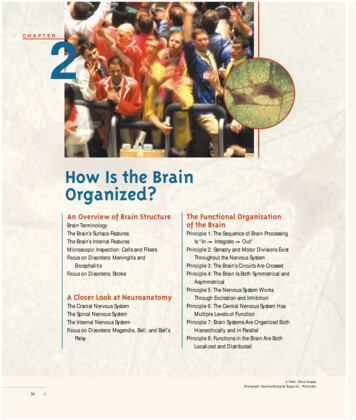
CHAPTER2How Is the BrainOrganized?An Overview of Brain StructureBrain TerminologyThe Brain’s Surface FeaturesThe Brain’s Internal FeaturesMicroscopic Inspection: Cells and FibersFocus on Disorders: Meningitis andEncephalitisFocus on Disorders: StrokeA Closer Look at NeuroanatomyThe Cranial Nervous SystemThe Spinal Nervous SystemThe Internal Nervous SystemFocus on Disorders: Magendie, Bell, and Bell’sPalsyThe Functional Organizationof the BrainPrinciple 1: The Sequence of Brain ProcessingIs “InIntegrateOut”Principle 2: Sensory and Motor Divisions ExistThroughout the Nervous SystemPrinciple 3: The Brain’s Circuits Are CrossedPrinciple 4: The Brain Is Both Symmetrical andAsymmetricalPrinciple 5: The Nervous System WorksThrough Excitation and InhibitionPrinciple 6: The Central Nervous System HasMultiple Levels of FunctionPrinciple 7: Brain Systems Are Organized BothHierarchically and in ParallelPrinciple 8: Functions in the Brain Are BothLocalized and DistributedA. Klehr / Stone ImagesMicrograph: Carolina Biological Supply Co. / Phototake36
hen buying a new car, people first inspect theIn many ways, examining a brain for the first time isoutside carefully, admiring the flawless finishsimilar to looking under the hood of a car. We have a vagueand perhaps even kicking the tires. Then theysense of what the brain does but no sense of how the partsopen the hood and examine the engine, the part of the carthat we see accomplish these tasks. We may not even beresponsible for most of its behavior — and misbehavior.able to identify many of the parts. In fact, at first glance theThis means gazing at a maze of tubes, wires, boxes, andoutside of a brain may look more like a mass of foldedfluid reservoirs. All most of us can do is gaze, becausetubes divided down the middle than like a structure withwhat we see simply makes no sense, except in the mostmany interconnected pieces. See what you can make of thegeneral way. We know that the engine burns gasoline tohuman brain in Figure 2-1. Can you say anything aboutmake the car move and somehow generates electricity tohow it works? At least a car engine has parts with regularrun the radio and lights. But this tells us nothing aboutshapes that are recognizably similar in different engines.what all the engine’s many parts do. What we need is in-This is not true of mammals’ brains, as shown in Figure 2-2.formation about how such a system works.When we compare the brain of a cat with that of a human,Wfor example, we see that there is an enormous differencenot just in overall size, but in the relative sizes of parts andin structure. In fact, some parts present in one are totally absent in the other. What is it that all these parts do that makesone animal stalk mice and another read textbooks?To make matters worse, even for trained research scientists, the arrangement of the brain’s parts does not justseem random, it really is haphazard. The challenge that weface in learning about the brain is to identify some regularities in its organization and to establish a set of principlesthat can help us understand how the nervous systemworks. After decades of investigation, we now have a goodidea of how the nervous system functions, at least in a general way. That knowledge is the subject of this chapter. Butbefore we turn our attention to the operation manual forthe brain and the rest of the nervous system, let us examinewhat the brain is designed to do. Knowing the brain’s functions will make it easier to grasp the rules of how it works.Perhaps the simplest statement of the brain’s functionsis that it produces behavior, as seen in Chapter 1. There ismore to this statement than is immediately apparent, however. In order for the brain to produce behavior, it musthave information about the world, such as informationabout the objects around us — their size, shape, movement, and so forth. Without such information, the brainFigure 2-1View of the human brain when the skull is opened. The gyri(bumps) and sulci (cracks) of the cerebral hemispheres are visible,but their appearence gives little information about their function.cannot know how to orient and direct the body to producean appropriate response. This is especially true when theresponse needed is some complex behavior, such as 37
38 CHAPTER ainstemCatCerebrumCerebellumBrainstemOlfactory ure 2-2BrainstemThis view of the brain’s primary purpose may seem ab-Inspection of the outside features of the brains of a cat, rat, monkey,and human shows them to differ dramatically in size and in generalappearance. The rat brain is smooth, whereas the other brains havefurrows in the cerebral cortex. The pattern of furrows differsconsiderably in the human, the monkey, and the cat. The cat brainand, to some extent, the monkey brain have long folds that appearto run much of the length of the brain, whereas the human brainhas a more diffuse pattern. The cerebellum is wrinkled in all speciesand is located above the brainstem. The brainstem is the route bywhich information enters and exits the brain. The olfactory bulb,which controls the perception of smells, is relatively larger in cats andrats but is not visible in monkeys and humans, because it is small andlies under the brain.stract to you, but it is central to understanding how thePhotos courtesy of Wally Welker, University of Wisconsin ComparativeMammalian Brain Collection.no such thing as sound. Rather, there is only the movementbrain functions. Consider the task of answering a telephone.The brain directs the body to pick up the receiver when thenervous system responds to vibrating molecules of air bycreating the subjective experience of a ring. We perceivethis sound and react to it as if it actually existed, when infact the sound is merely a fabrication of the brain. That fabrication is produced by a chain reaction that takes placewhen vibrating air molecules hit the eardrum. In the absence of the nervous system, especially the brain, there isof air molecules.The subjective nature of the experiences that the braincreates can be better understood by comparing the realitiescatching a ball. To perform complex behaviors, the ner-of two different kinds of animals. You are probably awarevous system has organs designed to receive informationthat dogs perceive sounds that humans do not. This differ-from the world and convert this information into biologi-ence in perception does not mean that a dog’s nervous sys-cal activity that produces subjective experiences of reality.tem is better than ours or that our hearing is poorer. Rather,The brain thus produces what we believe is reality in ordera dog brain simply creates a different world from that offor us to move. These subjective experiences of reality areour brain. Neither subjective experience is “right.” The dif-essential to carrying out any complex task.ference in experience is merely due to two different sys-
HOW IS THE BRAIN ORGANIZED?tems for processing physical stimuli. The same differences We can now identify the brain’s three primary functions:exist in visual perceptions. Dogs see very little color,whereas our world is rich with color because our brains1. to produce behavior;create a different reality from that of a dog’s brain. Such dif-2. to create a sensory reality; andferences in subjective realities exist for good reason: they3. to create knowledge that integrates information fromallow different animals to exploit different features of theirdifferent times and sensory domains and to use thatenvironments. Dogs use their hearing to detect the move-knowledge to guide behavior.ments of mice in the grass, whereas early humans probablyused color for such tasks as identifying ripe fruit in trees.Each of the brain’s three functions requires specific ma-Evolution, then, equipped each species with a view of thechinery. The brain must have systems to create the sensoryworld that would help it survive.world, systems to produce behavior, and systems to inte-These examples show how a brain’s sensory experi-grate the two.ences help guide an organism’s behavior. For this link be-In this chapter, we consider the basic structures andtween sensory processing and behavior to be made, thefunctions of those systems. First, we identify the compo-brain must also have a system for accumulating, integrating,nents of the nervous system. Then we look at what thoseand using knowledge. Whenever the brain collects sensorycomponents do. Finally, we look at how the parts work to-information, it is essentially creating knowledge about thegether and at some general principles of brain function.world, knowledge that can be used to produce more effec-Many of the ideas introduced in this chapter are devel-tive behaviors. The knowledge currently being created inoped throughout the rest of the book, so you may want toone sensory domain can be compared both with pastreturn to this chapter often to reconsider the basic princi-knowledge and with knowledge gathered in other domains.ples as new topics are introduced.AN OVERVIEW OF BRAIN STRUCTUREThe place to start our overview of the brain’s structure is to “open the hood” by opening the skull and looking at the brain snug in its home. Figure 2-1 shows a brainviewed from this perspective. The features that you see are part of what is called thebrain’s “gross anatomy,” not because they are ugly, but because they constitute a broadoverview. Zooming in on the brain’s microscopic cells and fibers is largely reserved forChapter 3, although this section ends with a brief introduction of some terms usedfor these tiny structures. Those terms are just a few of a great many new terms thatyou will encounter in this book, which is why we deal with brain terminology in general before moving on to a look at the brain itself. Because many of the words in thischapter will seem foreign to you, they will be accompanied by a pronunciation guideat their first appearance.Brain TerminologyThere are hundreds, even thousands of brain regions, making the task of masteringbrain terminology seem daunting. To make matters worse, many structures have several names, and many terms are often used interchangeably. This peculiar nomenclature arose because research on brain and behavior has spanned several centuries.When the first anatomists began to examine the brain with the primitive tools of theirtime, they made many erroneous assumptions about how the brain works, and thenames that they chose for brain regions are often manifestations of those errors. ForLink to an index listing the roots ofneuroanatomical terms atwww.worthpublishers.com/kolb/chapter2.39
CHAPTER 2Figure 2-3(A)Meaning “above,”sometimes referredto as superiorMeaning “middle”iorPosterMedialMeaning “front,”sometimes referredto as frontal or rostralMeaning “tail,”sometimes referredto as caudalLaterarAnteriolMeaning “side”VentralAnatomical terms are used to describeanatomical locations. (A) Anatomicaldirections relative to the head and brain.Because a human is upright, the termsposterior and caudal (both meaning“tail”) refer to a slightly differentorientation for the human headcompared with the head of a fourlegged animal. (B) Anatomical directionsrelative to the body.Dorsal Meaning “below” or“belly,” sometimesreferred to as ntralPosteriorVentral40Figure 2-4An afferent nerve carries informationinto the brain, and an efferent nervetakes information out of the brain andcontrols movement of a muscle.This afferent nervecarries informationfrom sensoryreceptors in skinto the brain.This efferent nervecarries informationfrom the brain tothe neuronscontrolling legmuscle, causinga response.Sensoryendingsinstance, they named one region of the brain the gyrus fornicatus because theythought it had a role in sexual function. In fact, most of this region has nothing to dowith sexual function. Another area was named the red nucleus because it appears reddish in fresh tissue. This name denotes nothing of the area’s potential functions,which turn out to be the control of limb movements.As time went on, the assumptions and tools of brain research changed, but thenaming continued to be haphazard and inconsistent. Early investigators named structures after themselves or objects or ideas. They used different languages, especiallyLatin, Greek, and English. More recently, investigators have often used numbers orletters, but even this system lacks coherence because the numbers may be Arabic orRoman numerals and are often used in combination with letters, which may be eitherGreek or Latin. When we look at current brain terminology, then, we see a mixture ofall these naming systems.Despite this sometimes confusing variety, many names do include informationabout a structure’s location in the brain. Table 2-1 summarizes these location-relatedterms, and Figure 2-3 shows how they relate to body locations. Structures found onthe top of the brain or on the top of some structure within the brain are dorsal. Struc-
HOW IS THE BRAIN ORGANIZED? 41tures located toward the bottom of the brain or one ofTable 2-1 Orientation Terms for the Brainits parts are ventral. Structures found toward the midTermMeaning with respect to the nervous systemdle of the brain are medial, whereas those located toAnteriorLocated near or toward the front or the headward the side are lateral. Structures located toward thefront of the brain are anterior, whereas those locatedCaudalLocated near or toward the tailtoward the back of the brain are posterior. SometimesDorsalOn or toward the back or, in reference to brain nuclei, locatedthe terms rostral and caudal are used instead of anteaboverior and posterior, respectively. And, occasionally, theFrontal“Of the front“ or, in reference to brain sections, a viewingterms superior and inferior are used to refer to strucorientation from the fronttures that are located dorsally or ventrally (these termsInferiorLocated belowdo not label structures according to their importance).LateralToward the side of the bodyIt is also common to combine terms. For example, aMedialToward the middle; sometimes written as mesialstructure may be described as dorsolateral, whichmeans that it is located “up and to the side.”PosteriorLocated near or toward the tailYou should also learn two terms that describe theRostral”Toward the beak”; located toward the frontdirection of information flowing to and from cells inSagittalParallel to the length (from front to back) of the skull; used inthe brain. Afferent refers to information coming intoreference to a planethe brain or a part of the brain, whereas efferent refersSuperiorLocated aboveto information leaving the brain or one of its parts,VentralOn or toward the belly or side of the animal in which the bellymeaning that efferent refers to brain signals that trigislocated or, in reference to brain nuclei, located belowger some response (Figure 2-4). These words are verysimilar, but there is an easy way to keep them straight.The letter “a” in afferent comes alphabetically before the “e” in efferent, and sensory information must come into the brain before an outward-flowing signal can trigger a response. Therefore, afferent means “incoming” and efferent means “outgoing.”On the CD, visit the module on theCentral Nervous System to better visualize the various planes of the brain.The Brain’s Surface FeaturesReturning to the brain in the open skull, you are now ready to examine its structuresmore closely. The first thing to notice is that the brain is covered by a tough materialknown as the meninges [men in jeez (the accented syllable is in boldface type)], whichis a three-layered structure, as illustrated in Figure 2-5. The outer layer is known as thedura mater (from Latin, meaning “hard mother”). It is a tough double layer of fibroustissue enclosing the brain in a kind of loose sack. The middle layer is the arachnoidlayer (from Greek, meaning “like a spider’s web”). It is a very thin sheet of delicateFigure 2-5SkullDuramaterArachnoidlayerMeningesPia materBrainSubarachnoid space(filled with CSF)The brain is covered by thick coveringsknown as the meninges and is cushionedby a fluid known as the cerebrospinalfluid (CSF).
42 CHAPTER 2Figure 2-6In these views of the human brain (fromthe top, bottom, side, and middle), thelocations of the frontal, parietal,occipital, and temporal lobes of thecerebral hemispheres are shown, as arethe cerebellum and the three major sulci(the central sulcus, lateral fissure, andlongitudinal fissure) of the cerebralhemispheres.Dorsal viewCentral sulcusParietallobeFrontallobePhotos courtesy of Yakolev ntral viewTemporal lobeCerebellumFrontallobeBrainstemCranial nervesLateral viewCentral llobeMedial viewOccipitallobeCentral lobeBrainstemCerebellum
HOW IS THE BRAIN ORGANIZED?connective tissue that follows the brain’s contours. The inner layer is the pia mater (fromLatin, meaning “soft mother”). It is a moderately tough membrane of connectivetissue fibers that cling to the surface of the brain. Between the arachnoid and piamater is a fluid, known as cerebrospinal fluid (CSF), which is a colorless solution ofsodium chloride and other salts. It provides a cushion so that the brain can move orexpand slightly without pressing on the skull. (Meningitis is an infection of themeninges. Its symptoms are described in “Meningitis and Encephalitis” on page 46.)If we remove the meninges, we can now remove the brain from the skull and examine its various parts. As we look at the brain from the top or the side, it appears tohave two major parts, each wrinkly in appearance. The larger part is the cerebrum[sa ree brum], which consists of two cerebral hemispheres, the left and the right, andthe smaller part is the cerebellum [sair a bell um]. Both the cerebrum and the cerebellum are visible in the brains shown in Figure 2-2. Each of these structures is wrinkled in large-brained animals because its outer surface is made of a relatively thinsheet of tissue, the cortex, that has been pushed together to make it fit into the skull.To see why the cortex is wrinkled, force a piece of writing paper, 81 2 by 11 inches, intoa cup. The only way is to crinkle the paper up into a ball. Essentially the same crinklingup has been done to the cortex of the cerebrum and the cerebellum. Like a crinkledpiece of paper, much of the cortex is invisible from the surface. All we can see fromthe surface are bumps and cracks. The bumps are known as gyri [jye rye; singular:gyrus (jye russ)], whereas the cracks are known as sulci [sul sigh; singular: sulcus(sul kus)]. Some of the sulci are very deep and so are often called fissures. The twobest-known fissures are the longitudinal fissure and the lateral fissure, both of whichare shown in Figure 2-6, along with the central sulcus.If we now look at the bottom of the brain, we see something completely different.The cerebrum is still the wrinkled part, but now there is also a whitish structure downthe middle with little tubes attached. This middle structure is known as the brainstem, and the little tubes are cranial nerves that run to and from the head.One final gross feature is obvious: the brain appears to be covered in blood vessels. As in other parts of the body, the brain receives blood through arteries and sendsit back through veins to the kidneys and lungs for cleaning and oxygenation. The arteries come up the neck and then wrap around the outside of the brainstem, cerebrum, and cerebellum, finally piercing the brain’s surface to get to its inner regions.Figure 2-7 shows the three major arteries that feed blood to the cerebrum — namely,the anterior, middle, and posterior cerebral arteries. Because the brain is very sensitiveto loss of blood, a blockage or break in a cerebral artery is likely to lead to the death ofthe affected region, a condition known as a stroke (see “Stroke” on page 48). Becausethe three cerebral arteries service different parts of the brain, strokes disrupt differentbrain functions, depending on the artery affected.Anterior cerebralarteryMiddle cerebralartery 43Cerebrum. The major structure of theforebrain, consisting of two equal hemispheres (left and right).Cerebellum. Major structure of the hindbrain specialized for motor coordination;in large-brained animals, it may also havea role in the coordination of other mentalprocesses.Brainstem. Central structures of thebrain including the hindbrain, midbrain,thalamus, and hypothalamus.Cranial nerve. One of a set of nervesthat control sensory and motor functionsof the head; includes senses of smell, vision, audition, taste, and touch on theface and head.Plug in the CD to examine, locate,and rotate the parts of the brain in the section on the subdivisions of the CNS in themodule on the Central Nervous System.Figure 2-7Each of the three major arteries of thecerebral hemispheres — the anterior,middle, and posterior — provides bloodto a different region of the cerebrum.Posterior cerebralartery
CHAPTER 2The Brain’s Internal FeaturesWhite matter. Those areas of the nervous system rich in axons, leading to awhite appearance.Gray matter. Those areas of the nervoussystem composed predominantly of cellbodies, leading to a gray appearance.Reticular matter. Area composed of intermixed cell bodies and axons that produce a mottled gray and white, or netlike,appearance.Look at the CD to examine a threedimensional model of the ventricular system in the section on subcortical structures in the module on the CentralNervous System.Figure 2-8This frontal section through the brainshows the internal features. The brain is(A) cut and then (B) viewed at a slightangle. This section displays regions thatare relatively white and gray. The whiteareas are largely composed of fibers,whereas the gray areas are composed ofcell bodies. The large bundle of fibersjoining the two areas is the corpuscallosum. Each ventricle is a fluid-filledtube.(A)The simplest way to examine what is inside something — be it an engine, a pear, or abrain — is to cut it in half. The orientation in which we cut makes a difference in whatwe see, however. Consider what happens when we slice through a pear held in different orientations. If we cut a pear from side to side, we cut across the core; whereas, ifwe cut it from top to bottom, we cut parallel to the core. Our impression of what theinside of a pear looks like is clearly influenced by the way in which we slice it. Thesame is true of the brain.We can begin by cutting the brain in half, slicing it downward through the middle. The result is shown in Figure 2-8. This view of the brain is known as a frontal section because we can now see the inside of the brain from the front.Several features of the brain’s interior are immediately apparent. First, it containsfour cavities, known as ventricles [ven trik uls], which are shown in Figure 2-9. Cellsthat line the ventricles make the cerebrospinal fluid that fills them. The ventricles areconnected, so CSF flows from the two lateral ventricles to the ventricles that lie on thebrain’s midline, eventually flowing into the space between the lower layers of themeninges as well as into the spinal-cord canal. Although the function of the ventriclesis not well understood, they are thought to play an important role in maintaining thebrain. The CSF may allow certain compounds access to the brain, and it probablyhelps the brain excrete metabolic wastes. Very likely, too, the CSF produced in theventricles acts as a kind of shock absorber. The CSF surrounds the brain; so, if there isa blow to the head, this fluid cushions the movement of the brain within the skull.A second feature apparent in our frontal section of the brain is that the brain’s interior is not homogeneous. There are both light and dark regions. These light anddark regions may not seem as distinct as the different parts of a car’s engine, but nevertheless they represent different components. The light regions, called white matter,are mostly fibers with fatty coverings. The fatty coverings produce the white appearance, much as fat droplets in milk make it appear white. The dark regions, called graymatter because of their gray-brown color, are areas where capillary blood vessels andcell bodies predominate. Some regions of the brain have a mottled gray and white, ornetlike, appearance. These regions, which have both cell bodies and fibers mixed together, are called reticular matter (from the Latin word rete, meaning “net”).Another way to cut the brain is from front to back. The result is a side view, calleda sagittal [sadj i tal] section. If we make our cut down the brain’s midline, we dividethe cerebrum into its two hemispheres. Figure 2-10 shows such a sagittal section.(B)(C)White matterGray TemporallobeGlauberman / Photo Researchers44
HOW IS THE BRAIN ORGANIZED?One feature seen from this viewing angle is a long band of white matter thatruns much of the length of the cerebral hemispheres. This band is called thecorpus callosum [ka loh sum]. The corpus callosum contains about 200 millionfibers that join the two hemispheres and allow communication between them. It isalso clear in Figure 2-10 that the cortex covers the cerebral hemispheres above thecorpus callosum, whereas below the corpus callosum are various internal structures of the brain. Owing to their locationbelow the cortex, these structures are known as subcorticalregions.We can see the internal structures of the brain in much moredetail by coloring them with special stains. For example, if we usea dye that selectively stains cell bodies, we can see that the distribution of cells within the gray matter is not homogeneous, asshown in Figure 2-11. In particular, it becomes apparent that thecerebral cortex is composed of layers, each of which containssimilarly staining cells. Furthermore, subcortical regions are nowseen to be composed of clusters, known as nuclei, of similarlystained cells. Although layers and nuclei are very different in appearance, they both form functional units within the brain.Whether a particular brain region has layers or nuclei is largelyan accident of evolution.If you were to compare the two sides of the brain in sagittalsection, you would be struck by their symmetry. The brain, infact, has two of nearly every structure, one on each side. The fewstructures that are one of a kind are found along the brain’s midline. Examples are the third and fourth ventricles and the pinealgland, mentioned in Chapter 1 in reference to Descartes’s theoryabout how the brain works.Figure 2-9(A)Figure 2-10(B)CortexCorpus callosumPlaneof cut 45There are two lateral cerebral ventricles,one in each hemisphere, and a third andfourth cerebral ventricle, each of whichlies in the midline of the leIn this sagittal section through the brain,the brain is (A) cut and then (B) viewedfrom the side. This particular plane of cutseparates the hemispheres, allowing aview of the midline structures of thebrain, including the corpus callosum,which connects the two hemispheres.You can see the subcortical structures(ventricle, brainstem, and cerebellum)that lie below the corpus callosum.VentricleBrainstemCerebellumSubcortical regions. All of the regionsMicroscopic Inspection: Cells and FibersAlthough the parts of a car engine are all large enough to be seen with the naked eye,the fundamental units of the brain — its cells — are so small that they can be viewedonly with the aid of a microscope. By using a microscope, we quickly discover that theof the brain that are located beneath theneocortex; the term is usually used to distinguish regions of the brain that controlbasic functions from those regions controlling cognitive functions, which aremediated by the neocortex.
46CHAPTER 2Figure 2-11When brain sections are stained, variousregions become clearly demarcated.These brain sections from the lefthemisphere of a monkey (midline is tothe left in each photograph) are stainedwith (A) a selective cell-body stain,known as a Nissl stain, and (B) a selectivefiber stain, staining for myelin. It isimmediately apparent that the twostains reveal a very different picture ofthe brain. (C and D) Higher-powermicrographs through the Nissl- andmyelin-stained sections show differentcortical regions. Notice the difference inappearance.(A)(B)(C)(D)Meningitis and Encephalitisor meninges, of the brain, particularly the pia mater and theBiophoto Associates / Science Source / Photo ResearchersFocus on DisordersA large number of harmful organisms can invade the linings,arachnoid layer, as well as the cerebrospinal fluid betweenthem. Such infections are called meningitis. One symptomis inflammation, which, because the skull is solid, placespressure on the brain. This pressure often leads to deliriumand, if the infection progresses, to drowsiness, stupor, andeven coma.Meningitis usually begins with severe headache and astiff neck (known as cervical rigidity). Head retraction is anextreme form of cervical rigidity. Convulsions are a commonsymptom in children. They indicate that the brain also is affected by the inflammation.In this photograph of the right hemisphere of a braininfected with meningitis, there is pus visible over the surfaceof the brain.Infection of the brain itself is called encephalitis. Thereare many forms of encephalitis, some of which have greathistorical significance. In World War I, a form of encephali-symptoms is death of the brain nucleus known as the sub-tis referred to as sleeping sickness (encephalitis lethargica)stantia nigra (black substance). Other forms of encephalitisreached epidemic proportions. The first symptoms were dis-may have different effects on the b
eral before moving on to a look at the brain itself. Because many of the words in this chapter will seem foreign to you, they will be accompanied by a pronunciation guide at their first appearance. Brain Terminology There are hundreds, even thousands of brain regions, making the task of ma