Transcription
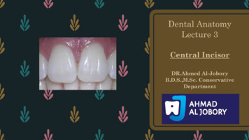
Dental anatomyIntroductionDR.Ahmed Al-JoboryB.D.S.,M.Sc. Conservative Department
Dental anatomy : is a field of anatomy dedicated to the study ofhuman tooth structures.
The Deciduous Teeth– At birth there are no teeth present in the mouth, but many teeth in variousstages of development are found in the jaws.– After birth (post natal period) the eruption of deciduous teeth starts at sixmonths and lasts until two and half years (28 4 months).– The deciduous teeth stay until the permanent teeth erupt at about six years ofage when the transition to the permanent dentition begins.– The deciduous teeth are 20 in number. They have the following formula:– I2\2C1\1M 2\2 10(For each side)– I Incisors (central and lateral).– C Canine.– M Molars (first and second).
The permanent teeth– The transition to permanent dentition begins with the emergence and eruptionof the first permanent molars at the age of six years, followed by shedding ofthe deciduous teeth, emergence and eruption of the remaining permanentteeth.– This process requires about 20 years to be completed.– The number of permanent teeth including third molars when present is 32.– I 2\2 C1\1P 2\2M 3\3 16 (For each side)– I Incisors (central and lateral).– C Canine.– P Premolars (first and second).– M Molars (first, second and third).
Anterior and posterior teeth– Teeth are grouped into:1. Anterior teeth which include the incisors and the canines.2. Posterior teeth which include the premolars and molars.
The jaw– The jaw is the bone which carries the teeth. There are two jaws:1. The upper jaw which is fixed, and is called Maxilla.2. The lower jaw which is movable and is called Mandible.
Crown and Roots
– Each tooth has a crown and root.– The crown is covered with enamel. The root is coveredwith cementum and they join at the cemento-enameljunction (CEJ) or cervical line.– The four tooth tissues are enamel, dentin, cementum andpulp. The first three are known as hard tissues, the last assoft tissues. The major bulk of the tooth is dentin.
Dental pulp: is the soft tissue of the tooth and present in the pulpchamber and pulp canal.Pulp chamber: is the part of dental pulp in the crownPulp canal: is the part of dental pulp in the root.The pulp chamber is continuous with the pulp canal andcollectively called as the "pulp cavity".Anatomical crown: is the portion of the tooth that covered byenamel.Clinical crown: is the portion of the tooth which is visible in themouth. In a healthy person the anatomical crown is larger than theclinical crown.
The number of roots1. Single root: in all anterior teeth, mandibular premolars and maxillarysecond premolar.2. Two roots with bifurcation: in mandibular molars and maxillary firstpremolar. Division of the tooth root is known as furcation.3. Three roots with trifurcation: in maxillary molars.
Surfaces and ridges– The crowns of incisors and canines have four surfaces and ridge,while the crowns of premolars and molars have five surfaces
The surfaces are:1. Labial surface: is the surface which is toward the lip in incisors and canines (in anterior teeth).2. Buccal surface: is the surface which is toward the cheek in premolars and molars (posteriorteeth).The labial and buccal surfaces could be termed as the "facial surfaces".3. Lingual surface: is the surface which is facing the tongue (all teeth).4. Occlusal surface: is the surface of the posterior teeth coming in contact with the teeth in theopposite jaw during closing the mouth.In anterior teeth, this surface is called "incisal ridge".5. Proximal surface: is the surface of the tooth facing toward adjacent teeth in the same dentalarch.a. Mesial surface: is the surface which is facing toward the median line.b. Distal surface: is the surface which is facing away from the median line. All teeth have their mesial surfaces touching the distal surfaces of the adjacent tooth except themaxillary and mandibular central incisor (both permanent and deciduous).The area of themesial and distal surface that touch its neighbor in the arch is called the "contact area".
Division of the crown intothirds– For description, the crown and the root are divided into thirds according to theposition of the surface.– Line angle: it is formed by the junction of two surfaces and gets its name fromthese surfaces. Example: mesio-labial line angle.– Point angle: it is formed by junction of three surfaces and get its name fromthese surfaces. Example: mesiolinguo-incisal point angle
Anatomical landmarks
In order to study an individual tooth intelligent, we must be able to recognize all landmarks ofimportance by name, these include: 1. Cusp: it is an elevation or mound on the crown portion of a tooth making up apart of the occlusal surface. It contributes to a significant portion of the tooth's surface.divisional
2. Tubercle: it is a smaller elevation on some portion of the crown produced by an extraformation of enamel. These are deviations from the typical form.3. Cingulum (Latin word for girdle): it’s the lingual lobe of the anterior teeth. It makes upthe bulk of the cervical third of the lingual surface. Its convexity mesiodistally resembles agirdle encircling the lingual surface at the cervical third. It is frequently identifiable as aninverted V-shaped ridge.
4. Ridge: it is any linear elevation on the surface of a tooth and is named according to itslocation ( e.g. buccal ridge, incisal ridge, marginal ridge).a.Marginal ridge: these are rounded borders of enamel that formed the mesial and distalmargins of the occlusal surfaces of premolars and molars and the mesial and distalmargins of the lingual surfaces of the incisors and canines.
5. Fossa : it is an irregular depression or concavity.a. Lingual fossa: it is located on the lingual surface of anterior teethb. Cenrtal fossa: it is located on the occlusal surface of molar
c. Triangular fossa: it is located on the occlusal surfaces of molars and premolars, mesial ordistal to marginal ridges6. Sulcus: it is a long depression or valley in the surface of a tooth between ridges and cusps, theinclines of which meet at an angle. A sulcus has a developmental groove at the junction of itsinclines.
7. Developmental groove: it is a shallow groove or line between the primary parts of thecrown or root.8. Pit: it is a small pinpoint depression located at the junction of developmental grooves or atterminals of those grooves, e.g. central pit is a term used to describe landmark in the centralfossa of molars where developmental grooves join.
Thank You
Dental anatomy Introduction DR.AhmedAl-Jobory B.D.S.,M.Sc. Conservative Department Dental anatomy : is a field of anatomy dedicated to the study of human tooth structures. The Deciduous Teeth – At birth there are no teeth present in the mouth, but many teeth in various stages of development are found in the jaws. – After birth (post natal period) the eruption of deciduous teeth starts .