Transcription
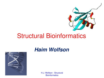
Structural BioinformaticsHaim WolfsonH.J. Wolfson - StructuralBioinformatics
Lecture overview Introduction and Motivation. Protein Folding – the RAPTORthreading algorithm. Modeling of protein-protein interactions– the PatchDock docking algorithm.H.J. Wolfson - StructuralBioinformatics
Why 3D Structures?1. 3D Structure (shape) is better preserved thansequence (text).2. Structural motifs may predict similar biologicalfunction.3. Drug Design. Example, identification of a person: a verbaldescription via a picture.Mid-aged man, black hearH.J. Wolfson - Structuraleyes and moustacheBioinformatics
Shape to functionMacromolecules, like many everyday objects,have been shaped (by evolution) to get their job done.Elucidation of macromolecular shape can supply insight onthe function of the molecules involved."It hasnot escapedour– noticethat thespecificClassicalexamplethe doublehelixshapepairingof DNAwehave postulateda possiblesupplied immediatelyinsight on thesuggestsreplicationprocess.copyingmechanism for the genetic material."H.J. Wolfson - StructuralBioinformatics
Structural Bioinformatics akaComputational Structural Biology Deals with Structural data of molecules. Exploits (and develops) algorithms forinterpretation and handling of 3D (spatial data) –Geometric Computing. Sister computational disciplines – ComputationalGeometry, Computer Vision, Computer Graphics,Medical Image Interpretation, PatternRecognition.H.J. Wolfson - StructuralBioinformatics
Recommended Web Sites: Proteopedia http://proteopedia.org/ Protein Data Bank (PDB)http://www.rcsb.org/pdb/H.J. Wolfson - StructuralBioinformatics
The Central DogmaRNA is aninformation carrierfrom DNA to ProteinH.J. Wolfson - StructuralBioinformatics
H.J. Wolfson - StructuralBioinformatics
The Biological Role(Robots of the Cell)1. Catalysis (enzymes).2. Signal propagation:– transmit nerve impulses– control cell growth and differentiation.3. Transport (of electrons ormacromolecules).4. Immune system (e.g. antibodies whichbind to specific foreign particles such asbacteria and viruses).5. Structural proteinsH.J. Wolfson(hair,- Structural skin, nails).Bioinformatics
Amino Acids and the Peptide BondH.J. Wolfson - StructuralBioinformatics
H.J. Wolfson - StructuralBioinformatics
Protein StructureH.J. Wolfson - StructuralBioinformatics
Primary StructureH.J. Wolfson - StructuralBioinformatics
Secondary StructureAlpha HelixesBeta StrandsH.J. Wolfson - StructuralBioinformatics
Alpha-Helix3.6residues,5.4ALength 4-40 residues.Main-chain atoms N and O arecolored red and blue respectively.H.J. Wolfson - StructuralThe hydrogen bonds between themare red and striated.Bioinformatics
Beta Strands and Beta SheetBeta strand. TypicalLength 5-10 residues.Beta sheets. Backbone NH and O atomshydrogen bonded to each other. O, N, Hand C atoms are colored red, blue, whiteand black respectively. Side chains areH.J.Wolfsonshownas- Structuralpurple circles.Bioinformatics
Tertiary structure Full 3D folded structureof the polypeptide chain.H.J. Wolfson - StructuralBioinformatics
Quaternary structure The interconnections and organization ofmore than one polypeptide chain.H.J. Wolfson - StructuralBioinformatics
Different RepresentationsAminoacidsFunctionalgroupsH.J. Wolfson - StructuralBioinformaticsSurface
Degrees of Freedom in ProteinsBond length12Dihedral angle3142Bond angle H.J. Wolfson - StructuralPatrice Koehl, 01/files/ECS229 Lecture2.pptBioinformatics
Backbone and Side-ChainsH.J. Wolfson - StructuralBioinformatics
Determination of ProteinStructureX-ray crystallographyNMR (Nuclear Magnetic Resonance)EM (electron microscopy)H.J. Wolfson - StructuralBioinformatics
Size of protein molecules(diameter) cell ribosome protein(1x10⁻⁶ m) µ microns(1x10⁻⁹ m) nanometers(1x10⁻¹⁰ m) angstromsH.J. Wolfson - StructuralBioinformatics
X-ray Crystallography Microscope is not suitable for distance smaller than thewavelength of the light you are using. X-rays get us in the right wavelength range. Each proteinhas a unique X-ray diffraction pattern.CrystallizationConversion of Diffraction Datato Electron Density and ImageH.J. Wolfson - Structural reassembleDiffractionFigure from: ourse/Overview/Overview.html
Nuclear Magnetic Resonance(NMR) Is based on the quantum mechanical propertiesof atoms (spin) and it determines informationabout atoms from the their response to appliedmagnetic fields. Provides the interatomic distances, and featuresof the spectrum that can be interpretedin terms of torsion angles. Solved by Distance Geometrymethods.H.J. Wolfson - StructuralBioinformatics
An NMR result is an ensembleof modelsCystatin (1a67)H.J. Wolfson - StructuralBioinformatics
H.J. Wolfson - abteilungen/103/single part.htm
EM vs. Crystallography & NMREMCrystallographyNMRPhysicallimitsFrozen (mostly Crystal (difficult)easy)In solution(easy)Metal atomscauseproblemsTimeFastMediumResolutionHigh to low (3 High ( 3A)-30 A)High ( 3A)PossibleStructureSizeBig (structures Smallcontainingmany proteins)Very small(singleproteins) 300aaSlowH.J. Wolfson - StructuralBioinformatics
High Resolution to LowResolutionHigh resolutionSpace-filling modelLow resolution4Å10ÅCrystallographyH.J. Wolfson - StructuralBioinformatics20ÅEM
Features as a Function ofResolutionLow Resolution15 ÅSizeShapeIntermediate Resolution9ÅDomainsHigh Resolution 4 Å6Åα Helicesβ sheets2BTV VP3AStrandsConnectivityH.J. Wolfson - StructuralBioinformaticsSidechains
Proteins work together Vital cellular functions are performed bycomplexes of proteins. Structures of single proteins are usually notinformative about function if taken out ofcontext.GlutamineSynthetaseRhinovirusH.J. Wolfson- StructuralBioinformaticsChaperonThe figures are adapted from http://www.rcsb.org/pdb/molecules/molecule list.html
The Protein Data Bank (PDB) International repository of 3D molecular data. Contains x-y-z coordinates of all atoms of themolecule and additional data.H.J. Wolfson - StructuralBioinformatics
Jan 8, 2017H.J. Wolfson - StructuralBioinformatics
H.J. Wolfson - StructuralBioinformatics
Major Protein StructureClassification /CATHhttp://www.biochem.ucl.ac.uk/bsm/cath/H.J. Wolfson - StructuralBioinformatics
Major Algorithmic Tasks : Structural Alignment of Proteins and theirClassification. Functional Annotation. Protein Structure Modelling Prediction of Protein Interactions and theStructure of Complexes. Computer Assisted Drug Design. Protein Design. Alignment and modeling of RNA structures. Modeling of DNA 3D structure (HiC).H.J. Wolfson - StructuralBioinformatics
Protein Structure PredictionFolding Given only the amino-acid sequence of aprotein, deduce its native tertiary structure.Target sequenceMA A G Y AV L Sstructural model
Protein structure Most proteins will fold spontaneously in water– amino acid sequence should be enough to determineprotein structure However, the physics are daunting:– 20,000 protein atoms, plus equal amounts of water– Many non-local interactions– Can take seconds (most chemical reactions takeplace 1012 --1,000,000,000,000x faster)
Levinthal Paradox Cyrus Levinthal, Columbia University, 1968 Levinthal's paradox– If we have only 3 rotamers (α,β,λ) per residue a 100residue protein has 3100 possible conformations.– To search all these takes longer than the time of theuniverse, however, proteins fold in less than a second. Resolution: Proteins have to fold through some directedprocess Goal - to understand the dynamics of this processArne Elofsson(arne@bioinfo.se)
Protein Folding vs StructurePrediction Protein folding investigates the process ofthe protein acquisition of its threedimensional shape.– The role of statistics is to support or discreditsome hypotheses based on physical principles. Protein structure prediction is solelyconcerned with the final 3D structure of theprotein– use theoretical and empirical means to get tothe end result.
Methods of Structure Prediction Homology modeling– Easy cases– high seq. identity to known structures Fold recognition– No discernable sequence identity to a knownstructure– a similar fold is (probably) known but hard toidentify Ab initio (de novo) methods– Most difficult– No similar folds are known
Fold Recognition – ThreadingThe RAPTOR Algorithm Jinbo Xu’s Ph.D. thesis work. J. Xu, M. Li, D. Kim, Y. Xu, Journal ofBioinformatics and Computational Biology,1:1(2003), 95-118.
There are not too manycandidates! There are only about 1000 – 1500 topologically differentdomain structures. Fold recognition methods aim toassign the correct fold to a given sequence and to alignthe sequence to the chosen fold.
UniqueSuperFamiliesannual histogramH.J. Wolfson - StructuralBioinformatics
Protein Threading Make a structure prediction through finding an optimal placement(threading) of a protein sequence onto each known structure(structural template)– “placement” quality is measured by some statistics-based energyfunction– best overall “placement” among all templates may give astructure predictiontarget YTEtemplate library
Threading Example
Formulating Protein Threading by LP Protein Threading Needs:1. Construction of a Structure TemplateLibrary2. Design of an Energy Function3. Sequence-Structure Alignment algorithm4. Template Selection and ModelConstruction
Assumptions :1. Each template sequence is parsed a linear series of(conserved) cores connected by (variable) loops.Each core is a conserved part of an α helix or βsheet.2. Alignment gaps are confined to loops.3. Only interactions between residues in cores areconsidered. An interaction is defined btwn tworesidues, if they are at least 4 positions apart in thesequence and the distance btwn their Cβ atoms isless than 7A.4. An interaction is defined btwn two cores if there is atleast one residue-residue interaction btwn thecores.
Threading Energy Functionhow preferable toput two particularresidues nearby: Ephow well a residuefits a structuralenvironment: Es(Pairwise potential)(Fitness score)sequence similaritybetween query andtemplate proteins: Emalignment gappenalty: Eg(gap score)(Mutation score)Consistency with the secondary structures: EssE Ep Es Em Eg EssMinimize E to find a sequence-structure alignment
Contact Graphtemplate1.2.3.Each residue as a vertexOne edge between tworesidues if their spatialdistance is within a givencutoff.Cores are the mostconserved segments in thetemplate: alpha-helix, betasheet
Simplified Contact Graph
Contact Graph and AlignmentDiagram
Contact Graph and AlignmentDiagram
Variables x(i,l) denotes core i is aligned to sequence position l y(i,l,j,k) denotes that core i is aligned to position l and core j isaligned to position k at the same time. D[i] – valid alignment positions for c(i). R[i,j,l] – valid pos. of c(j) given that c(i) is aligned to s(l).
Formulation 1MinimizeE ai ,l xi ,l b( i ,l )( j ,k ) y( i ,l )( j ,k )s.t.xi ,l xi 1,k 1y(i ,l )( j ,k ) xi ,l x j ,kEncodesscoring system xl D[ i ]i ,lEg , EpEs , Ess , Em 1xi ,l , y(i ,l )( j ,k ) {0,1}Encodes interaction structures: thefirst makes sure no crosses; thesecond is quadratic, but can beconverted to linear: a bc isequivalent to: a b, a c, a b c-1
Formulation used in RAPTORMinimizeE ai ,l xi ,l b(i ,l )( j ,k ) y(i ,l )( j ,k )Eg, Eps.t.xi ,l Encodesscoring system, l D[i] y, k D[ j ]( i ,l )( j , k )k R[ i , j ,l ]x j ,k xl D[ i ] y( i ,l )( j , k )l R[ j , k ,i ]i ,l 1xi ,l , y(i ,l )( j ,k ) {0,1}Es, Ess, EnEncodes interactionstructures
Solving the Problem Practically1. More than 99% threading instancescan be solved directly by linearprogramming, the rest can be solvedby branch-and-bound with only severalbranch nodes2. Relatively efficient3. Easy to extend to incorporate otherconstraints
Docking עגינה הגדרת הבעיה : בהינתן שני מבנים מצא טרנספורמציה במרחב שתביא למקסימום את אינטראקציה ביניהם )תן חיזוי למבנה המשותף( . H.J. Wolfson - Structural Bioinformatics
Docking ProblemGiven 2 input molecules in theirnative conformation, the goal is tofind their correct association as itappears in nature.T H.J. Wolfson - StructuralBioinformatics?
Docking Problem: H.J. Wolfson - StructuralBioinformatics ?
Detection of a Lead Drug Compound: The Key-in-Lock PrincipleH.J. Wolfson - StructuralBioinformatics
Docking - Motivation Computer aided drug design – a new drugshould fit the active site of a specific receptor. Understanding of the biochemical pathways many reactions in the cell occur throughinteractions between the molecules. Crystallizing large complexes and findingtheir structure is difficult.H.J. Wolfson - StructuralBioinformatics
The Docking Problem Input: A pair of molecules representedby their 3D structures. Tasks :– Decide whether the molecules will form acomplex (interact / bind).– Determine the binding affinity.– Predict the 3D structure of the complex.– Deduce function.H.J. Wolfson - StructuralBioinformatics64
Forces Governing BiomolecularRecognitionDepend on the molecules and the solvent. Van der Waals. Electrostatics. Hydrophobic contacts. Hydrogen bonds Salt bridges . etc.All interactions act at short ranges.Implies that a necessary condition for tightbinding is surface complementarity.H.J. Wolfson - StructuralBioinformatics65
Shape ComplementarityH.J. Wolfson - StructuralBioinformatics66
Necessary Condition for Docking Given two molecules find significantsurface complementarity.T H.J. Wolfson - StructuralBioinformatics67
Geometric Docking Algorithms Based on the assumption of shapecomplementarity between the participatingmolecules. Molecular surface complementarity –protein-protein, protein-drug.Remark : usually “protein” here can bereplaced by “DNA” or “RNA” as well.H.J. Wolfson - StructuralBioinformatics68
Issues to be examined whenevaluating docking methods Rigid docking vs. Flexible docking :– If the method allows flexibility: Is flexibility allowed for ligand only, receptor only or both ? Number of flexible bonds allowed and the cost of addingadditional flexibility. Does the method require prior knowledge of theactive site? Speed - ability to explore large libraries. Performance in “unbound” dockingexperiments.H.J. Wolfson - StructuralBioinformatics69
Bound Docking In the bound docking we are given a complex of 2molecules. After artificial separation the goal is to reconstruct thenative complex. No conformational changes are involved. Used as a first test of the validity of an algorithm.DockingAlgorithmH.J. Wolfson - StructuralBioinformatics70
Unbound Docking In the unbound docking we are given 2molecules in their native conformation. The goal is to find the correct association. Problems: conformational changes (side-chainand backbone movements), experimentalerrors in the structures.H.J. Wolfson - StructuralBioinformatics71
Bound vs. UnboundReceptor surface10 highly penetrating residuesLigandUnbound ligand and receptorsuperimposed on the complexH.J. Wolfson - StructuralKallikrein A/trypsin inhibitorBioinformaticscomplex (PDB codes 2KAI,6PTI)72
The PatchDock Algorithm Based on local shape feature matching. Focuses on local surface patches dividedinto three shape types: concave, convex andflat. The geometric surface complementarityscoring employs advanced data structures formolecular representation: DistanceTransform Grid and Multi-ResolutionSurface.H.J. Wolfson - StructuralBioinformatics73
Docking Algorithm Scheme Part 1: Molecular surface representation Part 2: Feature selection Part 3: Matching of critical features Part 4: Filtering and scoring of candidatetransformationsH.J. Wolfson - StructuralBioinformatics74
1. Surface Representation Dense MS surface Sparse surface(Connolly)(Lin et al.)H.J. Wolfson - StructuralBioinformatics75
1. Surface Representation Dense MS surface Sparse surface(Connolly)(Lin et al.)82,500 points4,100 pointsH.J. Wolfson - StructuralBioinformatics76
Sparse Surface Graph - Gtop Caps (yellow), pits(green), belts (red): Gtop – Surface topology graph:V surface pointsE {(u,v) u,v belong to the same atom}H.J. Wolfson - StructuralBioinformatics77
Docking Algorithm Scheme Part 1: Molecular surface representation Part 2: Feature selection Part 3: Matching of criticalfeatures2.1 Coarse curvaturecalculation2.2 Division to surfacepatches of similarcurvature Part 4: Filtering and scoring of candidatetransformationsH.J. Wolfson - StructuralBioinformatics78
2.1 Curvature Calculationknob Shape function is a measure of localcurvature.hole ‘knobs’ and ‘holes’ are local minima andmaxima ( 1/3 or 2/3), ‘flats’ – the rest ofthe points.flat Problems: sensitivity to molecularmovements, 3 sets of points withdifferent sizes.knobsflatsholes Solution: divide the values of theshape function to 3 equal sizedsets: ‘knobs’, ‘flats’ and ‘holes’.H.J. Wolfson - StructuralBioinformatics79
2.2 Patch DetectionGoal: Divide the surface into connected, nonintersecting, equal sized patches of criticalpoints with similar curvature. connected – the points of the patchcorrespond to a connected sub-graph of Gtop. similar curvature – all the points of the patchcorrespond to only one type: knobs, flats orholes. equal sized – to assure better matching wewant shape features of almost the same size.H.J. Wolfson - StructuralBioinformatics80
Patch Detection bySegmentation Technique Construct a sub-graph for each type ofpoints: knobs, holes, flats.Example: Gknob will include all surface pointsthat are knobs and an edge between two‘knobs’ if they belong to the same atom. Compute connected components of everysub-graph. Problem: the sizes of the connectedcomponents can vary. Solution: apply ‘split’ and ‘merge’ routines.H.J. Wolfson - StructuralBioinformatics81
Split and Merge Geodesic distance between two nodes is aweight of the shortest path between them insurface topology graph. The weight of eachedge is equal to the Euclidean distancebetween the corresponding surface points. Diameter of the component – is thelargest geodesic distance between the nodesof the component. Nodes s and t that givethe diameter are called diameter nodes.sH.J. Wolfson - StructuralBioinformaticst82
Split and Merge (cont.) The diameter of every connected componentis computed using the APSP (All pairsshortest paths) algorithm (O(n3)).1. low patch thr diam high patch thr validpatch split3. diam low patch thr merge2. diam high patch thrlow patch thr 10Åhigh patch thr 20ÅH.J. Wolfson - StructuralBioinformatics83
Split and Merge (cont.) Split routine: compute Voronoi cells of the diameternodes s,t. Points closer to s belong to newcomponent S, points closer to t belong to newcomponent T. The split is applied until the newcomponent has a valid diameter.Merge routine: compute the geodesic distance ofevery component point to all the patches. Merge withthe patch with closest distance.Note: the merge routine maymerge point with patch ofdifferent curvature type.H.J. Wolfson - StructuralBioinformaticsst84
Examples of Patches for trypsinand trypsin inhibitorYellow – knob patches, cyan – hole patches, green – flat patches, theproteins are in blue.H.J. Wolfson - StructuralBioinformatics85
Complementarity of the Patches:Interface holepatches of thereceptorInterface knobpatches of theligandH.J. Wolfson - StructuralBioinformatics86
Cox-2 cavity represented by asingle hole patch:Indomethacininside the COX2 hole patchIndomethacininside its knobpatchesH.J. Wolfson - StructuralBioinformatics87
Shape Representation PartH.J. Wolfson - StructuralBioinformatics88
Focusing on Active SiteThere are major differences in the interactions of different types ofmolecules (enzyme-inhibitor, antibody-antigen, protein drug).Studies have shown the presence of energetic hot spots in theactive sites of the molecules.Enzyme/inhibitor –Select patches with high enrichment of hot spot residues(Ser, Gly, Asp and His for the enzyme;Arg, Lys, Leu, Cys and Pro for the inhibitor).Antibody/antigen –1. Detect CDRs of the antibody.2. Select hot spot patches(Tyr, Asp, Asn, Glu, Ser and Trp for antibody;and Arg, Lys, Asn and Asp for antigen)Protein/drug – Select large protein cavitiesH.J. Wolfson - StructuralBioinformaticsStructural Bioinformatics 200889
Docking Algorithm Scheme Part 1: Molecular surfacerepresentation Part 2: Feature selection Part 3: Matching ofcritical features Part 4: Filtering and scoringof candidate transformationsH.J. Wolfson - StructuralBioinformatics90
3. Matching of patchesThe aim is to align knob patches with holepatches, and flat patches with any patch. Weuse two types of matching: Single Patch Matching – one patch from thereceptor is matched with one patch from theligand. Used in protein-drug cases. Patch-Pair Matching – two patches from thereceptor are matched with two patches from theligand. Used in protein-protein cases.H.J. Wolfson - StructuralBioinformaticsStructural Bioinformatics 200891
Creating Transformations in 3D Space A correspondence between a pair of 3 points isnecessary to compute a 3D transformationAb3D transformationaBCc A correspondence between a pair of 2 points isenough in case their normals are given3D TransformationH.J. Wolfson - StructuralBioinformaticsnaanbb92
Single Patch MatchingReceptor hole patchLigand knob patchTransformation Base: a pair of critical points with their normals fromone patch.Match every base from a receptor patch with all thebases from complementary ligand patches.Compute the transformation for each pair of matchedbases.H.J. Wolfson - StructuralBioinformatics93
Base CompatibilityThe signature of the baseis defined as follows:dE, dG, α, β, ω1. Euclidean and geodesic distancesbetween the points: dE, dG2. The angles α, β between the [a,b]segment and the normals3. The torsion angle ω between theplanesTwo bases are compatible if their signatures match.H.J. Wolfson - StructuralBioinformatics94
Patch Matching Preprocessing: the bases are built for allligand patches (single or pairs) and storedin hash table according to base signature. Recognition: for each receptor baseaccess the hash-table with base signature.The transformations set is computed for allcompatible bases.H.J. Wolfson - StructuralBioinformatics95
Docking Algorithm Scheme Part 1: Molecular surfacerepresentation Part 2: Feature selection Part 3: Matching of criticalfeatures Part 4: Filtering and scoringof candidate transformationsH.J. Wolfson - StructuralBioinformatics96
Distance Transform GridDense MS surface(Connolly)-10 1H.J. Wolfson - StructuralBioinformatics97
Filtering Transformations withSteric Clashes Since the transformations were computed by local shape featuresmatching they may include unacceptable steric clashes. Candidate complexes with slight penetrations are retained due tomolecular flexibility.Steric clash test:For each candidate ligand transformationtransform ligand surface pointsFor each transformed pointaccess Distance Transform Grid and check distance valueIf it is more than max penetrationDisqualify transformationH.J. Wolfson - StructuralBioinformatics98
Scoring Shape Complementarity The scoring is necessary to rank the remaining solutions. The surface of the receptor is divided into five shells according to thedistance function: S1-S5[-5.0,-3.6), [-3.6,-2.2), [-2.2, -1.0), [-1.0,1.0), [1.0 ). The number of ligand surface points inevery shell is counted. Each shell is given a weight: W1-W5-10, -6, -2, 1, 0. The geometric score is a weighted sum ofthe number of ligand surface points Ninside every shell:score N Si Wii99
Docking Algorithm Scheme Part 1: Molecularsurface Representation Part 2: FeaturesselectionThe correct solution isfound in 90% of thecases with RMSD under5A.The rank of the correctsolution can be in therange of 1 – 1000. Part 3: Matching ofcritical features Part 4: Filtering andscoring of candidateH.J. Wolfson - StructuraltransformationsBioinformaticsRefinement andRescoring minimizingan Energy Function !100
Example 1: Enzyme-inhibitor docking(unbound case)trypsininhibitor from complexdocking solutionΑ-chymotrypsin (5CHA) withEglin C (1CSE(I)). RMSD 1.46Å,rank 10H.J. Wolfson - StructuralBioinformatics101
Example 2: Antibody-antigen docking(unbound case)antibodytissue factor fromcomplexdocking solutionAntibody Fab 5G9 (1FGN)with tissue factor (1BOY).RMSD 2.27Å, rank 8H.J. Wolfson - StructuralBioinformatics102
Example 3: Protein-DNA docking(semi-unbound case)DNA strandendonucleasedocking solutionEndonuclease I-PpoI (1EVX)with DNA (1A73).RMSD 0.87Å, rank 2H.J. Wolfson - StructuralBioinformatics103
Example 4: Protein-drug docking(bound case)Estrogen receptorEstradiol fromcomplexdocking solutionEstrogen receptor withestradiol (1A52). RMSD0.9Å, rank 1, running time: 11secondsH.J. Wolfson - StructuralBioinformatics104
References (PatchDock): D. Duhovny, R. Nussinov, H.J. Wolfson, EfficientUnbound Docking of Rigid Molecules, 2’nd Workshop onAlgorithms in Bioinformatics (WABI’02 as part ofALGO’02), 2002, Lecture Notes in Computer Science2452, pp. 185-200, Springer Verlag. D. Schneidman-Duhovny, Y. Inbar, R. Nussinov and H.J. Wolfson, PatchDock and SymmDock: servers for rigidand symmetric docking, Nuc. Acids Res., 33, W363—W367, (2005). SERVER URL : http://bioinfo3d.cs.tau.ac.il/PatchDock/H.J. Wolfson - StructuralBioinformaticsStructural Bioinformatics 2008105
Bioinformatics "It has not escaped our notice that the specific pairing we . R[i,j,l] – valid pos. of c(j) given that c(i) is aligned to s(l). . programming, the rest can be solved by branch-and-bound with only several branch nodes 2. Re