Transcription
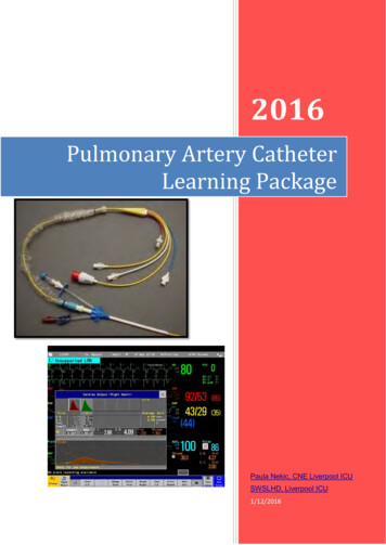
2016Pulmonary Artery CatheterLearning PackagePaula Nekic, CNE Liverpool ICUSWSLHD, Liverpool ICU1/12/2016
Liverpool HospitalIntensive Care: Learning PackagesIntensive Care UnitPulmonary Artery Catheter Learning PackageCONTENTS1. Objectives32. Pulmonary Artery Catheter4 Indications Contraindications Complications3. Sheath64. Lumens75. Insertion86. Waveforms137. Wedging218. Cardiac output studies239. Nursing management3310. Learning Questions3511. Reference List36LH ICU2016 Learning Package Pulmonary-Artery Catheter Learning Package2 Page
Liverpool HospitalIntensive Care: Learning PackagesIntensive Care UnitPulmonary Artery Catheter Learning PackageOBJECTIVESThe aim of this package is to provide the nurse with a learning tool which can be used inconjunction with clinical practice under supervision of a CNE and or resource person for themanagement of a pulmonary artery catheter.After completion of the package the RN will be able to:1. State the indications, contraindications and complications of a PA catheter2. Identify the lumens and their uses3. Identify normal and abnormal waveforms4. Perform all routine and safety checks5. Identify normal ranges for haemodynamic values measured from a PA catheter6. Identify the position and waveforms of PA catheter7. Perform a wedge procedure safely8. Perform cardiac output studies9. Interpret cardiac output studies10. Identify the risks and complications associated with the insertion and managementof a PA catheter11. Discuss nursing management of a PA catheterLH ICU2016 Learning Package Pulmonary-Artery Catheter Learning Package3 Page
Liverpool HospitalIntensive Care: Learning PackagesIntensive Care UnitPulmonary Artery Catheter Learning PackagePulmonary Artery Catheter HistoryThe first introduction of a catheter into a human pulmonary artery was in 1929 by Forsmann.He inserted a urinary catheter into his own cubital vein and into his right heart.In 1954 a catheter was developed by Lategola and Rann and used in dogs.It wasn’t until 1970 that Dr Swan was on an outing with his family and noticed how easy itwas for a sailboat to move even in the slightest breeze. Up until this point no one had beenable to float the catheter into the pulmonary artery. Dr Swan then invented the balloon tippedcatheter. Around the same time Dr Ganz was working on thermo dilution methods tocalculate cardiac outputs. So the pulmonary artery catheter was named Swan Ganz.The Pulmonary Artery CatheterThe pulmonary artery catheter (PAC) is a balloon tipped thermo dilution catheter 110cmslong, that is inserted via a large vein and floated into the pulmonary artery. It is used toobtain haemodynamic measurements which together with clinical observations indicate howefficiently the heart is functioning.The PAC directly measures: Right pulmonary systolic and diastolic pressures (PAP), Pulmonary Artery Wedge Pressure (PAWP), Cardiac Index (CI), Systemic and Pulmonary Vascular Resistance (SVR & PVR), Core body temperature Mixed venous oxygen saturation.These will be discussed later in this package.IndicationsA pulmonary artery catheter is indicated for assessment of: Shock statesCardiovascular functionPulmonary functionHaemodynamic function peri, intra and post cardiac surgeryFluid requirements and the effectiveness of therapyMultiorgan failureContraindications Coagulation defectsTricuspid or pulmonary valve replacementsRight heart mass / thrombus /tumorTricuspid or pulmonary valve endocarditisHigh risk of dysrhythmiasCaution with LBBB (5% risk of complete heart blockComplications Atrial and ventricular dysrhythmiasRBBB (0.1-5%)LH ICU2016 Learning Package Pulmonary-Artery Catheter Learning Package4 Page
Liverpool Hospital Intensive Care: Learning PackagesIntensive Care UnitPulmonary Artery Catheter Learning PackagePulmonary infarctPulmonary artery ruptureInfectionThrombusInsertion of introducer sheath Pneumothorax Arterial puncture Air emboluswww.yalemedicalgroup.orgLH ICU2016 Learning Package Pulmonary-Artery Catheter Learning Package5 Page
Liverpool HospitalIntensive Care: Learning PackagesIntensive Care UnitPulmonary Artery Catheter Learning PackagePULMONARY ARTERY CATHETERThe sheathPulmonary artery cathetersealed with plastic to maintainsterilitySheathSide arm of sheath for infusionof fluidsThe pulmonary artery catheter is inserted via an 8.5F sheath which is usually place in Subclavian veinJugular veinFemoral veinThe sheath has a removable seal on the end which allows the PAC to pass through it andforms a water tight seal around it. It also allows the PAC to be removed and the sheath toremain in place. A cap is then placed on the end of the sheath to maintain patency.A plastic protective sleave covers the PAC and attaches to the end of the sheath to maintainsterility of the PA while it is being advanced and withdrawn.The sheath may also have a side arm for infusion of fluids and drugs.LH ICU2016 Learning Package Pulmonary-Artery Catheter Learning Package6 Page
Liverpool HospitalIntensive Care: Learning PackagesIntensive Care UnitPulmonary Artery Catheter Learning PackageThe lumensCoretemperaturecableWedge syringe:when notperforming wedgeprocedure shouldalways be leftwith no air & tapopenYellow / distal lumen:for transducing PAP& specimens formixed venous gasesMedication lumenonly for sedation &low dose GTNCardiac output syringeand temperature cableBlue/ProximalLumen: fortransducing CVP &attaching CO syringeWedge port: For performing wedge procedure which inflates the balloon on the end of thePAC and allows the catheter to float into the pulmonary artery. Special syringe only allows 1.5mls of air to be injected to prevent balloon rupture After wedge procedure syringe should always be left with no air in it and tap opento prevent accidental wedgingDistal / yellow lumen: Located on tip of the catheter and sits in pulmonary artery therefore monitors thepulmonary artery pressures Blood gas specimens can be taken from this port for measuring patients mixedvenous gas Drugs or infusions are NEVER to be injected in this lumenProximal / blue lumen: Located in right atrium therefore monitors CVP Cardiac output syringe connected here to perform cardiac output studies Infusions or drugs are not to be connected to this lumenProximal /medication lumen:LH ICU2016 Learning Package Pulmonary-Artery Catheter Learning Package7 Page
Liverpool Hospital Intensive Care: Learning PackagesIntensive Care UnitPulmonary Artery Catheter Learning PackageThis lumen is usually utilised for sedation and low dose GTN not for inotropes.Temperature port: Temperature cable from the cardiac output cable is connected here to obtainpatients core temperature and assists in computing cardiac output studiesInsertion of pulmonary artery catheterThe PAC is inserted by a trained Registrar, with an ICU nurse assisting. Post –opcardiothoracic patients will have the PAC inserted in theatres.Equipment Large dressing pack Sterile gown, drapes, gloves, protective eyewear Pulmonary Artery Catheter Thermodilution Catheter Introducer Set (Sheath)Cardiac output injectate set 500 ml 5% Glucose Cardiac Output module & cable ECG and 2 pressure modules & cables Inflatable pressure bag 2 Pressure Transducers 2 Pressure line extension tubing 500 ml 0.9% Sodium chloride 5ml & 10ml syringes 2 x 10 mls 0.9% Sodium chloride 5 mls Lignocaine 1% Scalpel Blade Gauze Sponges 2/0 Silk Suture Large and Small Opsite Dressing 2 Three Way Taps 21g,23g,25g, needles Transducer holderPreparation for PA Catheter Insertion: Observe universal precautions.Insert an ECG, Pressure Module and a Cardiac Output Module into the monitor.Connect the 2 pressure cables, and the cardiac output cable.Prime both transducers & pressure extension lines with 0.9% sodium chloridefrom the same flask. Ensure that there are no bubbles in the lines ortransducers.Place the 0.9% sodium chloride in the inflatable pressure bag, and inflate to300mmHg.Label transducer as “CVP” and the other as “PA’’ & then place them in thetransducer holderLH ICU2016 Learning Package Pulmonary-Artery Catheter Learning Package8 Page
Liverpool Hospital Intensive Care: Learning PackagesIntensive Care UnitPulmonary Artery Catheter Learning PackageConnect PA transducer line to PA (yellow /distal) port. As seen in picture below.This needs to be connected so as you have a pressure waveform for ve/doc/RNSH/PA Catheter management rnsh 2007.pdf Connect one of the pressure cables to the “PA” transducer.Turn the stopcock on the (PAP) pulmonary artery pressure transducer off to thepatient (open to air).Transducer holder with transducers should be placed level with the patient’s 4thintercostal space in the midaxillary line.Select the “0- Zero” soft key. Press “ZERO PAP”Once calibrated, turn the PAP transducer stopcock back to the patient andresume tracing.Check that the scale of the PAP trace is set at optimal.If the PAP trace is dampened, flush the line well.Always ensure that the PAP trace is visible at all times and that the alarms areon.Check the alarm limits at the beginning of each shift.Prime the enclosed injectate delivery system set with 5% GlucoseSet up the basic tray with the requirements for catheter insertion.PA Catheter Insertion: (ICU Registrar) Observe universal precautions - Surgical scrub. Don gown, gloves and mask.Prep and drape the insertion area.Position’s patient to maximise access to desired area of insertion i.e.Trendelenberg position if requiredAttaches monitoring (ECG or SpO2) if availableFlush/prime each lumen with 0.9% normal salineZero, level and Transducer each lumen on monitor for correct waveformsInfiltrate the skin with 1% Lignocaine.Position the patient as appropriate for the insertion site chosen.Attach the long 18G needle to the 10ml syringe, and puncture the desired vein.LH ICU2016 Learning Package Pulmonary-Artery Catheter Learning Package9 Page
Liverpool Hospital Intensive Care: Learning PackagesIntensive Care UnitPulmonary Artery Catheter Learning PackageRemove the syringe, and confirm that a free flow of non-pulsatile blood isreturned.Insert the guide wire through the needle, maintaining control of the proximal end.An assistant should observe the monitor for arrhythmia caused by irritation of themyocardium with the wire. If arrhythmias occur, withdraw the wire slightly.Remove the needle. Make a small skin incision with the scalpel blade, at thepoint of entry of the guide wire.Insert the dilator through the haemostasis valve, and into the sheath.Thread the dilator/sheath/haemostasis valve assembly over the guide wire.Holding skin taut at the insertion site, advance the assembly into the vessel witha slight twisted motion, maintaining control of the proximal end of the guide wire.Remove the guide wire.Attach a 10ml syringe to the side point of the sheath, and aspirate blood toconfirm that the sheath is in position. Flush the port with 10mls N/Saline, andapply the cap.Firmly suture the sheath in position.Check the balloon on the pulmonary artery catheter by inflating it inside thetesting chamber with 1.5mls air. Use only the special syringe supplied for thispurpose.Allow the balloon to passively deflate.Check the patency of each lumen by flushing with 0.9% sodium chloride.Taking care not to contaminate the sterile field, pass the “PA (DISTAL) LUMEN”to an assistant, for connection to the already zeroed PA transducer. Thetransducer is positioned 4th intercostal space in the midaxillary line.Insert the catheter through the haemostasis valve on the sheath.The balloon is clear of the sheath when the catheter is inserted to 18cm (note the10cm graduations along its length).Inflate the balloon with 1.5mls, and close the red tap on the balloon inflation port.Insert the catheter until a CVP waveform appears on the monitor.Rapidly feed the catheter through the sheath while observing for thecharacteristic waveforms which indicate transition from: CVP RV PA PCWPLH ICU2016 Learning Package Pulmonary-Artery Catheter Learning Package10 P a g e
Liverpool Hospital Intensive Care: Learning PackagesIntensive Care UnitPulmonary Artery Catheter Learning PackageWhen a PCWP trace is obtained, deflate the balloon and confirm that a PA tracereturns on the monitor.If the trace remains wedged, the catheter will need to be withdrawn slightly.Usually, the correct insertion distance is:- 50 to 60cm for subclavian or internal jugular approachNote and document on flow chart the cm marking on the sheath at which aPCWP trace is obtained when the balloon is inflated. As the catheter warmsto body temperature, it lengthens slightly and can spontaneously wedge. If thisoccurs, it will need to be withdrawn slightly.Place the blue adapter on the Cath Guard over the haemostasis valve on thesheath.Clean the insertion site of all blood.Apply OpSite dressings to sheath siteConnect the CVP transducer to the 3 way tapConnect the cardiac output injectate syringe to CVP portConnect thermistor cable to cardiac output syringe port and temperature cable tored temperature port as shownCardiac output cableTemperature cableCardiac output syringe,primed with 5% dextroseThermistor cable. Confirm the transducer holder with PA and CVP transducers, are level with the4th intercostal space in the midaxillary line.Turn the tap on the cardiac output injectate set “off” to the syringe.Connect a pressure cable to the CVP transducer.Turn the stopcock on the CVP transducer off to the patient (open to air).Select the “0- Zero” soft key. Press “ZERO CVP”Once calibrated, turn the CVP transducer stopcock back to the patient andresume tracing.Press the alarms limit key on the Philips monitor.Press select parameter to highlight the parameter that you want to adjust e.g.PAPUse low limit and high limit keys if you wish to adjust the limits.You can alter the systolic, diastolic and mean alarm limits individually.LH ICU2016 Learning Package Pulmonary-Artery Catheter Learning Package11 P a g e
Liverpool Hospital Intensive Care: Learning PackagesIntensive Care UnitPulmonary Artery Catheter Learning PackageLeave the PAP trace displayed on the monitor at all times, so that inadvertentwedging of the catheter can be detected. Set PAP alarm as diastolic to alertinadvertent wedgingThe spare “infusion” lumen can be used for sedation and low dose GTN (no morethan 10mls/hr) only.Measure the PCWP and attend cardiac output studies.Obtain CXR to ensure correct placement – 2cm left of mediastinal borderPulmonary artery catheterMonitoring pulmonary artery pressures: Just the factsLH ICU2016 Learning Package Pulmonary-Artery Catheter Learning Package12 P a g e
Liverpool HospitalIntensive Care: Learning PackagesIntensive Care UnitPulmonary Artery Catheter Learning PackageWAVEFORMSWhen a right atrial pressure waveformdemonstrates that the catheter hasreached the right atrium the balloon isinflated.The catheter will then float through tothe right ventricle, then into thepulmonary artery. As the blood flowmakes the catheter float through theheart the waveforms will changeaccording to the position of the catheterin the heart. See picture on left.http://www.anaesthesiauk.com/articleCentral Venous Pressure (CVP):The normal range for CVP is 5 -15 mmHg.CVP measures right ventricular function and systemic fluid status.a wave atrial contractionc wave bulging of tricuspid valveinto atrium at start of ventricularcontractionx descent atrium relaxes andtricuspid valve is pulled htmv wave passive filling of rightatrium and vena cava whentricuspid valve closesy descent tricuspid valve opensand blood flows into right ventricleRight Ventricular Pressure (RVP):Once the catheter passes through the Right Atrium it then floats into the Right Ventricle.The normal value for RVP is 25/0 mmHg.Dangers of the catheter being in the ventricle are that the catheter could rupture the ventriclewall or irritate the ventricle causing VT.Monitor PA waveforms at all times to ensure catheter is not sitting in ventricle.LH ICU2016 Learning Package Pulmonary-Artery Catheter Learning Package13 P a g e
Liverpool HospitalIntensive Care: Learning PackagesIntensive Care UnitPulmonary Artery Catheter Learning PackagePulmonary Artery Pressure (PAP):The catheter should then float into the pulmonary artery.The normal range for PAP is 15 - 25 / 5-15 mmHg.The dicrotic notch is the usual feature of the PA waveform and represents aortic valveclosure.Systolic PA pressure indicates the pressure in the pulmonary artery as blood is beingejected from the right ventriclePulmonary artery diastolic pressure (PADP) indicates the pressure in thepulmonary artery as blood moves from the artery into the lungs capillaries.It can be used to assess preload as it is an indirect measurement of Left ventricular enddiastolic pressure (LVEDP)The PADP can be utilised as a wedge pressure if a wedge is not able to be obtainedMean pulmonary artery pressure is the average pressure in the pulmonary vasculaturethroughout the cardiac cycleOnce the catheter is inserted and zeroed the CVP and PAP readings will be displayed asseen in picture below:PAP traceArterialPressureCVP tracePulmonary Artery Wedge Pressure (PAWP):When the balloon is inflated, the catheter then floats through the pulmonary circulation until itwedges in a small artery. The balloon is effectively “wedged” against the artery.When the catheter is wedged, there is no blood flow to this part of the pulmonary arterytherefore the balloon should only be wedged long enough to obtain a reading, (usually 15secs or 2 respiratory cycles) as the pulmonary artery could rupture.The benefits of measuring PAWP are to optimise filling of the patient without overloading thelungs.LH ICU2016 Learning Package Pulmonary-Artery Catheter Learning Package14 P a g e
Liverpool HospitalIntensive Care: Learning PackagesIntensive Care UnitPulmonary Artery Catheter Learning PackageThe normal range for PAWP is 8 -12mmHgWedge trace: represented by achange in pressure tracing fromPA trace to a flattened wedgetracingPAWP represents LVEDP and is used to assess the function and workload of the leftventricle.As shown below, the balloon occludes the pulmonary artery creating a back pressure fromthe right atrium. The transducer on the tip of the catheter only sees what is happening in theleft atrium and left ventricle. So the PAWP measures filling pressures on the left side of theheart.http://www.ispub.com/ispub/ija/volume 11 number 2 1/paradigm shift in hemodynamicmonitoring/hemodynamicsLH ICU2016 Learning Package Pulmonary-Artery Catheter Learning Package15 P a g e
Liverpool HospitalIntensive Care: Learning PackagesIntensive Care UnitPulmonary Artery Catheter Learning PackageINTERPRETING PAP WAVEFORM CHARACTERISTICS: a, c, and v WAVESWaveform interpretation can be simplified by remembering that electrical activity (ECG)comes before mechanical activity (pressure waveform).The table below summarises the electrical and mechanical events in relation to pressurewaveforms.Monitoring pulmonary artery pressures: Just the facts Elizabeth J Bridges. Critical Care Nurse. AlisaVeijo: Dec 2000. Vol. 20, Iss. 6The LAP or PAWP waveform has the same characteristics as the RAP waveform, with threepositive and two negative deflections.The c wave, however, is often not visible as it may be "hidden" in the x descent of the awave.If only two positively deflected waves are seen, The first is the a wave, which represents left atrial systole The second is the v wave which represents left atrial filling against a closed mitral valve.The peak of the v wave marks the end of ventricular systole" and occurs after the T waveof the ECGLH ICU2016 Learning Package Pulmonary-Artery Catheter Learning Package16 P a g e
Liverpool HospitalIntensive Care: Learning PackagesIntensive Care UnitPulmonary Artery Catheter Learning PackageThe timing of the PAWP waveform is slightly delayed compared to the timing of the RAPwaveform relative to the ECG. This occurs because the left atrial waveform must travel backthrough the pulmonary vasculature to the distal tip of the PA catheter, whereas the RApressure is measured directly through the proximal port in the right atrium.Keep in mind that being able to recognize this time delay in relation to the ECG will help youdifferentiate the RAP from the PAWP waveform.The picture below illustrates this difference in timing of the RAP and PAWP waveformsrelative to the ECG.Identifying a wave in PAWP traceAlign the end of at least two QRS complexes at the point of end-expiration with the PAWPwaveform. Draw a vertical line from the end of the QRS down to the PAWP waveform, whichidentifies the a wave.Electricalevent (ECG)MechanicaleventPulmonary arterywedge pressure(normal PAWP is6 – 12 mm Hg)Approximately20millisecondsafter P waveLeft atrial(LA) systolea waveLA diastolex descentLAfilling/mitralvalveclosedv waveLAemptying atopening ofmitralvalve/onsetof leftventriclediastoley descentT-P intervalPA catheters: What the waveforms reveal. Sally Beattie,RN 2003LH ICU2016 Learning Package Pulmonary-Artery Catheter Learning Package17 P a g e
Liverpool HospitalIntensive Care: Learning PackagesIntensive Care UnitPulmonary Artery Catheter Learning PackageIt is important to recognise these waves as The shape of the v wave is primarily determined by the relationship between pressureand volume in the left atrium; that is, with decreased atrial compliance (e.g., ischemia orhypertrophy), any change in volume is associated with a greater increase in pressure asindicated by increased v wave amplitude.large influx of blood into the atrium (e.g., mitral valve regurgitation or a ventricular septaldefect) may cause a large v wave (known as a V wave)."ABNORMAL WAVEFORMSAltered a waves: Large a waves can occur when atria contract against stenotic mitral or tricuspid valves Giant a waves (or cannon waves) can occur with junctional and AV dissociative rhythms.The a wave produced by the simultaneous contraction of the atria and ventricle isenlarged and occurs later in the cardiac cycle usually where the v wave would occur Cannon waves can also occur with premature ventricular contractions or re- entranttachycardias.Large v waves: They are produced by the increase in blood volume entering the atria during the cardiaccycle The shape of the v wave is determined by the relationship between pressure and volumein the left atrium On the monitor the v wave will be taller than the a wave, followed by an exaggerated ydescent that reflects the release of atrial pressure with the opening of the tricuspid ormitral valve This is the most frequent wave abnormality They are commonly caused by tricuspid and mitral insufficiency due to a large influx ofblood into the atrium Others causes include ventricular failure, increased pulmonary or systemic resistanceand ventricular septal defect The importance of recognising a large v wave is being able to obtain an accurate PAWP.The large v wave maybe mistaken for the systolic PAP wave. This can be avoided bymeasuring pressures and waves in relation to the ECG. Large v waves in a case of mitral regurgitationPulmonary Artery Catheterization: Treatment & Medication Author: Bojan Paunovic, MDLH ICU2016 Learning Package Pulmonary-Artery Catheter Learning Package18 P a g e
Liverpool HospitalIntensive Care: Learning PackagesIntensive Care UnitPulmonary Artery Catheter Learning PackageAbnormal wedge trace Over wedging is when:The wedge trace rises dramatically with the balloon inflated. The balloon should beimmediately deflated and the catheter withdrawn 1 – 2cms with help from a Registrar, CNEor CNSCat:het:er too dist:alOverdamping oft:racing.Catheter spontaneous wedging dge type tracing withballoon deflaudFull inflation with 1.5 ccinflation volume. Appropriate'a" and v" waves noted.Overin.flation ofballoon.Note waveform rise on screen.LH ICU2016 Learning Package Pulmonary-Artery Catheter Learning Package19 P a g e
Liverpool HospitalIntensive Care: Learning PackagesIntensive Care UnitPulmonary Artery Catheter Learning PackageTable 3 Examples of abnormal waveformsConditionDiagnostic cues/interpretationTricuspidregurgitation(from apatient withlong-standingpulmonaryhypertension)The A wave (a) and the x descent (x) are normal.The X·prime wave. which is the downslope of the awave after the c wave. is obliterated. The result isa broad positive CV wave (cv) that is higher thanthe A wave.Note that inspiration (INSP) magnifies the nadir ofthe I) descent and the peak of the CV wave, sothere is little change in the mean right atrialpressure.An exaggerated Y descent is the key diagnosticfeature.Interpretation: The mean right atrial pressure iselevated at 22 mm Hg.ExampleFigure reprinted from Sharkey," with p tery (PAlpressuretracingAt end·diastole. when the tricuspid valve is open,the mean right atrial pressure (RAP) isapproximately equal to the right ventricular enddiastolic pressure (RVEDP).As the catheter is advanced from the right ventricleinto the PA, the end-diastolic pressure increases.The right ventricular systolic (RVS) pressure is equalto the PA systolic (PAS) pressure.PAEDP indicates PA end-diastolicpressure.Interpretation: If the waveform visualized from thedistal port has an end-diastolic pressure (measured 0.08 seconds after the onset of the QRS) that is equalto the RAP and RVS pressure - PAS pressure, catheter malposition should be suspected.PA pressure orPAwedge(PAW)pressure?Comparison of the PA pressure and the PAWpressure relative to electrocardiographic findings:The v wave of the PAW pressure waveformoccurs during the TP interval, whereas theinitial systolic upstroke of the PA pressurewaveform is closely related to the end of theQRS complex, with the PAS peak occurring:-- .Aduring the T wave.The regurgitant V wave can be observed as aI iil l/second peak or on the PA pressure waveforml 1\ \ I\(may also occur on the downslope). and hasI 1the same temporal relation to the QRS as theIV wave in the PAW pressure tracing.Interpretation: PAW pressure 30 mm Hg (read at the nadir of the waveform).Cannon AwavesOverwedgeCentral venous pressure tracing andelectrocardiogram of a patient in ventriculartachycardia.No P waves can be seen for certain on theelectrocardiogram.Central venous pressure tracing showsexaggerated A waves when the right atriumcontracts against the closed tricuspid valveduring atrioventricular dissociation.Interpretation: Cannon A waves; supportsdiagnosis of ventricular tachycardia.\ '-\Figure replinted from Jacobson, with permission.Interpretation: Overwedge as indicated by (1lprogressive increase in pressure off the screenand (2) loss of waveform components (a and vwaves).IN THIS SITUATION, STOP INFLATIONIMMEDIATELY AND DEFLATE TH E BALLOON.Monitoring Pulmonary Artery Pressures: Just the Facts. CCN Vol20, No6, Dec 2000LH ICU2016 Learning Package Pulmonary-Artery Catheter Learning Package20 P a g e
Liverpool HospitalIntensive Care: Learning PackagesIntensive Care UnitPulmonary Artery Catheter Learning PackagePULMONARY ARTERY WEDGE PROCEDURE(Adapted from Pulmonary Artery Catheter Guideline LHS ICU 2014) Ensure the configuration of the PA trace appears normal.Ensure the SaO2 is greater than 90%,Position the patient supine with the backrest elevated to 45 , unless contraindicated.Whatever position is used, the same position should be adopted for each reading.Ensure the PA transducer is positioned level with 4th ICS, midaxillary line, and has beenzeroed.In the main screen window inflate the balloon and ensure that catheter wedges and largev waves are not seen (see above explanation on v wavesGo to wedge screen then press Wedge to get into the “required wedge task window”.Inflate the balloon slowly and carefully watching the screen for a wedge traceThe waveform changes from PAP to PAWP and the message “wedging” will appear.Inflate the balloon only for the time required to see end expiration on the screen. Usually2 respiratory cycles.Press store trace.Deflate the balloon once a wedge trace is stored. Allow the balloon to deflate passively.Disconnect wedge syringe from balloon port, expel air and reattach syringe to port withtap open to the syringe. This prevents any accidental wedging of the catheterSee below for examples of a wedge trace.Normalwedge tracea wave: 20 millisecondsafter p wavev wave : T-P intervalLH ICU2016 Learning Package Pulmonary-Artery Catheter Learning Package21 P a g e
Liverpool HospitalIntensive Care: Learning PackagesIntensive Care UnitPulmonary Artery Catheter Learning PackageRespiratory Cycle and WedgingThe effect of the respiratory cycle on PCWP measurements is important in obtaining anaccurate PAWP. The timing of PCWP measurement is critical because intrathoracicpressures can vary widely with inspiration and expiration and are transmitted to thepulmonary vasculature. During spontaneous inspiration, the intrathoracic pressuresdecrease (more negative); during expiration, intrathoracic pressures increase (morepositive). Positive pressure ventilation (e.g., in an intubated patient) reverses this situation.To minimize the effect of the respiratory cycle on intrathoracic pressures, measurements areobtained at end-expiration, when intrathoracic pressure is closest to zero.Spontaneous ventilation:cursor would be on endexpirationMechanical ventilation:cursor would be on endexpirationEditing wedge trace.Press “edit wedge” soft key to open the next task window.A horizontal line (cursor) appears in the PAWP waveform in the position of the mean valuefor PAWP. A numerical value for PAWP appears on the screen, entitled cursor. If aprevious value is stored, it is also shown along with the time.Positivepressureventilation;cursor one
Prime the enclosed injectate delivery system set with 5% Glucose Set up the basic tray with the requirements for catheter insertion. PA Catheter Insertion: (ICU Registrar) Observe universal precautions - Surgical scrub. Don