Transcription
SRC KINASE ACTIVATION IN PULMONARY ARTERIAL HYPERTENSIONByAllison Renee PrewittDissertationSubmitted to the Faculty of theGraduate School of Vanderbilt Universityin partial fulfillment of the requirementsfor the degree ofDOCTOR OF PHILOSOPHYinCell and Developmental BiologyAugust, 2015Nashville, TennesseeApproved:Ethan Lee, Ph.D.Matthew Tyska, Ph.D.Anne Kenworthy, Ph.D.David Harrison, M.D/Ph.D.Mark de Caestecker, M.B., B.S., Ph.D.
To my parents, Dennis and Robin, my ever-growing family, and dear friends. I could nothave completed this journey without your endless love and support.ii
ACKNOWLEDGMENTSThis work was supported by numerous funding sources including the BiomedicalResearch Education and Training Grant, the American Heart Association, andNIH/NIHLBI grants R01HL093057 and R03HL115112. I would also like to thank theVanderbilt University Department of Medicine’s Division of Nephrology for their staffingsupport.I would like to acknowledge the Vanderbilt School of Medicine’s Department ofCell and Developmental Biology for designing and coordinating my training. Inparticular, I would like to thank Elaine Caine, Kristi Hargrove, and Susan Walker for theirsupport and guidance through all steps of my graduate career. I am appreciative toour students for allowing me to serve as the Graduate Student Association Presidentand Social Committee member during my last two years in the department. It has beena pleasure serving you all.I am grateful for the guidance and advice of my dissertation committee whohave pushed me to grow and mature as a scientist. Thank you to Drs. Ethan Lee,Matthew Tyska, Anne Kenworthy, and David Harrison. I would also like to thank themembers of the Pulmonary Hypertension interest group for their unabashed insight intothe depths of academia and the inner workings of research.I am eternally grateful to my mentor Mark de Caestecker. Without his enduringpatience and encouragement, I would not be where I am today. I cannot imaginehaving survived this process without Mark on my side. Mark encouraged me to breakout of my comfort zone and tolerated me when I invariably panicked my way throughit. Thank you for all your support.iii
I would be remiss if I did not thank my friends. Innumerable times have I relied onmy friends for coffee therapy sessions and jovial happy hours to get through the roughpatches and celebrate the good times. You have made this journey all the morememorable.And finally and most importantly, I would like to thank my family. To Mom andDad, I cannot thank you enough for all that you have provided me. You have goneabove and beyond to support me both emotionally and financially. Dad, I will forevercherish the time we have spent together during your travels through Nashville. Mom, Iam immeasurably appreciative of all your sacrifices and endless love. To my siblings, Icould not have made it through my days without your humor, snark, and moral support.You are all truly my best friends.iv
TABLE OF CONTENTSPageDEDICATION . .iiACKNOWLEDGMENTS . . .iiiLIST OF TABLES . .viiLIST OF FIGURES .viiiLIST OF ABBREVIATIONS xChapterI.Introduction .1Part I: Pulmonary Hypertension 1Definition of Pulmonary Hypertension . . 1Classification of Pulmonary Hypertension . 1Pathobiology of Pulmonary Hypertension . .4Incidence, diagnosis, and treatment of Pulmonary Hypertension .9Part II: BMPR2 mutations in Heritable Pulmonary Arterial Hypertension 12Part III: Caveolae and Pulmonary Hypertension .15Part IV: Src kinase and Pulmonary Hypertension .20Summary .27II.BMPR2 /- Mutations Promote Src Kinase Dependent Caveolar Trafficking Defectsand Endothelial Dysfunction .28Introduction .28Materials and Methods 29Results . .36Discussion . 55III.BMPR2 Regulation of Src Kinase Activation .59Introduction .59Materials and Methods 60Results . .62Discussion . 69v
IV.Discussion and Future Directions . 78Src Kinase Activation and Endothelial Dysfunction . .78Src-Mediated Mitochondrial Dysfunction in PAH .82Mechanisms of Src Activation . .84Summary .85REFERENCES . .86vi
LIST OF TABLESTablePage1. WHO Current Clinical Classification of Pulmonary Hypertension 22. Phosphoregulation of Src kinase .233. BMPR2 PCR amplification primers . .75vii
LIST OF FIGURESFigurePage1. Structural changes associated with pulmonary arterial hypertension 52. Right heart hypertrophy in pulmonary arterial hypertension .63. Diagram of right heart catheterization . .104. Germline BMPR2 mutational spectrum in primary pulmonary hypertension .145. Caveolar structure 176. Src-mediated caveolar endocytosis .187. Src kinase structure . 218. Non-catalytic Src kinase inhibitors 259. Mis-localization of caveolar structural proteins in Bmpr2 /- PECs 3710. Increased caveolar structures in Bmpr2 /- PECs and lungs .3911. Dynamin-2 inhibition restores Cav-1 localization to the plasma membrane inBmpr2 /- PECs. 4112. Bmpr2 /- PECs have increased caveolar endocytosis but exhibit normalexocytosis .4213. Increased Src and Cav-1 phosphorylation in Bmpr2 /- PECs. , . 4514. BMP2 stimulation reduces Src activity in Bmpr2 /- PECs . 4615. Increased Src activity in HPAH patient-derived late-outgrowth endothelialprogenitor cells (LO-EPCs) .4816. Src inhibition with PP2 reduces pY14-Cav-1 and restores Cav-1 localization to theplasma membrane in Bmpr2 /- PECs . 5117. Impaired endothelial barrier function in Bmpr2 /- PECs .53viii
18. Src inhibition with PP2 improves endothelial barrier function in Bmpr2 /- PECs .5419. Src kinase activation in distal, resistance endothelial cells from IPAH patients .6420. Src kinase is activated in Bmpr2 /- PECs but not in Bmpr2Δex2/ PECs . 6521. Bmpr2 /- PECs exhibit no change in pY530-Src expression . .6622. Bmpr2-flox cells express endothelial specific markers 6723. Increased Src activation in Bmpr2-flox PECs .6824. BMPR2 PiggyBac expression constructs .7325. Linearized BMPR2-Piggybac constructs .7426. Schematic of CymRepressor configuration .7627. BMPR2-PiggyBac schematic .77ix
LIST OF ABBREVIATIONSBMPBone Morphogenetic ProteinBMPR2Bone Morphogenetic Protein Receptor Type 2Cav-1Caveolin-1CSDC-term caveolar scaffolding domainECEndothelial celleNOSEndothelial Nitric Oxide SynthaseEPCEndothelial progenitor cellHPAHHeritable Pulmonary Arterial HypertensionIPAHIdiopathic Pulmonary Arterial HypertensionNONitric oxidePAHPulmonary Arterial HypertensionPHPulmonary HypertensionPVRPulmonary vascular resistanceRHCRight heart catheterizationRVRight ventricleSFKSrc family kinaseSMCSmooth muscle cellVEGFVascular Endothelial Growth FactorVEGFR2Vascular Endothelial Growth Factor Receptor Type 2x
CHAPTER IINTRODUCTIONPart I: Pulmonary HypertensionDefinition of Pulmonary HypertensionPulmonary hypertension (PH) is a progressive, fatal disease of the lungvasculature. In its simplest definition, PH is described as abnormal and sustainedelevation of pulmonary arterial pressures during resting conditions. Traditionally, PH isdefine as mean pulmonary arterial pressures (mPAP) greater than 25mmHg at rest, asmeasured by right heart catheterization. However, recent advanced measures ofpulmonary capillary wedge pressure (Ppcw), pulmonary vascular resistance (PVR), andcardiac output (CO) enable clinicians to effectively categorize PH heamodyanimicsubtype as either pre-capillary (Groups 1, 3, 4, 5) and post-capillary (Group 2- PH due toleft heart disease)(Galie, Hoeper, et al., 2009a).Classification of Pulmonary HypertensionFirst discovered by Dresdale in the 1950’s (Dresdale et al., 1954), PH has gonethrough a series of classification systems. Most recently in 2008, the World HealthOrganization (WHO), at the Fourth World Symposium of Pulmonary Hypertension inDana Point, California, categorized PH into five major groups listed in Table 1 (Galie,Hoeper, et al., 2009a; PAH-info.com, 2015; Simonneau et al., 2009). We will discuss thesegroups below.1
Table 1: WHO Current Clinical Classification of Pulmonary HypertensionGroup 1Pulmonary Arterial Hypertension1.1Idiopathic pulmonary hypertension1.2Heritable pulmonary arterial hypertension (BMPR2, ALK1, ENDOGLIN,unknown)Drug and toxin-induced pulmonary arterial hypertensionAssociated with: connective tissue diseases, HIV infection, portalhypertension, congenital heart disease, schistosomiasis, chronichemolytic anemiaPersistent pulmonary hypertension of the newbornPulmonary veno-occlusive disease and/or pulmonary capillaryhemangiomatosisPulmonary Hypertension as a result of left heart diseases1.31.41.51’Group 22.12.22.3Group 4Systolic dysfunctionDiastolic dysfunctionValvular diseasePulmonary Hypertension associated with lung disease and/orhypoxiaChronic obstructive pulmonary diseaseInterstitial lung diseaseOther pulmonary diseases with mixed restrictive and obstructivepatternSleep-disordered breathingAlveolar hypoventilation disordersChronic exposure to high altitudeDevelopmental abnormalitiesChronic thromboembolic pulmonary hypertensionGroup 5Pulmonary Hypertension with unclear multifactorial mechanismsGroup 33.13.23.33.43.43.63.75.1Hematologic disorders5.2Systemic disorders5.3Metabolic disorders5.4Others: tumor obstruction, chronic renal failureAdapted from (PAH-info.com, 2015)2
Group 1, which the bulk of this work will discuss, is composed of patients withpulmonary arterial hypertension (PAH). The term PAH differs from PH in that PAH isdefined as the presence of pre-capillary PH that is not due to an accompanying lungdisease; however, this manuscript will use both terms interchangeably. This groupincludes the following: 1) heritable forms of the disease associated with germlinemutations, predominantly in the Bone Morphogenetic Protein Receptor Type 2 (BMPR2)gene, Activin-like Kinase Type 1 (ALK1) gene, and Endoglin and familial cases with noknown germline mutations; idiopathic, or sporadic, PAH (IPAH); 2) drug and toxininduced PAH, notably the Fen-Phen epidemic of the late 20th century (Brenot et al.,1993); 3) PAH associated with disorders/diseases including but not limited to connectivetissue diseases(Hachulla et al., 2005), HIV (Degano et al., 2010), and portalhypertension(Rodriguez-Roisin et al., 2004); 4) persistent pulmonary hypertension of thenewborn (Teixeira-Mendonca et al., 2013); 5) and pulmonary veno-occlusive disease(Montani et al., 2008).Group 2 makes up the largest population of PH patients and has a high rate ofmorbidity and mortality. It is the only post-capillary PH group and is composed ofpatients with PH as a result of left heart disease. Group 2 PH is suspected to occur inaccordance with left heart disease due to “passive” pressures emanating from the leftheart, though many suspect it is a combination of left heart pressures and preexistingpulmonary pathologies (Guazzi et al., 2012).Group 3 PH patients develop PH in association with other lung diseases and/orhypoxia. Development of this classification most often arises from alveolar hypoxia, apotent stimulant of vasoconstriction. Perhaps the most well known co-morbidity in thisgroup is chronic obstructive pulmonary disease (COPD); however this group includes a3
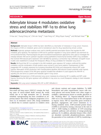
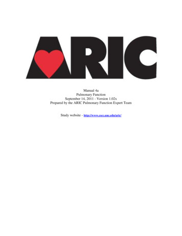
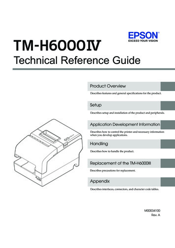
variety of factors including interstitial lung disease, alveolar hypoventilation disorders,chronic exposure to high altitude (hypoxia), and developmental abnormalities (Sajkovet al., 2013).Making up Group 4 PH are patients with chronic thromboembolic pulmonaryhypertension (CTEPH). Development of CTEPH is the result of increased artery pressuresdue to obstruction of the pulmonary arteries with thromboembolic materials. Whilethese patients exhibit high morbidity and mortality rates, CTEPH is the only severe PH inwhich medical intervention other than lung transplant may cure patients (McNeil et al.,2007).Finally, Group 5 PH is composed of patients with unclear multifactorialmechanisms, including hematologic disorders, systemic disorders, metabolic disorders,tumor obstruction, and chronic renal failure. This group is predominantly a catchall forPH that does not fall into other categories. Given the heterogeneity of this group, Group5 is the least characterized and studied (Galie, Hoeper, et al., 2009a).Pathobiology of Pulmonary HypertensionSeveral structural and functional abnormalities contribute to the developmentand progression of PH. Chief among these are structural changes in the pulmonaryarteries [loss of the distal pulmonary vasculature (vascular pruning), development ofneointima and plexiform lesions, and remodeling of the distal pulmonary arteries (Fig 1)]abnormal vascular reactivity, and right ventricle (RV) hypertrophy due to chronic RVpressure overload (Fig 2). It is this chronic RV overload that eventually results in patientdeath due to right heart failure (cor pulmonale) (Voelkel et al., 2012).4
Figure 1: Structural changes associated with pulmonary arterial hypertension.Abnormalities throughout the pulmonary circulation: abnormal muscularization of distaland medial arteries, loss of distal arteries, thickening of large arteries, occlusiveneointimal formation and plexiform lesions. From (Rabinovitch, 2012), CopyrightPermission: License #36565907552895
Figure 2: Right heart hypertrophy in pulmonary arterial hypertension. Adaptiveremodeling of the PH heart includes thickening right ventricle and reduction of the leftventricle.6
Current thinking is that endothelial insult and/or molecular changes withinendothelial cells (ECs) initiates a cascade of cellular and environmental changes withinthe pulmonary structure(Rabinovitch, 2012). The same processes that contribute tovascular lesion formation play a role in the generalized remodeling phenotype andvascular pruning phenomenon. This includes excessive proliferation of apoptoticresistant ECs (thought to be of monoclonal origins), recruitment of endothelialprogenitor cells (EPCs) and inflammatory cells, and the differentiation and proliferationof smooth muscle cells (SMCs)(Cool et al., 1999; S.-D. Lee et al., 1998; Masri et al., 2007;Montani et al., 2011; Perros et al., 2012; Perros et al., 2007; Rabinovitch, 2012;Taraseviciene-Stewart et al., 2002). These events result in remodeling of all three arteriallayers (intima, media, and adventitia) and in most cases are not reversible (Sakao et al.,2010).Exactly how these changes occur within the vascular bed is unclear; however,recent evaluation has prompted comparison to cancer-like mechanisms(Guignabert etal., 2013). Indeed hallmarks of cancer such as increased proliferation and decreasedapoptosis are clearly evident in the pulmonary vasculature of PH patients and can beattributed to the increased production and activity of pro-proliferative factors, such asFGF-2 and p130-CAS, and anti-apoptosis factors including Bcl-xI, Bcl-2, and Survivin(McMurtry et al., 2005; Tu et al., 2012; Tu et al., 2011). The result of this excessiveproliferation and reduced apoptosis is the development of obstructive and obliterativelesions that prevent free blood flow and contribute to increased pulmonary pressures.In addition to expansion of existing cell populations within the arterial wall,muscularization of normally non-muscularized distal arteries occurs. Muscularizationrefers to the addition of SMCs to the normally bare EC layer. Extensively studied, this7
process involves the migration of SMCs to the distal arteries, differentiation of precursorcells, and possibly endothelial-to-mesenchymal transition (EMT), all of which results inthe thickening of the arterial wall and contributes to abnormal vasoreactivity (Frid et al.,2006; Jones et al., 1998; Meyrick et al., 1980; Morrell et al., 2009; Sakao et al., 2009, 2010;Sheikh et al., 2014).Further contributing to increased pulmonary artery pressures is paradoxicalpulmonary artery pruning. The reduced surface area forces the steady blood volume topass through fewer blood vessels thereby increasing pressure. It is unclear why the lungsof PH patients undergo this process as there are an abundance of pro-angiogenicfactors circulating in the blood of these patients; however, it seems that PH ECs nolonger respond to angiogenic factors as normal ECs do (Guignabert et al., 2013;Hernndez et al., 2013).In addition to the expansive vascular remodeling that occurs in PH patients, thepulmonary arteries also exhibit abnormal vascular reactivity. These abnormalitiesinclude altered production of key vascular tone modulators including but not limited to,prostacyclin, endothelin-1, nitric oxide, and serotonin (Budhiraja et al., 2004; Farber etal., 2004), as well as altered responses to vasoactive stimuli (Frank et al., 2008), Togetherthese defects lead to increased vasoconstriction and reduced vasodilation.Finally, RV hypertrophy and subsequent right heart failure is the main cause ofdeath in PH patients. Despite this, little is known about the mechanisms that drive RVhypertrophy and failure; however, it is appreciated that increased afterload may bethe predominant factor driving the progression to right heart failure. Collectively, theabove remodeling events and aberrant vasoreactivity cause excessively highpulmonary pressures, forcing the heart to work harder (increase afterload) to push the8
same blood volume through reduced vascular space. Due to its inherent compliance,the RV is able to adapt to these changes in volume by increasing contractility. Initially,the RV adapts to increased contractility, undergoing concentric remodeling byincreasing muscle mass (hypertrophy). Once the RV has reached its limit of adaptivehypertrophy there is a switch to dilation, which increases wall tension, and therebyoxygen demands. Ultimately, the heart is unable to maintain normal function in themidst of remodeling, and suffering from low oxygen, eventually succumbs to ischemia(Voelkel et al., 2012; Vonk Noordegraaf et al., 2011).Incidence, Diagnosis, and Treatment of Pulmonary HypertensionThere are an estimated 2.4 to 7.6 new cases of Group 1 PH (PAH) per millionpeople annually, with a markedly higher incidence in high-risk populations (Archer,Weir, et al., 2010; Humbert et al., 2006; Peacock et al., 2007). This large range reflectsthe difficulty of diagnosing patients, as well as missing data. The difficulty diagnosingPAH stems in part from the nondescript symptoms which patients present, includingdyspnea (difficultly breathing), fatigue, syncope (fainting), and chest pain. Given therisks associated with invasive right heart catheterization (RHC) that is required foraccurate diagnosis, physicians must play close attention to previous medicalconditions, drug use, and family history when pursuing PAH as a diagnosis (Nauser et al.,2001). A series of tests are performed to rule out PH Groups 2-5 and can include anyand all of the following: electrocardiograph (ECG), Doppler electrocardiography, ch
Increased Src activity in HPAH patient-derived late-outgrowth endothelial . (Ppcw), pulmonary vascular resistance (PVR), and . connective tissue diseases, HIV infection, portal hypertension, congenital heart disease, schistosomiasis, chronic hemolytic anemia 1.5 Persistent pulmonary hy