Transcription
O ri g i nalResearchA Practical MRI Technique for Detecting Abdominal AortaAneurysm and Peripheral Arterial DiseaseAbdominal Aorta Anevrizması ve Periferik Arter HastalığıTanısında Pratik MR Görüntüleme TekniğiA Practical MRI Technique for Detecting AAA and PAHAykut Recep Aktas1, Birsen Unal2, Simay Kara2, Gokhan Kemal2, Omer Yılmaz1, Mustafa Kayan1, Sevda Yılmaz2, Mustafa Kara1, Bumin Degırmencı1, Meltem Çetin11Süleyman Demirel University Faculty of Medicine Dept. of Radiology, Isparta, 2Kırıkkale University Faculty of Medicine Dept. of Radiology, Kırıkkale, TurkeyÖzetGiriş: Anevrizmatik ve aterosklerotik damar patolojilerinde kontrast maddeverilmesi sakıncalı olan hastalarda çok kısa süreli koronal Balans Turbo Field Echo (B-TFE) manyetik rezonans (MR) görüntüleme sekansının etkinliğini, kontrastlı MR anjiografi (MRA) ve konvansiyonel Dijital Çıkarımlı Anjiografi (DSA) ile karşılaştırmalı olarak değerlendirmek. Gereç ve Yöntem: Etik kurul onayı alındıktan sonra, DSA planlanan hastalara inceleme öncesi onamları da alınarak 1,5 Tesla MR (Philips İntera, Netherland) cihazında BTFE görüntüleme yapıldı. Philips DICOM Viewer R2.2 kullanılarak, B-TFE, kontrastlıMRA ve DSA görüntüleri üzerinden, infrarenal aorta, renal arterler, aorta bifurkasyonu, ana, yüzeyel, derin femoral ve iliak arterlerin proksimal kesimlerinden ölçümler alındı. İstatistiksel analiz için SPSS 11.5 programı kullanılmıştır. B-TFE, MRA ve DSA tekniklerinin birbirleri yerine kullanılabilirlik ve güvenilirliklerinin saptanması için sınıf-içi korelasyon katsayısı ve güvenirlik değerleri, iki yöntemle ölçülen değerler arasında farklılığı incelemek için de bağımlı gruplarda t-testi uygulandı. Bulgular: Aorta, iliak ve femoral arterlerde B-TFE sekans ölçümleri ile MRA ve DSA ölçümleri arasında yüksek korelasyon mevcuttu. Ancak renal ve popliteal arterlerde B-TFE ölçümleri, DSA veMRA kadar güvenilir bulunmadı. Tartışma: Periferik arter hastalığının tanısında, DSA altın standart görüntüleme yöntemi olmakla birlikte işlem esnasında kontrast madde ve X-ışını kullanılır. DSA’da tespit edilen aorta, iliak ve femoral arterlerdeki anevrizmatik ve aterosklerotik patolojilerin takibinde vekontrast madde verilmesi sakıncalı olan hastalarda koronal B-TFE sekansıgüvenle kullanılabilir.AbstractAim: Peripheral Arterial Disease(PAD) and abdominal aorta aneurysm(AAA)are frequent problems in geriatric population. In DSA, CTA or MRA techniquescontrast agents has to be used for diagnosis that can be nephrotoxic forelderly patients. Magnetic resonans imaging (MRI) is the most powerful, nonionising radiological diagnostic tool that has the highest soft tissue contrastresolution. The aim of our study was to investigate the effectivity of MRI bythe means of detecting the AAA and PAD in comparison with DSA. Materialand Method: After getting ethical commitee approvel and informed consent,we have performed Balanced turbo field echo(B-TFE) MRI technique withoutcontrast agent in 1.5 Tesla MRI device before DSA examination. The luminaldiameters of renal arteries, infrarenal abdominal aorta, iliac and femoralarteries was measured by using Philips DICOM Viewer R2.2 application. Theintraclass corelation coefficient and reliability used to check if the techniquescould be used for each other and the t-test was used to measure the differences between them. Results: There has been a high relationship betweenB-TFE and DSA in detecting the pathologies of larger arteries like aorta. Inthe case of small arterial pathologies, there is relatively lower relationshipbetween BTFE and DSA. Discussion: For the diagnosis of AAA and PAD, DSAis the gold standart technique but it is invasive and patients have radiationexposure. In the follow up of geriatric patients with larger arterial pathologies B-TFE can be used instead of contrast enhanced MRA and invasive DSA.Anahtar KelimelerMRI; Anjiografi; Dijital Çıkarım; Periferik Arter Hastalığı; AnevrizmaKeywordsMRI; Angiography; Digital Subtraction; Peripheral Arterial Disease; AneurysmDOI: 10.4328/JCAM.2725Received: 14.08.2014 Accepted: 01.09.2014 Printed: 01.05.2016J Clin Anal Med 2016;7(3): 295-9Corresponding Author: Aykut Recep Aktas, Department of Interventional Radiology, Medical Faculty, Suleyman Demirel University, Isparta, Turkey.T.: 90 2462112050 F.: 90 2462112050 GSM: 905324747281 E-Mail: aykutraktas@gmail.comJournal of Clinical and Analytical Medicine Journal of Clinical and Analytical Medicine 1 295
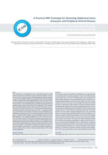
A Practical MRI Technique for Detecting AAA and PAHIntroductionAbdominal Aorta Aneurism (AAA) and Peripheral Artery Disease(PAD) are widespread diseases especially in elderly patients.Digital subtraction angiography (DSA), is widely accepted asgold standard method for evaluation of abdominal, iliac andperipheral arteries. Magnetic resonance imaging (MRI), is theradiological diagnostic method that has highest soft-tissuecontrast resolution while having no ionizing radiation. DSA isan invasive procedure and sometimes contrast agents havehazardious effects on the kidneys. Three dimensional magneticresonance angiography (3DMRA) development has emergedas a non-invasive technique. MRA has the advantages whilehaving no X-ray exposure and arteries can be studied in threedimensions and excellent soft tissue contrast can be provided.DSA in the front-rear projection, in the assessment of stenosisis sometimes inadequate because of superposition. This cancause treatment planning mistakes. In addition to DSA andMRA, coronal Balanced-Turbo Field Echo (B-TFE) was used as afast and non contrast enhanced sequence. This technique havedifferent names under different brands. B-TFE (Balanced TurboField Echo-Philips), FIESTA (Fast Imaging Employing SteadyState Acquisition-GE), True FISP (True Fast Imaging with SteadyState Precession-Siemens). In SSFP, RF signals are applied tothe body in very short time intervals that has a great SNR independent from the blood stream and excellent blood-tissue contrast (T2 * / T1) was obtained. With a shorter examination timeSSFP has allowed us to examine patient without breath holding.Today, this sequence is being used in cardiac examinations(1,2).Material and MethodThe diagnosis of peripheral vascular disease was investigatedwith MRI (1.5T, Philips Medical Systems, Netherlands) as coronal B-TFE and three dimensional contrast enhanced MRA priorto digital subtraction angiography (DSA) (Allura, Philips MedicalSystems, Netherland).We studied the differences between examinations without gender and age discrimination, and correlations between differentsequences, were evaluated statistically using SPSS program.This study was approved by the institutional review board ofour university hospital. Written informed consent was obtainedfrom all patients.Imaging parametersMagnetic resonance imagingMRI was performed using sense body coil and the localizationsequences were taken from aorta, iliac and femoral arteries inthree plans.Getting started with examination first coronal B-TFE sequenceof 40 sections with a total of 24 seconds, matrix 192x256, flipangle 80 º, FOV 430 mm, slice thickness 4mm, gap 2 mm, TR3.5 ms and TE 1.7 ms which is a Steady State Free Precession(SSFP) technique.Before MRA sequences has been taken 2D TIMING (TR 13.7 ms,TE 2 ms, 128x128 Matrix, 450 FOV, 10 mm slice thickness) sequence were performed by 2ml of contrast agent and inspectingthe arrival time at the level of pre-aortic bifurcation was detected. Contrast agent dose and injection rate; was calculatedaccording to the duration of 2D TIMING sequence and patient’s Journalof Clinicaland AnalyticalMedicine2296 Journalof Clinicaland AnalyticalMedicineweight. 20 ml of 0.9% saline was injected after the contrastmaterial in all examinations.MRA sequences which has matrix 512x512, slice thickness 3mm, flip angle 40 º, TR 5.1 ms, TE of 1.5 ms was launched withthe raw data and reconstruction time lasted about 60 seconds.3DMRA images were obtained in the 9-20 plan with the help ofmultiplanar intensity projection (MIP) software.Digital Subtraction AngiographyThe dynamic peripheral vascular examination protocol was usedfor each patient.The active ingredient N, N’-bis (2,3-dihidroksipropil) -5 [(hidroksiasetil) methylamino] -2,4,6-triiodo-1 ,3-benzendicarboxamide (Iomeprol) was used as contrast agent.5F intraducer sheet is inserted in to right or left common femoral artery with the use of Seldinger technique. Pigtail catheterwas advanced in the vessel lumen to the level to be viewedthrough the suprarenal aorta.DSA device was connected to and synchronized working automatic injector (MEDRAD Inc, Warrendale, PA) and injection ofcontrast medium through the pigtail catheter at the level ofrenal arteries was performed. By the way renal arteries, distalsegment of abdominal aorta, aortic and iliac bifurcations, iliacand femoral arteries were examined.Image assessmentPhilips DICOM Viewer R2.2 was used to ases the DICOM imagesof B-TFE, MRA, 3DMRA sequences and DSA. The arterial treewas divided into 16 segments as the right and left renal arteries, infrarenal aorta, aortic bifurcation, iliac bifurcation, rightand left main iliac artery, right and left external and internaliliac artery, right and left common superficial and deep femoralartery. Total of 704 diameter measurements were made in 11patients and 44 sequences.Statistical AnalysisData analysis was made using SPSS 11.5 (Chicago, IL) package program. Our goal is to evaluate the similarity betweensequences. The intraclass corelation coefficient was used tocompare the values obtained in measurements and reliabilityused to check if the techniques could be used for each other ,the t-test was used to measure the differences between them.Results11 out of 13 patients agreed to participate in the study. Patients were between 48-69 years old and the average was 57years. 1 (9%) patient was female, 10 (91%) patients were male.Vessel diameters from each diagnostic method compared withB-TFE sequence; intra-class correlation coefficient has been calculated statisticaly (Table 1). We found any corelation in unitmeasurements of vessel diameters between, B-TFE (Figure 1a),MRA (Figure 1b) 3DMRA (Figure 1c) and DSA(Figure 1d). Thiscompare values presented at Table 2.We found that in most large vessel diameter mesurements, BTFE has a perfect harmony with the MRA, 3DMRA and DSA. Butin small vessels, B-TFE has a moderate correlation with MRAand 3DMRA and low correlation with DSA diameter measurements.
A Practical MRI Technique for Detecting AAA and PAHA Practical MRI Technique for Detecting AAA and PAHTable 1. Mesurements of the diameter of the arterial segments in BTFE, MRA, 3DMRA sequences and DSAB-TFEMRA3DMRADSAHasta NoSeg. NoArterial segmentsA1B1C1A2B2C2A3B3C3A4B4C411INFRARENAL AORTA DIAMETER9.812.9723.812.382412.788.312.932R RENAL ARTERY .683L RENAL ARTERY 54BIFURCATION 962.835R COMMON ILIAC R EXTERNAL ILIAC A.4.20.431.278.20.340.828.70.360.9839.10.441.37R INTERNAL ILIAC A.3.30.3418.90.370.898.20.340.9235.80.411.198L COMMON ILIAC L EXTERNAL ILIAC A.4.10.421.2410.60.451.069.40.391.06380.431.2610L INTERNAL ILIAC A.30.310.919.20.390.929.40.391.0637.70.431.2511R COMMON FEMORAL 2R SUPERFICIAL R DEEP FEMORAL A.2.30.230.760.250.64.60.190.5226.60.30.8814L COMMON FEMORAL 5L SUPERFICIAL 6L DEEP FEMORAL A.3.30.341100.4218.90.37130.10.341ACBDFigure 1. Transvers diameter measurement 1 cm above aortic bifurcation fromcoronal section. B-TFE sequence(A), MRA sequence(B), 3DMRA sequence(C), DSAimage(D).Table 2. Comparison of sequencesİnterclass Corelation Coefficient (ICC)A B-TFE & A MRA0,3946B B-TFE & B MRA0,8861C B-TFE & C MRA0,7517A B-TFE & A 3DMRA0,4769B B-TFE & B 3DMRA0,8776C B-TFE & C 3DMRA0,7222A B-TFE & A DSA-0,6796B B-TFE & B DSA0,8618C B-TFE & C DSA0,6808Another means B-TFE could be as reliable as MRA, and DSA inlarge vessels like aorta, iliac, femoral arteries. But this is notthe case for small arteries like renal arteries.Also difference between measurements of B-TFE and DSA was3 Journal of Clinical and Analytical Medicineexamined by t-test’ ‘on dependent groups and no significant difference was found(p 0.05). Details summurized on Table 3.DiscussionToday, AAA rupture and PAD increase the morbidity and mortality in many healthcare facilities among the world. Approachto this issue, as in every disease is to provide, early screeningmethods before the illness has emerged and is to use minimallyinvasive and easy methods for diagnosis (3). In addition to conventional surgical procedures endovascular minimally invasiveattempts are also considered for treatment (4).When evaluating vascular disease, there are some basic rulesin the selection of methods. Because there are many methodsand many applications in the diagnosis of a disease. In thisapproach some criteria as utility, harmlessness, the economyare considered. The most important point in selection of themethod is that has the most accurate results. Diagnostic activity is the basic criteria in determining the value of a radiologicaldiagnostic method. It is required to compare with a standard, toevaluate the value of any diagnostic examination. This standardreference method based analysis is called “gold standard”.Benefit of non-invasive test is that it can be used as a screeningtest for arterial disease, and the treatment can be performed ina short time before the progress of pathology. Another benefitis the power of saving time and money in the intensive clinicalwork (5).Today MRA, pushing the limits and approaching to DSA which isconsidered as the gold standard.In the diagn
Philips DICOM Viewer R2.2 was used to ases the DICOM images of B-TFE, MRA, 3DMRA sequences and DSA. The arterial tree was divided into 16 segments as the right and left renal arter-ies, infrarenal aorta, aortic bifurcation, iliac bifurcation, right and left main iliac artery, right and left external and internal iliac artery, right and left common superficial and deep femoral artery. Total of .Author: Aykut Recep AktasPublish Year: 2016