Transcription
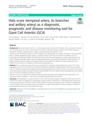
Sebastian et al. BMC Rheumatology(2020) Y PROTOCOLBMC RheumatologyOpen AccessHalo score (temporal artery, its branchesand axillary artery) as a diagnostic,prognostic and disease monitoring tool forGiant Cell Arteritis (GCA)Alwin Sebastian1,2, Kornelis S. M. van der Geest3, Fiona Coath4, Prisca Gondo5, Abdul Kayani1, Craig Mackerness5,Bernard Hadebe5, Sue Innes6, Jo Jackson6 and Bhaskar Dasgupta1,2*AbstractBackground: Giant cell arteritis (GCA) is a common large vessel vasculitis of the elderly, often associated with sightloss. Glucocorticoids (GC remain the mainstay of treatment, although biologic treatments have been approved.Biomarkers predicting disease severity, relapse rates and damage are lacking in GCA.EULAR recommends ultrasound (US) as the first investigation for suspected GCA. The cardinal US finding, a non-compressiblehalo, is currently categorised as either negative or positive. However, the extent and severity of this finding may vary.In this study, we hypothesise whether the extent and severity of the halo sign [calculated as a single composite Halo score(HS)] of temporal and axillary arteries may be of diagnostic, prognostic and monitoring importance; whether baseline HS islinked to disease outcomes, relapses and damage; whether HS can stratify GCA patients for individual treatment needs;whether HS can function as an objective monitoring tool during follow up.Methods: This is a prospective, observational study. Suspected GCA Participants will be selected from the GCA FTC at theparticipating centres in the UK. Informed consent will be obtained, and patients managed as part of standard care. Patientswith GCA will have HS (temporal and axillary arteries) measured at baseline and months 1,3,6 and 12 long with routine clinicalassessments, blood sampling and patient-reported outcomes (EQ5D). Non-GCA patients will be discharged back to the referralteam and will have a telephone interview in 6 months.We aim to recruit 272 suspected GCA referrals which should yield 68 patients (25% of referrals) with confirmed GCA. Therecruitment will be completed in 1 year with an estimated total study period of 24 months.Discussion: The identification of prognostic factors in GCA is both timely and needed. A prognostic marker, such as the HS,could help to stratify GCA patients for an appropriate treatment regimen. Tocilizumab, an IL-6R blocking agent, switches offthe acute phase response (C-Reactive Protein), making it difficult to measure the disease activity. Therefore, an independentHS, and changes in that score during treatment and follow-up, maybe a more objective measure of response compare topatient-reported symptoms and clinical assessment alone.Keywords: Outcomes in GCA, Risk stratification, Prognostic factors, Halo score, GCA probability score, Clinical severity index,Glucocorticoid toxicity* Correspondence: Bhaskar.dasgupta@southend.nhs.uk1Rheumatology, Mid and South Essex University Hospital Groups, SouthendUniversity Hospital, Westcliff-On-Sea, Essex, UK2University of Essex, Colchester, UKFull list of author information is available at the end of the article The Author(s). 2020 Open Access This article is licensed under a Creative Commons Attribution 4.0 International License,which permits use, sharing, adaptation, distribution and reproduction in any medium or format, as long as you giveappropriate credit to the original author(s) and the source, provide a link to the Creative Commons licence, and indicate ifchanges were made. The images or other third party material in this article are included in the article's Creative Commonslicence, unless indicated otherwise in a credit line to the material. If material is not included in the article's Creative Commonslicence and your intended use is not permitted by statutory regulation or exceeds the permitted use, you will need to obtainpermission directly from the copyright holder. To view a copy of this licence, visit http://creativecommons.org/licenses/by/4.0/.The Creative Commons Public Domain Dedication waiver ) applies to thedata made available in this article, unless otherwise stated in a credit line to the data.
Sebastian et al. BMC Rheumatology(2020) 4:35BackgroundGiant cell arteritis (GCA) is a common form of systemicvasculitis characterised by granulomatous inflammation oflarge and medium-sized arteries [1]. GCA predominantlyaffects Caucasian, older people ( 50 years), with a peakincidence among those 70–80 years old [1, 2]. The incidence of GCA rises with increasing age, ranging from 2.6per 100,000 in patients aged 50–59 to 44.6 per 100,000 inpatients over the age of 80 [3]. GCA predominantly involves branches of the external carotid arteries such as thetemporal arteries and the aorta and its large branches,including the subclavian and axillary arteries. Commonpresenting symptoms include new headache, scalp tenderness, jaw claudication, diplopia and amaurosis fugax [1, 2,4]. GCA can cause significant morbidity and ischaemiccomplications, including irreversible sight loss. Othercomplications include aortitis, myocardial infarction andstroke. The 1990 American College of Rheumatology(ACR) classification criteria were not intended for diagnosis [5] and may not be accurate, particularly for cases withophthalmic involvement [6]. The criteria have low specificity and predictive values [2, 7, 8]. Screening tests are vitalas the GCA symptoms can be often non-specific and missing the diagnosis can be devastating [9].Glucocorticoids (GC) have remained the cornerstoneof treatment for GCA [10], although cohort studies showonly 15–20% sustained remission with glucocorticoidsalone Glucocorticoid-sparing treatments in GCA arealso needed due to the harmful effects of long-termglucocorticoid use. This includes hypertension, hyperglycaemia, osteoporosis, cushingoid changes, mood disturbance and electrolyte imbalance, but this is not anexhaustive list [11, 12]. It is recommended to start GCimmediately in strongly suspected GCA pending investigations [13]. Targeted treatments have recentlybeen introduced, but heterogeneity in disease outcomes has still been observed. In GiACTA, the landmark trial of Tocilizumab in GCA that provides theevidence base for its current use, 42% of participantsrandomised to weekly Tocilizumab still did notachieve sustained remission [14]. Currently, validatedbiomarkers predicting disease severity, relapse ratesand damage are lacking in GCA.A positive temporal artery biopsy (TAB) has been thegold standard for histological diagnosis of GCA [15–17].However, a biopsy is invasive, and it lacks sensitivity.This is particularly true in extra-cranial involvement,termed large-vessel GCA (LV-GCA), where access tosample material has obvious practical constraints and isusually identified incidentally following cardiovascularsurgery [18]. Non-invasive imaging techniques, includingultrasound (US), Magnetic resonant image (MRI) andposition emission tomography (PET-CT) are increasinglybeing used to identify these patients [19–21].Page 2 of 10Ample evidence now indicates that US of temporal arteries can promptly diagnose cranial forms of GCA, aswell as screening for LV-GCA at the axillary arteries[22]. US is a safe, non-invasive and higher sensitivity,particularly in extra-cranial disease. It is a relativelyquick procedure [23], often delivered as a point of caretest, well tolerated by patients and is suitable for followup examinations. Timely diagnosis of GCA by ultrasound in GCA fast track clinics has resulted in a significant reduction in permanent visual loss [24–26].The EULAR recommendations for imaging in LargeVessel Vasculitis recommend US of temporal and/or axillary arteries as the first imaging modality, where thereis adequate expertise and equipment, particularly in patients with suspected predominantly cranial GCA [27].Estimation of GCA probability has become importantgiven recent EULAR recommendations suggesting different diagnostic strategies in patients with low, intermediate or high GCA probability. In patients where there is ahigh clinical suspicion of GCA and an initial positive imaging test e.g. US, the diagnosis of GCA may be madewithout additional investigations (e.g. biopsy or furtherimaging). In patients with a low clinical probability and anegative imaging result, the diagnosis of GCA can be considered unlikely, and the patient reassured [18]. There isalso a report from Southend suggesting that the ‘pre-testGCA Probability Score’ may be a useful tool for rating thepre-test probability of GCA, stratifying patients into ‘low’or ‘not-low’ probability groups [28]. This score may alsoreflect clinical severity and extent of disease.The main finding on US in GCA patients is the halo sign:non-compressible hypoechoic wall swelling [29, 30]. Severalstudies have been conducted to investigate the accuracy,construct and criterion validity of US in the diagnosis ofGCA [31–34]. The latest meta-analysis of prospective studies has shown a pooled sensitivity of 77% and a pooled specificity of 96% for temporal artery US when compared tothe final clinical diagnosis of GCA [35]. US allows measurement of the arterial intimal media complex (IMC). Studiesshow that at the age of 70 years the temporal artery has anormal IMC diameter of about 0.2 mm, whilst inflamedtemporal arteries have a diameter of 0.5–0.9 mm [27, 36].Axillary arteries of patients aged about 70 have a normalIMC diameter of 0.6 mm, whilst patients with extra-cranialGCA have an average diameter of 1.6–1.7 mm [36, 37]. Acut off value was determined at 1.0 mm [36]. Currently, thetemporal artery US of GCA patients are categorised as either negative or positive. However, variation in extent andseverity of these findings on temporal and axillary arteryUS in GCA is observed [38]. We have recently developedan ultrasonographic halo score that correlates with arterialinflammation in GCA [39]. In the current study, will furtherinvestigate the novel halo score as a diagnostic, prognosticand disease monitoring tool for GCA.
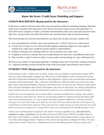
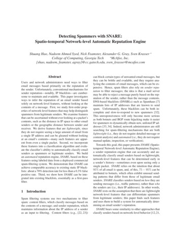
Sebastian et al. BMC Rheumatology(2020) 4:35Page 3 of 10Fig. 1 Diagram demonstrating the six temporal artery segments for calculating the temporal artery halo scoreWe will systematically measure the extent and severityof the halo sign. Bilateral US assessment of the commontemporal artery, the parietal branch, the frontal branchand axillary arteries will be performed (Figs. 1 and 2).The halo sign at each branch of the common temporal,parietal and frontal arteries will be scored 0–4 points,giving a maximum possible halo score (HS) score of 24(Table 1). At the axillary arteries, the IMT will be scored0–4 points on each side, allowing a maximum total score8, which will be multiplied by 3 (Fig. 2). A total haloscore (THS) will be constructed by adding the scores ofthe temporal artery branches with the axillary artery score.Subsequently, the HS and THS will be assessed for anycorrelation to disease outcomes in GCA, as characterisedFig. 2 Diagram demonstrating the six temporal artery segments and two axillary arteries for calculating the total artery halo score
Sebastian et al. BMC Rheumatology(2020) 4:35Page 4 of 10Table 1 Halo Score GradingStudy designThis is a pragmatic, prospective, observational study.This study will involve two specific phasesHaloGradingCommonsuperficial TAhalo thickness(mm)Parietal TAhalo thickness(mm)Frontal TAhalo thickness(mm)Axillaryartery halothickness(mm)Grade 00.3 or less0.2 or less0.1 or less0.5 or lessGrade 10.40.30.20.6Grade 20.50.40.30.7–0.8Grade 30.6–0.70.5*0.40.9–1.5Initial presentation and diagnosisGrade 40.8 or more0.6 or more0.5 or more1.6 or moreThis phase will involve recruiting patients from theGCA FTP at participating sites. Patients recruited willbe subject to inclusion/exclusion criteria detailedbelow. Their General Practitioner, Emergency Department or other specialities refer patients to the FTP.Patients will be invited to participate in this study bythe Rheumatology Research team, who will providethem with information about the study. Patients willbe informed of the phases of participation, the voluntary nature of the study, and their right to withdrawat any stage. Written consent to participate will beobtained by the researcher prior to the commencement of the screening assessments.In this phase following will be assessed: (Additional file 1)by responsiveness to therapy - remitting, relapsing or refractory disease. Other outcome measures that may bereviewed include the development of large vessel diseaseand vascular damage (as assessed by cross-sectional scanning such as PET-CT), accumulation of glucocorticoid related adverse events and need for additional conventional(e.g. leflunomide, methotrexate) or biologic (Tocilizumab)DMARDs. Remitting disease in GCA is defined as a diseaseunder sustained satisfactory control with minimum oneflare during standard GC taper. The relapsing disease iswhere the condition initially comes under control but thenflares on GC tapering. Refractory GCA patients are thosewho do not respond to GC at all.The identification of prognostic factors in GCA is bothtimely and needed. The GiACTA trial has shown thatIL-6R blocking therapy may help to sustain glucocorticoid free remission [14]. In addition, the GiACTA trialhas shown that a subset of GCA patients can be quicklywithdrawn from glucocorticoid therapy without the development of relapses. A prognostic marker, as outlinedabove, could help to stratify GCA patients to an appropriate treatment regime. IL-6R blockade switches off the inflammatory marker response, making it difficult to usetraditional biomarkers such as CRP to measure disease activity. Therefore, an independent HS, and changes in thatscore during treatment and follow-up, maybe a more objective measure of response, rather than relying only onpatient-reported symptoms and clinical assessment.Methods/designStudy aims and hypothesisTo determine whether the severity of vessel wall oedema(halo/IMT) in the common temporal artery, its branchesand the axillary arteries, as measured by a compositeultrasound score (THS), is of prognostic value in predicting severity and outcomes in GCA.To determine the prognostic and monitoring value ofthe HS and THS in GCA, with regards to predicting outcomes (remission, refractory or relapsing disease) inGCA. We will also determine the diagnostic value of theHS and THS for discriminating GCA from non- GCA.1) Initial presentation and diagnosis of GCA or non-GCA.2) Follow-up over 12 months for GCA patients and 6months for non-GCA patients. Clinical historyClinical examinationRoutine bloods including biomarkersPatient-reported outcome (EQ5D)Starting dose of GCUS scan of the temporal artery including itsbranches (frontal and parietal) and the axillary arterybilaterally Probability score (Additional file 3) GCA Probabilityscore will be calculated on all the patients referredto the FTP to clinically stratify their risk of havingGCA and as a measure of severity of the diseaseA diagnosis of GCA will be based on revised classificationcriteria as proposed recently (Dejaco et al. Rheumatology2016) in the modified GiACTA criteria detailed below. Theaccuracy of the diagnosis will be evaluated after 6 months.Patients were classified as having GCA if all of thefollowing criteria were met Age 50 years with ESR 30 mm/hr. or CRP 10 mg/L Unequivocal cranial symptoms of GCA (i.e. newonset localised headache, scalp or temporal arterytenderness, ischemia-related vision loss, or otherwiseunexplained mouth or jaw pain upon mastication)or symptoms of polymyalgia rheumatica (PMR), defined as shoulder and/or hip girdle pain associatedwith inflammatory morning stiffnessa. Cranial symptoms defined as new localised headpain, generalised scalp tenderness, tender temporal
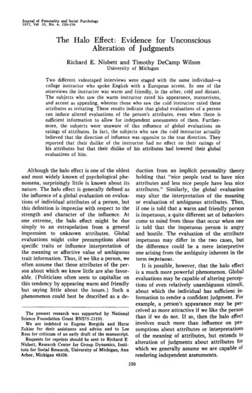
Sebastian et al. BMC Rheumatology(2020) 4:35artery, AION or PION, jaw claudication or tongueclaudication in the current studyb. PMR symptoms defined as morning stiffness 1 hwith bilateral shoulder pain and/or bilateral hippain or stiffness in the current study Temporal artery biopsy revealing features of GCA orevidence of GCA by imaging (i.e. ultrasound orcross-sectional imaging such as CTA or PET-CT)Page 5 of 10In this phase, following will be assessed: Clinical historyClinical examinationRoutine bloods including biomarkersUS scan of the temporal artery including its branches(frontal and parietal) and the axillary artery bilaterally Patient-reported outcomes (EQ5D) Cumulative GC requirementFollow-up period: (Additional file 1)Participants who are diagnosed with GCA will be seen forfollow-up visits at 1, 3, 6 and 12 months. Participants witha non-GCA diagnosis will be seen or through a telephoneinterview one further time at 6 months to confirm thenon-GCA diagnosis. At any time, point throughout thefollow-up period patients may require unscheduled visits ifthey have symptoms of relapse. Patients will be educated atbaseline as to the symptoms that might be expected withrelapse and guided to contact their clinician or Rheumatology Research team (if different) immediately (Fig. 3).For study purposes, relapse means those patientswhose GCA symptoms flare or return in response tocurrent standard treatment, that is a tapering regimen ofglucocorticoids. Refractory GCA patients are those whodo not respond from the outset.Fig. 3 Study Flow chartDEFINITION OF RELAPSE AND REMISSION1. Remission is defined as absence of clinical signs and symptoms ofGCA and normalization of ESR [ 30 mm/hr] and CRP [ 10 mg/L]2. Relapse is defined as recurrence of symptoms attributable toactive GCA, with or without ESR 30 mm/hr. and CRP 10 mg/L3. The refractory non-remitting disease subjects are those whohave had no remission within 6 weeks of initiation of high doseglucocorticoid treatment.Eligibility criteriaPatients with clinical suspicion of GCA referred to theFTC would be eligible for the study subjects to theinclusion and exclusion criteria below.
Sebastian et al. BMC Rheumatology(2020) 4:35Inclusion criteriaThe clinician responsible for the patient’s care will makethe diagnosis of GCA as part of the standard of careusing the modified GiACTA criteria. Age 50 years with ESR 30 mm/hr. or CRP 10 mg/L Unequivocal cranial symptoms of GCA (i.e. new-onset localised headache, scalp or temporal arterytenderness, ischemia-related vision loss, or otherwiseunexplained mouth or jaw pain upon mastication)or symptoms of polymyalgia rheumatica (PMR), defined as shoulder and/or hip girdle pain associatedwith inflammatory morning stiffnessa. Cranial symptoms defined as new localised headpain, generalised scalp tenderness, tendertemporal artery, AION or PION, jaw claudicationor tongue claudication in current studyb. PMR symptoms defined as morning stiffness 1h with bilateral shoulder pain and/or bilateralhip pain or stiffness in the current study Temporal artery biopsy revealing features of GCAor evidence of GCA by imaging (i.e. ultrasound orcross-sectional imaging such as CTA or PET-CT) Participants must have the capacity and willingnessto give informed written consentExclusion criteria Participants must not have a previous diagnosis ofGCA Participants must not have had a previous temporalartery biopsy i.e. as part of diagnostics for previouslysuspected GCA Participants must not be under 18 years Participants must not be on treatment with a highdose of steroids ( 7.5 mg) more than 2 weeks priorto the first review in the FTP Inability to give informed consentSamplingThe nature of this disease is rare; thus, the number ofparticipants will be collected up to 12 months and willbe followed up to 12 months as per the study protocol.Patients will be recruited after referral to theparticipating site FTP.Participants The cohort of patients for this study will berecruited from the fast track GCA clinics (FTC), which iscurrently the sta
and disease monitoring tool for GCA. Sebastian et al. BMC Rheumatology (2020) 4:35 Page 2 of 10. We will systematically measure the extent and severity of the halo sign. Bilateral US assessment of the common temporal artery, the parietal branch, the frontal branch and