Transcription
StructureArticleMechanism of Dengue VirusBroad Cross-Neutralizationby a Monoclonal AntibodyJoseph J.B. Cockburn,1,4,8 M. Erika Navarro Sanchez,1,4 Nickolas Fretes,1,4,9 Agathe Urvoas,2,5,10 Isabelle Staropoli,3,6Carlos M. Kikuti,1,4,11 Lark L. Coffey,4,7,12 Fernando Arenzana Seisdedos,3,6 Hugues Bedouelle,2,5,* and Felix A. Rey1,4,*1Unitéde Virologie Structurale, Département de Virologiede Recherche Prévention et Thérapie Moléculaires des Maladies Humaines, Département d’Infection et Epidémiologie3Unité de Pathogénie Virale, Département de VirologieInstitut Pasteur, 75724 Paris Cedex 15, France4CNRS, URA30155CNRS, URA30126INSERM, U81975724 Paris Cedex 15, France7Département de Virologie, Groupe à 5 ans Populations Virales et Pathogenèse, 75724 Paris Cedex 15, France8Present address: London Research Institute, Cancer Research UK, London WC2A 3LY, UK9Present address: University of Michigan Medical School, Ann Arbor, MI 48109, USA10Present address: Université Paris 11, Institut de Biochimie Biophysique Moléculaire et Cellulaire IBBMC, CNRS UMR 8619,91405 Orsay, France11Present address: Institut Curie, CNRS UMR144, 75005 Paris, France12Present address: Blood Systems Research Institute, University of California, San Francisco, San Francisco, CA 94118, USA*Correspondence: hugues.bedouelle@pasteur.fr (H.B.), felix.rey@pasteur.fr (F.A.R.)DOI 10.1016/j.str.2012.01.0012UnitéSUMMARYThe dengue virus (DENV) complex is composed offour distinct but serologically related flaviviruses,which together cause the present-day most important emerging viral disease. Although DENV infectioninduces lifelong immunity against viruses of thesame serotype, the antibodies raised appear tocontribute to severe disease in cases of heterotypicinfections. Understanding the mechanisms of DENVneutralization by antibodies is, therefore, crucial forthe design of vaccines that simultaneously protectagainst all four viruses. Here, we report a comparative, high-resolution crystallographic analysis of an‘‘A-strand’’ murine monoclonal antibody, Mab 4E11,in complex with its target domain of the envelopeprotein from the four DENVs. Mab 4E11 is capableof neutralizing all four serotypes, and our studyreveals the determinants of this cross-reactivity.The structures also highlight the mechanism bywhich A-strand Mabs disrupt the architecture of themature virion, inducing premature fusion loop exposure and concomitant particle inactivation.INTRODUCTIONThe four serotypes of dengue virus (DENV) constitute the largestvector-borne viral disease burden in the developing world(Monath, 1994), with 50–100 million cases reported yearly. Thereis neither approved vaccine nor specific therapy to fight theseinfections. Dengue fever (DF), the most common form of denguedisease, is an acute febrile illness and is generally nonlethal.However, about 1% of DF cases evolve into severe forms,such as life-threatening dengue hemorrhagic fever (DHF) ordengue shock syndrome (DSS) (Mackenzie et al., 2004). Infection by DENV raises lifelong immunity against the infecting serotype (Sabin, 1952), but only transient protection against the otherserotypes. In the long term, subsequent infections by virusesfrom a different DENV serotype are associated with a greaterrisk for DHF/DSS (Sangkawibha et al., 1984). A likely explanationis that weakly neutralizing, serotype cross-reactive antibodiespresent in such patients can mediate viral uptake by—andenhanced infection of—cells bearing Fcg receptors (Halsteadand O’Rourke, 1977), a phenomenon termed antibody-dependent enhancement (ADE) of infection.DENV is a member of the flavivirus genus of the Flaviviridaefamily, which includes several other important human pathogenssuch as Japanese encephalitis (JE), West Nile (WN), yellow fever(YF), and tick-borne encephalitis (TBE) viruses (Lindenbachet al., 2007). The virus particles are enveloped by a lipid bilayerthat is enclosed within an icosahedral scaffold formed by theenvelope glycoprotein E (Kuhn et al., 2002) (Figure 1). The virionsenter cells by receptor-mediated endocytosis followed by lowpH-induced fusion of the viral and endosomal membranes (vander Schaar et al., 2008). The E glycoprotein is both responsiblefor receptor binding and for inducing membrane fusion in theendosome, and is thus the main player in the flavivirus entryprocess. Flavivirus E is about 500 amino acids long and isanchored in the viral membrane by two C-terminal transmembrane (TM) helices. A soluble fragment of E, containing roughlythe 400 N-terminal amino acids of E, has been crystallizedStructure 20, 303–314, February 8, 2012 ª2012 Elsevier Ltd All rights reserved 303
StructureDeterminants of Dengue Virus Cross-NeutralizationFigure 1. Overview of the DENV Envelope Protein and Its Complexes with scFv 4E11(A) Crystal structure of the prefusion DENV-2 sE dimer (PDB accession number 1OKE). One E subunit is highlighted colored by domains (DI, red; DII, yellow; DIII,blue). The fusion loop (residues 100–108) is colored green. The arrows indicate the view zoomed in (B).(B) Close-up showing DIII from one subunit in the sE dimer and its orientation with respect to the viral membrane in the mature virion. Note the proximity of thefusion loop of the partner subunit (in the forefront, yellow and green).(C) The icosahedral glycoprotein shell of the mature DENV-2 virion formed by 90 E dimers, docked into a 9.5 Å resolution cryo-EM reconstruction (PDB accessionnumber 1THD). Each E subunit is colored according to domains, as in (A). View down an icosahedral 3-fold axis (central gray triangle). Icosahedral 5- and 2-foldaxes of the particle are marked with pentagons and ovals, respectively.(D) Amino acid sequence alignment of DIII from the four DENV strains used in this study (listed in Table 1). Residue numbers correspond to the full-length DENV-1E protein (the numbering for DENV-3 DIII is shifted by two residues). Strictly and semiconserved residues are highlighted in red and yellow, respectively. b Strandsare labeled above the sequence with blue arrows. Epitope residues are marked with asterisks.(E) Overview of the structure of DENV-1 DIII (dark blue) in complex with scFv 4E11. The heavy-/light-chain framework regions are dark/light gray, respectively,with the CDR loops color coded and labeled H1-H3 and L1-L3.(F and G) Superposition of the scFvs (F) and DIIIs (G) from the four complexes. The scFv 4E11 molecules were superposed using the framework region residues.Residues at the antibody-antigen interface are labeled according to the Kabat scheme (Kabat et al., 1991) and shown in ball and stick (oxygen, red; nitrogen, dark304 Structure 20, 303–314, February 8, 2012 ª2012 Elsevier Ltd All rights reserved
StructureDeterminants of Dengue Virus Cross-Neutralization(Modis et al., 2003, 2005; Rey et al., 1995; Zhang et al., 2004). Itsstructure features three distinct domains (Figure 1A). DI, whichcontains the N terminus, is at the center, with DII and DIII at eitherside. DII displays a hydrophobic fusion loop—the amino acidsequence of which is conserved across flaviviruses—at itsdistal-most tip. The fusion loop inserts into the endosomalmembrane during an acid-pH-driven membrane-fusogenicconformational change. This process is necessary to releasethe viral nucleic acid within the cytoplasm to initiate infection.DIII, the C-terminal portion of the ectodomain, is an eightstranded b sandwich (strands labeled A–G, Figure 1B) with animmunoglobulin superfamily fold. DIII is thought to be involvedin receptor binding (Crill and Roehrig, 2001), although the natureof the relevant receptor(s) at the surface of susceptible cells isnot known despite intensive studies (Lindenbach et al., 2007).Infectious dengue virions derive from proteolytic activationof noninfectious immature particles, the surface of which iscomposed of heterodimers of the viral glycoproteins prM and E(Lindenbach et al., 2007). Cleavage by furin or other proteaseseliminates the N-terminal half of prM (the ‘‘pr’’ domain’’). Thismaturation step is often incomplete, resulting in a significantfraction of only partially mature virions released from infectedcells (Junjhon et al., 2010). These virons are infectious andhave been shown to display both mature- and immature-likepatches on the same virion (Plevka et al., 2011), displaying antigenic characteristics of both types (Nelson et al., 2008).The human humoral immune response to DENV is dominatedby cross-reactive antibodies, many of which are non-neutralizingand target prM (Crill et al., 2009; Dejnirattisai et al., 2010; Laiet al., 2008; Oliphant et al., 2007). The resulting antigenic heterogeneity of DENV appears to increase the potential for ADE(Cherrier et al., 2009). Identifying the epitopes on the E proteinthat are targeted by potent, broadly neutralizing antibodies,ideally neutralizing simultaneously all four dengue serotypes, istherefore important for vaccine design.A recent study on memory B cells recovered from DENVinfected individuals revealed the presence of clones encodingMabs binding to DIII, which potently neutralize multiple serotypes (Beltramello et al., 2010). Cross-neutralizing Mabs targeting DIII are also a well-characterized component of the murineimmune response to DENV. Biochemical studies showed theirepitopes to be clustered on the DIII A strand (Gromowski et al.,2008, 2010; Lisova et al., 2007; Matsui et al., 2009; Shresthaet al., 2010; Sukupolvi-Petty et al., 2007; Thullier et al., 2001).These ‘‘A-strand’’ Mabs are believed to neutralize mainly byinterfering with attachment to host cells (Crill and Roehrig,2001; Thullier et al., 2001). The crystal structure of the Fab fragment of an A-strand antibody, Mab 1A1D-2, has been determined in complex with recombinant DIII to 3 Å resolution, andthe structure was docked into a low-resolution cryo-EM reconstruction of the DENV-2 particle in complex with the same Fab(Lok et al., 2008). This study indicated that binding of 1A1D-2disrupts the organization of the particle. Binding of Fab 1A1D-2to the mature DENV-2 virion was shown to be temperaturesensitive, reflecting the partially occluded nature of the epitope,and its transient exposure through a ‘‘breathing’’ motion of theparticle.Mab 1A1D-2 was raised against DENV-2 strain NGC andneutralizes DENV-2, DENV-1, and DENV-3 (in order ofdecreasing efficacy) but does not neutralize DENV-4 (Roehriget al., 1998). To our knowledge, the determinants of its crossreactivity have, however, not been investigated. We approachhere this issue by analyzing the murine Mab 4E11 (Megretet al., 1992), which also binds to the DIII A strand (Lisova et al.,2007) and neutralizes all four DENV serotypes (Thullier et al.,1999). We report the crystal structures of a recombinantsingle-chain variable fragment (scFv) of 4E11 in complex withDIII from all four serotypes to high resolution (1.6–2.1 Å). Ourstudy reveals the basis for serotype-dependent variations inbinding affinity and neutralization efficacy, and provides insightsinto the immunodominance of the DIII A strand in mice. In particular, our study, together with the studies on Mab 1A1D-2mentioned above, supports a neutralization mechanism forA-strand antibodies that explains how 4E11 can neutralize allfour serotypes with IC50 varying between 1 and 100 nM, despiteabout more than five orders of magnitude difference in Fab/DIIIbinding affinities across serotypes.RESULTS AND DISCUSSIONCharacterization of Mab 4E11Competition ELISA experiments were carried out to determinethe affinity, at equilibrium and in solution, between recombinantFab 4E11 and isolated DIII from each of the clinical isolates ofDENV listed in Table 1, spanning all four serotypes. The resultsshow that Fab 4E11 binds with nanomolar affinity to DIII fromDENV-1, DENV-2, and DENV-3 (in this order), and with onlymicromolar affinity to DENV-4 DIII. The ratio of affinities, in theorder DENV-1:DENV-2, DENV-2:DENV-3, and DENV-3:DENV-4,is 1:5, 1:20, and 1:500, respectively, resulting in a 1:50,000 ratiobetween DENV-1 and DENV-4. Table 1 also reports the Mab4E11 concentration corresponding to 50% neutralization(IC50) of each of the corresponding DENV isolates for infectionof Huh7.5 cells (see Experimental Procedures). The trend inIC50 values of the full-length, bivalent Mab roughly reflects theaffinity trend between the monovalent Fab and isolated DIIIsfrom each of the isolates but varies over a much narrower range(1–100 nM), with IC50 ratios of 1:1, 1:50, and 1:2 among DENV-1:DENV-2, DENV-2:DENV-3, and DENV-3:DENV-4, respectively.As expected, neutralization by Mab 4E11 was weakest withDENV-4, yet it was specific because an isotype-matched Mabcontrol at the same concentration had no effect (data notshown). The small difference in neutralization IC50 betweenDENV-3 and DENV-4 was unexpected, given the 500-fold ratioin affinities between the corresponding recombinant DIII andFab 4E11.blue) with carbon atoms following the main-chain color scheme. The relevant DIII b strands A, B, and G, as well as the D-E loop, are labeled. The AG patch (seetext) is highlighted in yellow. The views correspond approximately to an ‘‘open book’’ view of the complexes (see also Figure S1), with (G) corresponding roughlyto the orientation in (B).See also Table S1.Structure 20, 303–314, February 8, 2012 ª2012 Elsevier Ltd All rights reserved 305
StructureDeterminants of Dengue Virus Cross-NeutralizationTable 1. Binding and Neutralization DataFab:DIII Binding AffinitySerotypeStrainGenBank Accession No.KD (nM)aDG (kcal/mol)DENV-1Guiana/FGA89/1989Q9II010.082 0.00513.76 0.041.1 (0.88–1.3)DENV-2Jamaica/1409/1983P075640.43 0.0412.79 0.050.85 0 1.011.07 0.0754 (26–110)8.0DENV-4Burma/63632/1976Unpublished7.36 0.1100 (65–150)15.04,100 700IC50 (nM)bIC50 (mg/ml)b0.16KD values between the recombinant DIII and 4E11 Fab fragment at equilibrium in solution at 25 C, measured by competition ELISA.bConcentration of Mab 4E11 corresponding to 50% neutralization of infection as measured by flow cytometry on huh7.5 cells. Values in parenthesescorrespond to the 95% confidence range.aStructure DeterminationFor crystallization we used constructs directing periplasmicsecretion in E. coli (see Experimental Procedures) of scFv 4E11and the recombinant DIII from each of the DENV isolates characterized above (Figure 1D). The crystallization and structure determination of all four complexes are described in the ExperimentalProcedures, and the resulting statistics are listed in Table 2. Thecrystals of the DENV-1 DIII/scFv complex diffracted better thanthe others (1.6 Å resolution versus 2.0–2.1 Å, Table 2). We didnot obtain enough DIII from the DENV-3 isolate from the E. Coliconstruct, and therefore, we made cocrystallization trials usingthe dimeric DENV-3 sE ectodomain (residues 1–393, or DI-DIIDIII) produced in Drosophila S2 cells. Unexpectedly, the resultingcrystals contained only the scFv in complex with DENV-3 DIII,lacking the rest of the ectodomain. SDS-PAGE analysis of thematerial used for crystallization confirmed the presence, in addition to the scFv, of a protein at the expected molecular weight forDIII (Figures S1A and S1B available online), indicating that DIIIhad been cleaved from the sE dimer, presumably by a proteasecontaminant. Such cleavage was not observed in samples of sEnot mixed with the scFv preparation, even when left for a longtime at 4 C. Indeed, the DENV-3 sE dimer was stable and ledTable 2. Crystallographic Data Processing and Structure RefinementDENV 1DENV 2DENV 3DENV 4Space groupP3121P41212P21C2221Copies of complex per asymmetric unit1121a, b, c (Å)110.71, 110.71, 58.3260.46, 60.46, 207.4654.06, 74.67, 86.89106.60, 155.88, 55.91a, b, g ( )90, 90, 12090, 90, 9090, 104.49, 9090, 90, 90Crystal FeaturesUnit cell parametersData QualityResolution (Å)a47.95–1.60 (1.69–1.60)45.50–2.10 (2.21–2.10)42.86–2.04 (2.15–2.04)47.19–2.00 (2.11–2.00)Number of observations150,734186,284143,216211,889Number of unique reflections54,20522,89340,60231,933Completeness (%)99.6 (99.5)98.394.8 (76.0)99.8 (98.7)Redundancy2.8 (2.7)8.1 (8.2)3.5 (2.5)6.6 (6.7)I/s (I)13.9 (1.9)23.5 (3.3)10.8 (2.0)18.5 (4.2)Rsym (%)a,b4.5 (50.7)4.6 (56.4)8.4 (44.4)6.8 (44.1)Model QualityRwork/Rfree (%)c17.7/18.820.6/21.421.8/23.418.1/20.8Number of protein atoms2,56126534,8602,505Number of solvent atoms (of which water)354 (304)115 (112)342 (277)326 (277)Average atomic B factor for main chains/side chains and waters (Å2)30.8/38.657.4/64.433.2/41.034.1/43.8Rmsd in bond lengths/angles (Å, ran plotFavored (%)98.196.697.597.5Allowed (%)1.63.12.52.5Disallowed (%)0.30.30.00.0aValues in parentheses correspond to the highest resolution shell.P PP PbRsym 100 3 [ h ijIi(h) I(h) j]/ h iIi(h), where I(h) is the mean of all observations {Ii(h)} of reflection h.PPcR hjFo(h) Fc(h)j/ h Fo(h), where Fo(h) and Fc(h) are the observed and calculated structure factor amplitudes of reflection h.306 Structure 20, 303–314, February 8, 2012 ª2012 Elsevier Ltd All rights reserved
StructureDeterminants of Dengue Virus Cross-NeutralizationFigure 2. The Common Core of the 4E11 Epitopes(A) View down the AG patch, with the molecules colored as in Figure 1E.(B) Same as (A) but rotated by 90 about a vertical axis in the plane of the paper. The boxes in both top panels indicate the regions depicted in more detail in thelower panels, in which the main chain is in a ‘‘worm’’ representation. Shown are the coordinates of the complex for DENV-1. Hydrogen bonds and salt bridges arerepresented as black dashes (see also Figure S2.).to crystals of the intact dimeric ectodomain in the absence ofscFv, the structure of which was determined in parallel (unpublished data). Furthermore, the DENV-3 sE dimer was resistantto limited proteolysis in the absence of the antibody, indicatingthat binding of antibody to DIII destabilizes the dimer, likely byleading to dimer dissociation (see below) and exposinga segment of polypeptide N-terminal to DIII (in the DI-DIII linker),which becomes accessible to contaminating proteases.Overall Structure of the Immune ComplexesThe complexes of scFv 4E11 with DIII from each of the four DENVisolates have, as expected, a very similar overall structure. The4E11 variable domains are oriented with their complementarity-determining regions (CDRs) facing the A and G b strands,which make the edge of the DIII b sandwich. The VL domainlies above the G strand, and the VH domain rides atop the Astrand (Figure 1E). The residues in both paratope and epitopehave very similar conformations in all four complexes (Figures1F and 1G), showing that 4E11 cross-reactivity does not involvea significant induced fit. There are, however, differences in scFvpositioning between the complexes, especially for serotypes 3and 4 when compared to the DENV-1 complex (Figure S1C).The DENV-1, DENV-2, and DENV-4 DIIIs bury 900–950 Å2 ofaccessible surface area on binding the scFv, with surface complementarity coefficients in the range 0.7–0.75. The apparentburied area is smaller in the complex with DENV-3 DIII (780 Å2)because DIII residues 362–364 (DE loop) are disordered (andthus absent from the calculation), whereas these residues aresee contributing to the interface in the other three complexes.The Mab 4E11 variable domains of both the heavy and lightchains share 85% amino acid sequence identity with the counterparts from Mab 1A1D-2. Accordingly, the complexes ofDENV-2 DIII with scFv 4E11 and Fab 1A1D-2 (PDB accessionnumber 2R29; Lok et al., 2008) can be superposed witha root-mean-square deviation (rmsd) of 1.2 Å over 330 Ca atoms,confirming that the epitopes of these two antibodies aresuperimposable.Determinants of 4E11 Binding to and Affinity for the FourDENV SerotypesThe list of all the intermolecular contacts in the structures of theimmune complexes of each serotype is provided in Tables S1Aand S1B. The four different 4E11 epitopes analyzed here sharea common core, located around a hydrophobic patch betweenthe A and G b strands, which we term the ‘‘AG patch’’ (Figures1G and 2). This patch is part of the hydrophobic core of DIIIand consists of the side chains of residues 308, 312 (strand A)and 387, 389, and 391 (strand G). These side chains are directedtoward the internal hydrophobic core of the DIII b sandwich butare accessible because the A and G strands form the edge of theb sandwich (highlighted in yellow in Figure 1G). Residues Leu387and Trp391 are strictly conserved between DENV serotypesStructure 20, 303–314, February 8, 2012 ª2012 Elsevier Ltd All rights reserved 307
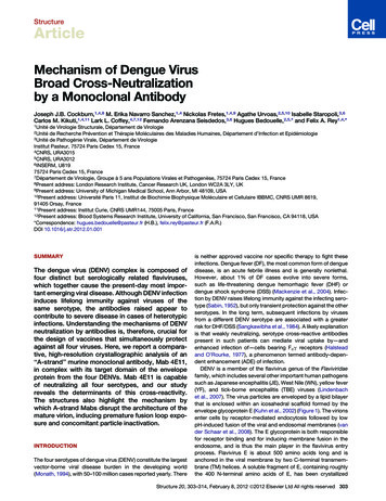
StructureDeterminants of Dengue Virus Cross-NeutralizationFigure 3. Serotype-Dependent Antibody-Antigen Interactions(A–D) Close-ups of the 4E11 paratope (left panel) and epitope (right panel) inthe complexes with DENV serotypes 1–4 as indicated. Views correspondapproximately to the boxed areas in the key in the top part of the figure, whichcontain the serotype-specific residues. Semitransparent surfaces of the scFvand DIII are colored according to their electrostatic potentials, calculatedseparately for each component in the complex. The surface electrostaticpotential is represented in a color gradient from blue (basic) through white(neutral) to red (acidic), between values of 2kBT (where kB is the Boltzmannconstant). Salt bridges between residues in the antibody and the antigen areshown as black dashes in the right-hand panels.(E) The corresponding views of the interface between Fab 1A1D-2 and theDENV-2 DIII (PDB accession number 2R29).(Figure 1D), whereas the other locations display conservativesubstitutions that preserve the short, aliphatic character of theside chains in the AG patch, as well as its shape (defined bythe abutting A and G strands). Indeed, the short aliphatic sidechains in the AG patch are responsible for the specificity ofMab 4E11 for the DENV group, especially at position 389, whichin other flaviviruses is either His or Tyr. The available structures ofDIII from TBE (PDB number 1SVB), WN (1ZTX), JE (1PJW), YF(2JV6), and Langat (2GG1) viruses show that the bulky sidechains at position 389 protrude from the patch in a way thatwould prohibit 4E11 binding via steric clashes with TyrL28(Figures 2A and S2).Strand A of DIII presents a prominent central bulge (conservedin all flaviviruses), located to one side of the AG patch, featuringthe conserved residues Lys310 and Glu311 (Figures 1D and 1G).The conserved ‘‘core’’ epitope is recognized by Mab 4E11in the same way in all four serotypes, as depicted in Figure 2:packing against the hydrophobic side chains of the AG patch,recognition of its shape via hydrogen bonding to the A- andG-strand main-chain groups defining the periphery, and makingsalt bridges with Lys310 and Glu311 from the central bulge. Anumber of the core epitope residues identified here were previously shown, through biochemical studies, to make importantcontributions to the affinity of Fab 4E11 for the DENV-1 DIII. Inparticular, Lys310 was found critical for binding (Lisova et al.,2007). Likewise, TrpH96 and GluH97 in the CDR H3 loop, whichinteract with the AG patch and A-strand main-chain groups atthe center of the antibody-antigen interface, were also shownto be essential for Fab 4E11 binding to DENV-1 DIII (Bedouelleet al., 2006).Serotype-Specific ContactsThe contacts of the antibody with residues in the 4E11 epitopethat are not conserved across DENV serotypes also contributeto binding. These E residues are located on the upper portionof DIII, which is exposed in the virion (Figure 3). The principal antibody residues involved in sensing these nonconserved E residues are ArgH94 (third heavy-chain framework region) andGluL55 (CDR L2 loop). Additionally, residues AspH31-ThrH32,TyrH102, and AsnL53 (Figure 1F) engage in a small number of interactions with DIII residues at the periphery of the antibodyantigen interfaces that vary between serotypes (Tables S1Aand S1B). ArgH94 and GluL55 create adjacent patches of positiveand negative electrostatic potential in the paratope. In thecomplex with DENV-1 DIII (the serotype against which Mab4E11 was raised), these residues of the paratope form saltbridges with DIII serotype-specific residues Glu309 and Lys307,respectively (Figures 1D and 3A). Binding to DIII from the otherserotypes is facilitated by conformational variability of theArgH94 side chain and changes in overall antibody positioning,allowing the partial conservation of charge complementaritydespite amino acid sequence variations between serotypesover this region (Figures 1D and 3B–3D). This is particularlystriking for the complex with DIII from the DENV-3 isolate.Here, the antibody shifts down such that GluL55 and ArgH94face the DIII B strand (which is antiparallel to strand A; seeFigures 1B and 1G) and interact with Lys327 and Glu325,mimicking the interactions with the DENV-1 A-strand residuesLys307 and Glu309 (Figure 3C). Finally, the absence of a basicresidue at positions 307 or 327 in DENV-4 DIII results in308 Structure 20, 303–314, February 8, 2012 ª2012 Elsevier Ltd All rights reserved
StructureDeterminants of Dengue Virus Cross-Neutralizationa much lower degree of charge complementarity across the antibody-antigen interface than in the complexes with the otherserotypes (Figure 3D), reflecting the much lower affinity of Fab4E11 for DENV-4 DIII (Table 1).Previous mutagenesis studies on the DENV-1 DIII A and Gstrands showed that mutation of residue Lys307 to alaninereduced the free energy of dissociation (DGd) of Fab 4E11 from13.8 to 9.7 kcal/mol (Lisova et al., 2007), which was thesecond-greatest reduction in affinity after mutation of theconserved Lys310. This suggests that the effect of truncatingthe Lys307 side chain in the DENV-1 DIII accounts for themajority of the difference in DGd of Fab 4E11 binding to DENV-1and DENV-4 DIIIs (Table 1). Likewise, the DGd value of12.8 kcal/mol for the DENV-2 DIII (Table 1), compared with12.5 kcal/mol for the DENV-1 DIII Glu309Ala mutant (Lisovaet al., 2007), shows that elimination of the negative charge atposition 309 appears to account for the affinity difference ofFab 4E11 for the DENV-2 DIII versus that of DENV-1. Residuechanges at positions 309 and 307 are thus the main determinants of the affinities of Fab 4E11 for the DENV-2 and DENV-4DIIIs compared with that of DENV-1. The ability of the B-strandresidues Glu325 and Lys327 of DENV-3 to mimic these interactions, despite having unfavorable A-strand residues at theselocations, is likely to be crucial in maintaining high-affinitybinding to this serotype. Previous biochemical studies haveshown that cross-reactive murine Mabs binding to A-strand residues on the DENV-2 DIII commonly have epitope determinantson the B strand of the DENV-3 DIII (Gromowski et al., 2008;Matsui et al., 2009). These observations are a strong indicationthat the strand-switching mechanism of 4E11 cross-reactivitywith DENV-3 that we describe here is a general feature of allA-strand Mabs.The contacts observed here between scFv 4E11 and theDENV-2 DIII are mostly conserved in the corresponding complexof Fab 1A1D-2. However, an important difference is that 1A1D-2has an aspartic acid at position H95 in its CDR H3 loop that formsan additional salt bridge with Lys307, whereas the correspondingresidue in 4E11 is glycine. As a result, the 1A1D-2 paratope issignificantly more negatively charged than that of 4E11 (Figure 3E). This presumably reflects the fact that 4E11 and 1A1D-2were raised against DENV-1 and DENV-2 strains, respectively,which have different charge distributions along the A strand(because the residue at position 309 is glutamate in DENV-1and valine in DENV-2). As an additional consequence, the1A1D-2 paratope displays an even lower charge complementarity with DENV-4 DIII than that of 4E11 (Figures 3D and 3E).These observations explain the differing serotype dependenceof neutralization between these two antibodies, and in particularwhy Mab 4E11 neutralizes DENV-4 strains at moderate antibodytiters, whereas Mab 1A1D-2 does not.Molecular Basis for Immunodominance of the DENV DIIIA Strand in MiceAnalysis of the murine germline shows that the 4E11 and 1A1D-2variable domains are both derived from the same heavy- andlight-chain variable (V) and junction (J) gene segments, butdifferent diversity (D) segments (see Experimental Procedures).In both antibodies, almost all residues that contact DIII are present in the germline sequences of the relevant gene segments(Figures 4A and 4B). In both cases, VDJ recombination hasresulted in CDR H3 loops with identical lengths (six residues),with a glutamate at H97 and an aromatic side chain at positionH96 (Trp in 4E11 and Tyr in 1A1D-2) that plug the center of theantibody-antigen interface (Figures 2, 4C,and 4D). The onlysignificant difference is the residue at position H95 (glycine in4E11 and aspartic acid in 1A1D-2), which dictates the differingserotype responses of these antibodies as discussed above.Of the 14 known murine D segments, 6 would be capable offorming 1A1D-2/4E11-like H3 loops (Figure 4E). This, in conjunction with the inherent capacity of the V gene segments utilized by4E11 and 1A1D-2 to form high-affinity interactions with DIII residues that are conserved among all four serotypes, may explainthe immunodominance of A-strand epitopes in mice. Aminoacid sequence variations at position H95, arising from N regionsor somatic hypermutations, could then generate a family ofcross-reactive Mabs with distinct avidity and neutralizationprofiles by serotype. In summary this analysis shows that themurine germline harbors gene segments with an inherentcapacity for high-affinity binding to DIII from all four DENV serotypes. This is in line with the observation that most stronglyneutralizing murine Mabs against DENV bind to DIII, whereashumans do not appear to make such antibodies as readily (Crillet al., 2
Structure Article Mechanism of Dengue Virus Broad Cross-Neutralization by a Monoclonal Antibody Joseph J.B. Cockburn,1,4,8 M. Erika Navarro Sanchez,1,4 Nickolas Fretes,1,4,9 Agathe Urvoas,2,5,10 Isabelle Staropoli,3,6 Carlos M. Kikuti,1 ,411 Lark L. Coffey, 7 12 Fernando Arenzana Seisdedos,3 6 Hugues Bedo