Transcription
VIROLOGICA SINICA 2017, 32 (1): 16–22DOI: 10.1007/s12250-016-3855-9REVIEWProgress towards understanding the pathogenesis of denguehemorrhagic fever11,21,2*Xiaojing Pang , Rudian Zhang , Gong Cheng1. Tsinghua-Peking Center for Life Sciences, School of Medicine, Tsinghua University, Beijing 100084, China2. School of Life Science, Tsinghua University, Beijing 100084, ChinaDengue virus (DENV) is a mosquito-borne virus belonging to the Flaviviridae family. There are 4serotypes of DENV that cause human disease through transmission by mosquito vectors. DENVinfection results in a broad spectrum of clinical symptoms, ranging from mild fever to denguehemorrhagic fever (DHF), the latter of which can progress to dengue shock syndrome (DSS) anddeath. Researchers have made unremitting efforts over the last half-century to understand DHFpathogenesis. DHF is probably caused by multiple factors, such as virus-specific antibodies, viralantigens and host immune responses. This review summarizes the current progress of studies onDHF pathogenesis, which may provide important information for achieving effective control ofdengue in the future.KEYWORDS DENV; dengue hemorrhagic fever (DHF); NS1; genome; antibody-dependentenhancement (ADE); T cellINTRODUCTIONDengue virus (DENV) is a mosquito-borne virus belonging to the Flaviviridae family. There are 4 serotypes ofDENV (DENV-1 through DENV-4) that cause humandisease through transmission via the mosquito vectorsAedes aegypti and Aedes albopictus (Gubler, 2011).There are more than 100 dengue-epidemic countries inthe world. Furthermore, approximately 2.5 billion peopleare at risk of dengue infection every year. It estimated390 million dengue infections per year, of which 96 million manifest apparently any level of disease severity(Bhatt et al., 2013). In 2015, 2.35 million cases of denguewere reported in America alone, and more than ten thousand of these cases involved severe hemorrhagic-relatedclinical symptoms. Dengue infection remains an important threat to public health and is responsible for a largeglobal disease burden.Received: 22 August 2016, Accepted: 12 October 2016,Published online: 14 November 2016*Correspondence:Phone: 86-10-62788494; Fax: 86-10-62788494,Email: gongcheng@mail.tsinghua.edu.cnORCID: 0000-0001-7447-548816 FEBRUARY 2017 VOLUME 32 ISSUE 1Most DENV infections are asymptomatic or present amild illness with flu-like symptoms, such as fever, headache, myalgia and decreased platelet counts and leucopenia. These symptoms are known as dengue fever(DF), which is an acute, self-limited, febrile illness. However, some DF patients develop a severe syndrome knownas Dengue Hemorrhagic Fever (DHF), in which patientsmay display hematomas with marked thrombocytopeniaor extremely low platelet counts. Even more severe clinical symptoms are produced when DENV infection develops Dengue Shock Syndrome (DSS), a disease similarto DHF that additionally includes coagulation abnormalities, increased vascular fragility, raised plasma leakage/heme concentration, loss of fluid due to capillary permeability that may progress to hypovolemic shock, andincreased risk of multi-organ failure (WHO/TDR,2009). The clinical hallmark of DHF is plasma leakage,which usually lasts for approximately 48 hours and leadsto reduced circulatory volume (Srikiatkhachorn, 2009).Bleeding is common following dengue infection, however it is more frequent in DHF/DSS than in DF. Mostdengue-associated deaths result from the symptoms associated with DHF/DSS (Hermann et al., 2015). There isno antiviral therapy against DENV infection. Dengvaxia, The Author(s) 2017. This article is published with open access at Springerlink.com
Progress towards pathogenesis of DHFa commercial vaccine developed by Sanofi Pasteur, waslicensed in several dengue-endemic countries (Hladish etal., 2016; Recker et al., 2016). DHF is the most lifethreatening consequence of DENV infection. Previousstudies have suggested that both viral and host factorsmight contribute to the pathogenesis of this disease, including Nonstructural protein 1 (NS1) viral antigen andits antibodies, virus variation and virulence, subgenomicRNA, antibody-dependent enhancement (ADE), and thepresence of memory cross-reactive T cells. The furtherresearch on the pathogenesis of DHF can provide a newtarget for the prevention and control of dengue infectious diseases.ROLE OF DENV NONSTRUCTURAL PROTEIN-1(NS-1) AND ITS ANTIBODIES IN DHFPATHOGENESISNS1, a nonstructural protein encoded by DENV, is expressed in multiple oligomeric forms and is present in avariety of cellular locations, including on intracellularmembranes, at the cell surface and extracellularly as asoluble, secreted lipoparticle (Chuang et al., 2016). During acute DENV infection, secreted NS1 protein is present in patient sera at high levels (Libraty et al., 2002),which might correlate with disease severity and contribute to the pathogenesis of DHF in a host (Chuang et al.,2013; Srikiatkhachorn and Kelley, 2014). Indeed, DENVNS1 might directly bind to the surfaces of host cells tocause tissue damage (Avirutnan et al., 2007; Beatty et al.,2015; Modhiran et al., 2015). A recent study suggestedthat DENV NS1 elicits inflammatory cytokine production by activating macrophages and human peripheralblood mononuclear cells (PBMCs) through Toll-like receptor 4 (TLR4), thus leading to disruption of endothelialcell monolayer integrity in blood vessels (Modhiran etal., 2015). Moreover, DENV NS1 might also triggercomplement activation, causing plasma leakage (Kurosuet al., 2007). During this process, soluble DENV NS1 isreleased from infected cells and independently activatescomplement factors present in the fluid phase (Nielsen,2009). A close correlation has been found betweenDENV NS1 concentration and C5b-C9 complex formation. The C5b-C9 complex can stimulate robust expression of inflammatory cytokines that are associated withDHF development (Suresh et al., 2016). Recently, it hasbeen reported that DENV NS1 may induce autophagy inthe HMEC-1 human endothelial cell line as well as inmice, and that this autophagy is mediated by NS1-associated secretion of a macrophage migration inhibitory factor(MIF) (Chen et al., 2016).In addition to the NS1 protein itself, NS1 antibodiesmight also contribute to DHF pathogenesis. For example,www.virosin.orgthe binding of anti-NS1 antibodies to GPI-anchored NS1proteins on cell membranes can activate cellular signaltransduction pathways, leading to protein tyrosine phosphorylation, which may enhance DENV replication withininfected cells (Jacobs et al., 2000). Moreover, proteinphosphorylation and NF-κB activation have been observed after stimulating human HMEC-1 cells with antiDENV NS1 antibodies. Furthermore, the expression ofseveral cytokines and chemokines, such as IL-6, IL-8,and MCP-1, is increased after treatment of endothelialcells with anti-DENV NS1 antibodies, suggesting thatDENV NS1 antibodies can stimulate the release of multiple inflammatory factors in an NF-κB-dependent manner (Lin et al., 2005). Disordered release of cytokines isconsidered as a major factor underlying DHF pathogenesis. Therefore, NS1 antibody-mediated activation of immune responses may play an important role in the development of Dengue-mediated thrombocytopenia and vascular leakage during the critical phase of DHF/DSS.Recently, the induction of autoimmune disorders byanti-NS1 antibodies has also been described as a contributing factor to DHF pathogenesis. Because there is sequence homology between DENV NS1 and several selfantigens, such as plasminogen and integrin, proteins onhuman endothelial and platelet cells, auto-antibodies induced by NS1 may cross-react with these self-antigens(Falconar, 1997; Liu et al., 2016; Rachman et al., 2013;Chuang et al., 2016). These reactions stimulate nitric oxide (NO) expression and cell apoptosis in endothelialcells and cause cell lysis and inhibition of aggregation inplatelets (Sun et al., 2007). Damage to endothelial cellsand platelets may contribute to thrombocytopenia, coagulopathy and vascular leakage in DHF (Sun et al.,2007; Wan et al., 2014).ROLE OF DENV GENOME IN DHFPATHOGENESISGenetic differences among DENV genotypes are associated with differential viral virulence that may contributeto the development of the severe diseases, such as DHFand DSS (Ubol et al., 2008; Mammen et al., 2014). SomeDENV genotypes have greater virulence and epidemicpotential than others. This viewpoint was primarily introduced based on epidemiological observations (Gubler etal., 1981). In the American Dengue outbreak that occurred in 1981, a Southeast Asian DENV-2 genotypeemerged more virulent than the indigenous Americangenotype (Rico-Hesse et al., 1997; Rico-Hesse et al.,1998). Indeed, evidence has indicated that a Southeastern Asian DENV-2 genotype virus replicate at highertiters than an American genotype virus in both humans andmosquitoes (Anderson and Rico-Hesse, 2006; VasilakisFEBRUARY 2017 VOLUME 32 ISSUE 1 17
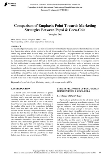
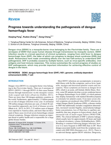
Xiaojing Pang et al.Figure 1. The mechanisms involved in DHF pathogenesis include nonstructural protein 1 (NS1) viral antigen and its antibodies, virus genome variation, sfRNA, antibody-dependent enhancement (ADE), and the presence of memory crossreactive T cells. NS1 and its antibodies contribute to DHF pathogenesis by triggering complement activation and theneliciting inflammatory cytokine production, inducing autophagy in endothelial cell, enhancing DENV replication, inducingautoimmune disorders; DENV genome variation evolve greater virulence, and sfRNA induce the enhancement of viralreplication; FcγR-mediated ADE can enhance viral replication and trigger cytokines and chemokines release; ActivatedDENV-specific cross-reactive T cells promote the release of cytokines and chemokines, and T cell apoptosis also contributes to DHF pathogenesis.et al., 2007). Besides, the intra-serotype evolution of epidemic DENV strains might be another cause of increasing viral virulence. The clinical manifestations and casefatality rates of DHF typically increase during the laterperiods of epidemics, a phenomenon that was observedin the Cuban epidemics of 1981 and 1997, the Townsville, Australia epidemic of 1992, and the Oaxaca, Mexico epidemic of 2005 (Guzman et al., 2000; Martina etal., 2009; Ritchie et al., 2013). An analysis of DENVgenomes suggested that circulating DENV might evolvegreater virulence through passage in patients during anepidemic (Chen et al., 2008). Thus, variations in theDENV genome serve to determine viral infectivity inboth hosts and vectors (Ritchie et al., 2013).Besides the genome variation, the subgenomic flavivirus RNA (sfRNA) may play a role in DENV replication in host cells, thus contributing to DHF pathogenesis.During DENV replication, a 11 kb RNA genome may beincompletely degraded from their 3’ untranslated region(UTR) by host exoribonuclease to generate a 0.3 to 0.5kb small RNA termed sfRNA (Pijlman et al., 2008; Funket al., 2010; Chapman et al., 2014; Roby et al., 2014).sfRNA may accumulate in the infected cells to suppress18 FEBRUARY 2017 VOLUME 32 ISSUE 1the host antiviral immune responses, such as type I interferon signaling (Manokaran et al., 2015; Chang et al.,2013). sfRNA may also facilitate DENV replicationthrough altering host mRNA stability (Moon et al., 2012;Schnettler et al., 2012), thereby acting as a player inDENV immune evasion and DHF pathogenesis.ROLE OF ANTIBODY-DEPENDENTENHANCEMENT (ADE) IN DHF PATHOGENESISEpidemiological evidence has shown that increasedrisk for DHF/DSS is associated with a secondary infection from a different DENV serotype than that causingthe initial infection (Guzman et al., 2013). Data gatheredfrom Cuba, Hawaii and Thailand have shown that individuals with circulating DENV 1 antibodies are at an enhanced risk for acquiring DHF during subsequent DENV2 or DENV 3 infections (Alvarez et al., 2006). Furthermore, an infant infected with a DENV serotype that isdifferent from the serotype that its infected mother is atan increased risk for DHF/DSS (Halstead et al., 2002).Based on assessments of hospital patients, the incidenceof DHF/DSS during primary dengue infections was 11 toVIROLOGICA SINICA
Progress towards pathogenesis of DHF12 per 1000 compared with 118 to 208 per 1000 duringsecondary dengue infections (Guzman et al., 2013). Therisk of developing DHF during a secondary infection isat least 10-fold greater than the risk of developing thecondition during a primary infection (Srikiatkhachorn,2009). This increased infection rate is caused by antibodies induced from the first infection and has beentermed antibody-dependent enhancement (Halstead,1970). Indeed, the ADE phenomenon has been repeatedly demonstrated in DENV infections of cell cultures and animals. When DENV is incubated with diluted anti-DENV sera or DENV-specific antibodies, viralreplication in human monocytes and some cell lines issignificantly promoted. Passive immunization withDENV antibodies/antisera prior to virus inoculation inrhesus monkeys enhanced viral replication more than100-fold over control animals (Goncalvez et al., 2007;Sasaki et al., 2013; Chaichana et al., 2014; Martinezgomez et al., 2016). Nonetheless, not all cases of DHF/DSS are associated with ADE. Evidence from a phase 3clinical trial of a Dengue vaccine (Sanofi Pasteur) foundno evidence of ADE generation in the study population(Sabchareon et al., 2012). Although ADE has beenprimarily described in relation to DENV infection, thephenomenon has also been associated with several otherviral species based on in vitro enhancements of infection(Takada et al., 2003; Martina et al., 2009; Chan-Hui andSwiderek, 2016).Previous studies have demonstrated that ADE are directed toward anti-E antibodies and anti-prM antibodies.Although more studies have been designed to evaluatethe role of E protein, prM have attracted more attentionsince 2010, when it was first proposed that anti-prM antibodies involved in the ADE phenomenon (Dejnirattisaiet al., 2010). Several groups have shown that anti-prMMAbs exhibit ADE properties in cell culture and in animal models of DENV infection (Smith et al., 2012; Luoet al., 2013). Thus, anti-prM antibodies are likely to playan important role in the pathogenesis of DHF in humans.The mechanism of ADE remains only partially understood. Fcγ receptor (FcγR)-mediated uptake of virionantibody complexes into permissive cells may be one explanation for the enhancement of DENV infection (Satoet al., 2015). FcγR is a multi-subunit complex that is distributed on the surfaces of many types of immune cells,such as dendritic cells, macrophages and mast cells, andrecognizes the Fc regions of immunoglobulins. DuringDENV infection, antibodies with infection-enhancingactivity are generated and subsequently form virion-antibody complexes. These complexes can rapidly internalize into FcγR-bearing cells via interactions with FcγR,which results in a higher number of infected cells in thepresence versus the absence of antibodies (Halstead,1982). Moreover, previous studies have also shown thatwww.virosin.orgFcγR-mediated DENV infection might enhance viral replication through suppression of intracellular antiviral innate immune responses, including interferon-mediatedantiviral responses, and enhancement of IL-10 production in DENV-infected cells. The suppression of host immune responses may promote the production of highnumbers of infectious DENV virions (Ubol and Halstead,2010).The clinical manifestations of DHF include endothelialcell damage, enhancement of vascular permeability,plasma leakage and liver injury (Halstead and Cohen,2015). However, these manifestations may be not necessarily a direct result of cell death induced by DENV replication because viral load peaks during the febrile phaseand rapidly declines by the time hemorrhagic fever andplasma leakage develop. In addition to enhancing viralentry, FcγR-mediated ADE directly triggers robust release of cytokines from mast cells and other immunecells, which may mediate vascular endothelial cell dysfunction and increase vascular permeability. The releaseof cytokines is considered a key factor that contributes toDHF pathogenesis (King et al., 2002; Brown et al., 2011).ROLE OF T CELLS IN DHF PATHOGENESISThe increases in cytokine production and CD8 T cellactivation that have been observed in severe dengue patients imply that cross-reactive T cells have a role in mediating the pathogenesis of DHF (Mathew and Rothman,2008). Memory T-cells from the primary infection wereactivated by a heterologous virus during second infection, and showed highly cross-reactivity and low affinityleading to inefficiency at killing infected cells and clearing the newly infective virus serotype. On the contrary,cross-reactive T cell contribute to DHF occurrence bysubstantial immunopathology. DHF patients have beenreported to demonstrate enhanced circulation of cytokines and chemokines, IFN-γ, IL-2 and TNF-α produced by activated DENV-specific cross-reactive T cells,compared to DF patients (Bozza et al., 2008; Malavige etal., 2012; Kurane et al., 2011), suggesting a positive correlation between cross-reactive T cell activation and theseverity of dengue-associated disease. Furthermore, Tcell apoptosis in DHF patients contributes to the severityof dengue-associated disease (Jaiyen et al., 2009;Midgley et al., 2011; Malavige et al., 2012). A previousstudy reported that the immunosuppressive cytokine IL10 could lead to T cell apoptosis in patients with acutedengue infection. IL-10 concentrations are elevated inthe sera of patients with severe dengue (Mathew andRothman, 2008). IL-10 blockade has been significantlyassociated with reduced T cell apoptosis in acute DENVinfections. In agreement, reduced T cell numbers haveFEBRUARY 2017 VOLUME 32 ISSUE 1 19
Xiaojing Pang et al.been found in DHF patients compared to DF patients(Green et al., 1999), suggesting that T cell apoptosisreduces viral clearance and consequently results in animpaired antiviral response, leading to severe clinicaldengue diseases.CONCLUSIONClinicians and scientists have increasingly focused onunderstanding the pathogenesis of DHF from severalperspectives; however, the mechanisms underlying thispathogenesis are not yet fully elucidated. Further studiesare urgently required to improve our understanding ofDHF pathogenesis, which is important for developing effective therapeutics for and vaccine strategies againstDENV infection.COMPLIANCE WITH ETHICS GUIDELINESThe authors declare that they have no conflict of interests. This article does not contain any studies with human or animal subjects performed by any of the authors.OPEN ACCESSThis article is distributed under the terms of the CreativeCommons Attribution 4.0 International License(http://creative commons.org/licenses/by/4.0/), whichpermits unrestricted use, distribution, and reproduction inany medium, provided you give appropriate credit to theoriginal author(s) and the source, provide a link to theCreative Commons license, and indicate if changes weremade.REFERENCESAlvarez M, Rodriguez-Roche R, Bernardo L, Vazquez S, MorierL, Gonzalez D, Castro O, Kouri G, Halstead SB, Guzman MG.2006. Dengue hemorrhagic Fever caused by sequential dengue1-3 virus infections over a long time interval: Havana epidemic,2001-2002. Am J Trop Med Hyg, 75: 1113–1117.Anderson JR, Rico-Hesse R. 2006. Aedes aegypti vectorial capacity is determined by the infecting genotype of dengue virus. AmJ Trop Med Hyg, 75: 886–892.Avirutnan P, Zhang L, Punyadee N, Manuyakorn A, Puttikhunt C,Kasinrerk W, Malasit P, Atkinson JP, Diamond MS. 2007.Secreted NS1 of dengue virus attaches to the surface of cells viainteractions with heparan sulfate and chondroitin sulfate E. PLoSPathog, 3: e183.Beatty PR, Puerta-Guardo H, Killingbeck SS, Glasner DR, Hopkins K, Harris E. 2015. Dengue virus NS1 triggers endothelialpermeability and vascular leak that is prevented by NS1 vaccination. Sci Transl Med, 7: 304ra141.Bhatt S, Gething PW, Brady OJ, Messina JP, Farlow AW, MoyesCL, Drake JM, Brownstein JS, Hoen AG, Sankoh O, MyersMF, George DB, Jaenisch T, Wint GR, Simmons CP, Scott TW,20 FEBRUARY 2017 VOLUME 32 ISSUE 1Farrar JJ, Hay SI. 2013. The global distribution and burden ofdengue. Nature, 496: 504–507.Bozza FA, Cruz OG, Zagne SM, Azeredo EL, Nogueira RM, Assis EF, Bozza PT, Kubelka CF. 2008. Multiplex cytokine profile from dengue patients: MIP-1beta and IFN-gamma as predictive factors for severity. BMC Infect Dis, 8: 86.Brown MG, Hermann LL, Issekutz AC, Marshall JS, Rowter D,Al-Afif A, Anderson R. 2011. Dengue virus infection of mastcells triggers endothelial cell activation. J Virol, 85: 1145–1150.Chaichana P, Okabayashi T, Puiprom O, Sasayama M, Sasaki T,Yamashita A, Ramasoota P, Kurosu T, Ikuta K. 2014. Lowlevels of antibody-dependent enhancement in vitro using viruses and plasma from dengue patients. PLoS One, 9: e92173.Chan-Hui PY, Swiderek KM. 2016. Immunological considerations for developing antibody therapeutics for Influenza A.Hum Vaccin Immunother, 12: 474–477.Chang RY, Hsu TW, Chen YL, Liu SF, Tsai YJ, Lin YT, ChenYS, Fan YH. 2013. Japanese encephalitis virus non-codingRNA inhibits activation of interferon by blocking nuclear translocation of interferon regulatory factor 3. Vet Microbiol, 166:11–21.Chapman EG, Costantino DA, Rabe JL, Moon SL, Wilusz J, NixJC, Kieft JS. 2014. The structural basis of pathogenic subgenomic flavivirus RNA (sfRNA) production. Science, 344: 307–310.Chen HL, Lin SR, Liu HF, King CC, Hsieh SC, Wang WK. 2008.Evolution of dengue virus type 2 during two consecutive outbreaks with an increase in severity in southern Taiwan in2001–2002. Am J Trop Med Hyg, 79: 495–505.Chen HR, Chuang YC, Lin YS, Liu HS, Liu CC, Perng GC, YehTM. 2016. Dengue Virus Nonstructural Protein 1 Induces Vascular Leakage through Macrophage Migration Inhibitory Factorand Autophagy. PLoS Negl Trop Dis, 10: e0004828.Chuang YC, Lin J, Lin YS, Wang S, Yeh TM. 2016. Dengue VirusNonstructural Protein 1-Induced Antibodies Cross-React withHuman Plasminogen and Enhance Its Activation. J Immunol,196: 1218–1226.Chuang YC, Wang SY, Lin YS, Chen HR, Yeh TM. 2013. Reevaluation of the pathogenic roles of nonstructural protein 1 andits antibodies during dengue virus infection. J Biomed Sci, 20:42.Dejnirattisai W, Jumnainsong A, Onsirisakul N, Fitton P, Vasanawathana S, Limpitikul W, Puttikhunt C, Edwards C,Duangchinda T, Supasa S, Chawansuntati K, Malasit P, Mongkolsapaya J, Screaton G. 2010. Cross-reacting antibodies enhance dengue virus infection in humans. Science, 328: 745–748.Falconar AK. 1997. The dengue virus nonstructural-1 protein(NS1) generates antibodies to common epitopes on humanblood clotting, integrin/adhesin proteins and binds to human endothelial cells: potential implications in haemorrhagic feverpathogenesis. Arch Virol, 142: 897–916.Funk A, Truong K, Nagasaki T, Torres S, Floden N, BalmoriMelian E, Edmonds J, Dong H, Shi PY, Khromykh AA. 2010.RNA structures required for production of subgenomic flavivirusRNA. J Virol, 84: 11407–11417.Goncalvez AP, Engle RE, St Claire M, Purcell RH, Lai CJ. 2007.Monoclonal antibody-mediated enhancement of dengue virusinfection in vitro and in vivo and strategies for prevention. ProcNatl Acad Sci U S A, 104: 9422–9427.Green S, Pichyangkul S, Vaughn DW, Kalayanarooj S, Nimmannitya S, Nisalak A, Kurane I, Rothman AL, Ennis FA. 1999.Early CD69 expression on peripheral blood lymphocytes fromchildren with dengue hemorrhagic fever. J Infect Dis, 180:1429–1435.Gubler DJ. 2011. Emerging vector-borne flavivirus diseases: areVIROLOGICA SINICA
Progress towards pathogenesis of DHFvaccines the solution? Expert Rev Vaccines, 10: 563–565.Gubler DJ, Suharyono W, Lubis I, Eram S, Gunarso S. 1981. Epidemic dengue 3 in central Java, associated with low viremia inman. Am J Trop Med Hyg, 30: 1094–1099.Guzman MG, Alvarez M, Halstead SB. 2013. Secondary infectionas a risk factor for dengue hemorrhagic fever/dengue shock syndrome: an historical perspective and role of antibody-dependentenhancement of infection. Arch Virol, 158: 1445–1459.Guzman MG, Kouri G, Halstead SB. 2000. Do escape mutants explain rapid increases in dengue case-fatality rates within epidemics? Lancet, 355: 1902–1903.Halstead SB. 1970. Observations related to pathogensis of denguehemorrhagic fever. VI. Hypotheses and discussion. Yale J BiolMed, 42: 350–362.Halstead SB. 1982. Immune enhancement of viral infection. ProgAllergy, 31: 301–364.Halstead SB, Cohen SN. 2015. Dengue Hemorrhagic Fever at 60Years: Early Evolution of Concepts of Causation and Treatment. Microbiol Mol Biol Rev, 79: 281–291.Halstead SB, Lan NT, Myint TT, Shwe TN, Nisalak A,Kalyanarooj S, Nimmannitya S, Soegijanto S, Vaughn DW,Endy TP. 2002. Dengue hemorrhagic fever in infants: researchopportunities ignored. Emerg Infect Dis, 8: 1474–1479.Hermann LL, Gupta SB, Manoff SB, Kalayanarooj S, GibbonsRV, Coller BA. 2015. Advances in the understanding, management, and prevention of dengue. J Clin Virol, 64: 153–159.Hladish TJ, Pearson CA, Chao DL, Rojas DP, Recchia GL,Gomez-Dantes H, Halloran ME, Pulliam JR, Longini IM. 2016.Projected Impact of Dengue Vaccination in Yucatan, Mexico.PLoS Negl Trop Dis, 10: e0004661.Jacobs MG, Robinson PJ, Bletchly C, Mackenzie JM, Young PR.2000. Dengue virus nonstructural protein 1 is expressed in aglycosyl-phosphatidylinositol-linked form that is capable of signal transduction. FASEB J, 14: 1603–1610.Jaiyen Y, Masrinoul P, Kalayanarooj S, Pulmanausahakul R, UbolS. 2009. Characteristics of dengue virus-infected peripheralblood mononuclear cell death that correlates with the severity ofillness. Microbiol Immunol, 53: 442–450.King CA, Anderson R, Marshall JS. 2002. Dengue virus selectively induces human mast cell chemokine production. J Virol,76: 8408–8419.Kurane I, Matsutani T, Suzuki R, Takasaki T, Kalayanarooj S,Green S, Rothman AL, Ennis FA. 2011. T-cell responses todengue virus in humans. Trop Med Health, 39: 45–51.Kurosu T, Chaichana P, Yamate M, Anantapreecha S, Ikuta K.2007. Secreted complement regulatory protein clusterin interacts with dengue virus nonstructural protein 1. Biochem Biophys Res Commun, 362: 1051–1056.Libraty DH, Young PR, Pickering D, Endy TP, Kalayanarooj S,Green S, Vaughn DW, Nisalak A, Ennis FA, Rothman AL.2002. High circulating levels of the dengue virus nonstructuralprotein NS1 early in dengue illness correlate with the development of dengue hemorrhagic fever. J Infect Dis, 186: 1165–1168.Lin CF, Chiu SC, Hsiao YL, Wan SW, Lei HY, Shiau AL, LiuHS, Yeh TM, Chen SH, Liu CC, Lin YS. 2005. Expression ofcytokine, chemokine, and adhesion molecules during endothelialcell activation induced by antibodies against dengue virus nonstructural protein 1. J Immunol, 174: 395–403.Liu JY, Liu Y, Nie KX, Du SY, Pang XJ, Wang PH, Cheng G.2016. Flavivirus acquisition by mosquitoes requires the secretion of nonstructural protein 1 from infected hosts. Nature Microbiology. 1: 16087.Luo YY, Feng JJ, Zhou JM, Yu ZZ, Fang DY, Yan HJ, Zeng GC,Jiang LF. 2013. Identification of a novel infection-enhancingwww.virosin.orgepitope on dengue prM using a dengue cross-reacting monoclonal antibody. BMC Microbiol, 13: 194.Malavige GN, Huang LC, Salimi M, Gomes L, Jayaratne SD, OggGS. 2012. Cellular and cytokine correlates of severe dengue infection. PLoS One, 7: e50387.Mammen MP, Lyons A, Innis BL, Sun W, McKinney D, ChungRC, Eckels KH, Putnak R, Kanesa-thasan N, Scherer JM,Statler J, Asher LV, Thomas SJ, Vaughn DW. 2014. Evaluationof dengue virus strains for human challenge studies. Vaccine,32: 1488–1494.Manokaran G, Finol E, Wang C, Gunaratne J, Bahl J, Ong EZ,Tan HC, Sessions OM, Ward AM, Gubler DJ, Harris E, GarciaBlanco MA, Ooi EE. 2015. Dengue subgenomic RNA bindsTRIM25 to inhibit interferon expression for epidemiological fitness. Science, 350: 217–221.Martina BE, Koraka P, Osterhaus AD. 2009. Dengue virus pathogenesis: an integrated view. Clin Microbiol Rev, 22: 564–581.Martinez Gomez JM, Ong LC, Lam JH, Binte Aman SA, LibauEA, Lee PX, St John AL, Alonso S. 2016. Maternal AntibodyMediated Disease Enhancement in Type I Interferon-DeficientMice Leads to Lethal Disease Associated with Liver Damage.PLoS Negl Trop Dis, 10: e0004536.Mathew A, Rothman AL. 2008. Understanding the contribution ofcellular immunity to dengue disease pathogenesis. ImmunolRev, 225: 300–313.Midgley CM, Bajwa-Joseph M, Vasanawathana S, Limpitikul W,Wills B, Flanagan A, Waiyaiya E, Tran HB, Cowper AE, Chotiyarnwong P, Grimes JM, Yoksan S, Malasit P, Simmons CP,Mongkolsapaya J, Screaton GR. 2011. An in-depth analysis oforiginal antigenic sin in dengue virus infection. J Virol, 85: 410–421.Modhiran N, Watterson D, Muller DA, Panetta AK, Sester DP,Liu L, Hume DA, Stacey KJ, Young PR. 2015. Dengue virusNS1 protein activates cells via Toll-like receptor 4 and disruptsendothelial cell monolayer integrity. Sci Transl Med, 7:304ra142.Moon SL, Anderson JR, Kumagai Y, Wilusz CJ, Akira S,Khromykh AA, Wilusz J. 2012. A noncoding RNA produced byarthropod-borne flaviviruses inhibits the cellular exoribonuclease XRN1 and alters host mRNA stability. RNA, 18:2029–2040.Nielsen DG. 2009. The relationship of interacting immunologicalcomponents in dengue pathogenesis. Virol J, 6: 211.Pijlman GP, Funk A, Kondratieva N, Leung J, Torres S, van derAa L, Liu WJ, Palmenberg AC, Shi PY, Hall RA, KhromykhAA. 2008. A highly structured, nuclease-resistant, noncodingRNA produced by flaviviruses is required for pathogenicity.Cell Host Microbe, 4: 579–591.Rachman A, Harahap AR, Widhyasih RM. 2013. The role of antidengue virus NS-1 and anti-protein disulfide isomerase antibodies on platelet aggregation in secondary dengue infection. ActaMed Indones, 45: 44–48.Recker M, Vannice K, Hombach J, Jit M, Simmons CP. 2016. Assessing dengue vaccination impact: Model challenges and future directions. Vaccine, 34: 4461–4465.Rico-Hesse R, Harrison LM, Nisalak A, Vaughn DW, Kalayanarooj S, Green S, Rothman AL,
an increased risk for DHF/DSS (Halstead et al., 2002). Based on assessments of hospital patients, the incidence of DHF/DSS during primary dengue infections was 11 to Figure 1. The mechanisms invol