Transcription
Nocturnal Efficiency and Tolerance of aDemand Oxygen Delivery System in COPDPatients With Nocturnal Hypoxemia*Antoine Cuvelier, Jean-François Muir, Pierre Czernichow, EmmanuelVavasseur, Florence Portier, Daniel Benhamou and DominiqueSamson-DolfussChest 1999;116;22-29DOI 10.1378/chest.116.1.22The online version of this article, along with updated information andservices can be found online on the World Wide Web /22.full.htmlChest is the official journal of the American College of ChestPhysicians. It has been published monthly since 1935.Copyright1999by the American College of Chest Physicians, 3300Dundee Road, Northbrook, IL 60062. All rights reserved. No part ofthis article or PDF may be reproduced or distributed without the priorwritten permission of the copyright c/reprints.xhtml)ISSN:0012-3692Downloaded from chestjournal.chestpubs.org by guest on February 18, 2011 1999 American College of Chest Physicians
Nocturnal Efficiency and Tolerance of aDemand Oxygen Delivery System inCOPD Patients With NocturnalHypoxemia*Antoine Cuvelier, MD; Jean-François Muir, MD, FCCP; Pierre Czernichow, MD;Emmanuel Vavasseur, MD; Florence Portier, MD; Daniel Benhamou, MD; andDominique Samson-Dolfuss, MDObjectives: We compared the efficacy of the standard nasal cannula and the demand oxygendelivery system (DODS) during sleep in patients with COPD.Subjects: Twenty patients with moderate or severe COPD were included in the study.Methods: Four consecutive polysomnographic recordings were performed under the following conditions: DODS powered by compressed air (night 1 [N1]); oxygen delivered with anasal cannula alone (night 2 [N2]); oxygen delivered through a DODS (night 3 [N3]); andoxygen delivered with nasal cannula alone (night 4 [N4]). Oxygen flow rates with and withoutDODS were adjusted the day before the first night so that the resulting transcutaneousarterial oxygen saturation (SaO2) was 95%. The following parameters were evaluated eachnight: apnea-hypopnea index, nocturnal SaO2, total oxygen saving, and several neurophysiologic parameters.Results: The oxygen saving with the DODS was, on average, 60%. All parameters obtainedduring N2 and N4 (oxygen alone) were identical. The percentage of total recording time spentat SaO2 95% was comparable between N2 ([mean 6 SD]; 69 6 32%) and N3 (61 6 31%)(difference is not significant [NS]), as was the time spent at SaO2 between 90% and 95% (N2,29.8 6 31%; N3, 35.9 6 27%; NS) and 90% (N2, 0.75 6 2.6%; N3, 2.5 6 8.6%; NS).Although the mean response time was not significantly different between N2 and N3, twopatients experienced a substantial increase in response time with an SaO2 90% on theDODS. The DODS device did not induce any difference in the percentage of time spent inrapid eye movement (REM) sleep (N2, 12.3 6 8.7%; N3, 16.4 6 7.8%; NS) or non-REM sleep(N2, 87.7 6 8.7%; N3, 83.7 6 7.9%; NS). Non-REM distribution in stage 1–2 sleep and in stage3– 4 sleep was comparable between N2 and N3. Similarly, no difference was observed for thesleep efficiency index (N2, 71 6 15%; N3, 69.6 6 14%; NS). Differences between sleep onsetlatency times were NS.Conclusions: In a majority of moderate to severe COPD patients, the use of a DODS devicedoes not induce any significant alteration of nocturnal neurophysiologic and ventilatoryprofiles. However, the presence of nocturnal desaturation in a few patients justifies the needto systematically perform a ventilatory polygraphic recording when prescribing a DODSdevice.(CHEST 1999; 116:22–29)Key words: COPD; demand oxygen delivery system; nocturnal hypoventilation; oxygen therapyAbbreviations: AHI 5 apnea-hypopnea index; AI 5 apnea index; DODS 5 demand oxygen delivery system;LTO 5 long-term oxygen therapy; NREM 5 non-rapid eye movement; NS 5 not significant; REM 5 rapid eyemovement; Sao2 5 arterial oxygen saturation; SOL 5 sleep onset latency; TST 5 total sleep time*From the Respiratory and Intensive Care Department (Drs.Cuvelier, Muir, Vavasseur, Portier, and Benhamou), Epidemiology Department (Dr. Czernichow), and Neurology Department(Dr. Samson-Dolfuss), Rouen University Hospital, Rouen,France.Manuscript received June 23, 1998; revision accepted January 28,1999.Correspondence to: Antoine Cuvelier, MD, Respiratory andIntensive Care Department, Rouen University Hospital, 76031Rouen cedex, France; e-mail: a-cuvelier@webmails.comlong-term oxygen therapy (LTO) adminB ecauseistered . 5 h/d has led to increased survival inhypoxemic COPD patients,1,2 it could be assumedthat correction of hypoxemia during rapid eye movement (REM) and non-REM (NREM) sleep is beneficial in the management of these patients. Oxygenavailability during the night and during the day hasbeen greatly improved with concentrators, but these22Clinical InvestigationsDownloaded from chestjournal.chestpubs.org by guest on February 18, 2011 1999 American College of Chest Physicians
devices are noisy and remain stationary. In contrast,administration of ambulatory oxygen is a mainstayprocedure to augment daily duration of LTO, thusimproving treatment efficacy3 either with compressed or liquid oxygen cylinders. To deliver anoptimal amount of oxygen during a 24-h period,liquid oxygen therapy has recently been developed;and, currently, portable gas cylinders may be obtained from a compressor linked to a concentrator.The use of these ambulatory devices raises the issueof oxygen preservation either from an economicpoint of view or to maximize patient autonomy.Demand oxygen delivery system (DODS) deviceshave been designed to increase oxygen autonomy withgas or liquid portable reservoirs. DODS devices deliveroxygen only during the inspiratory phase of the respiratory cycle and, therefore, permit oxygen leaks to bekept at a minimum and reduce oxygen costs. Becausethe use of a portable oxygen source (gas or liquid) isassociated with a better compliance with LTO,3 previous studies have focused on the efficiency of DODSdevices during ambulatory activities.4 – 6 However, consequences of the use of DODS devices on sleep qualityand quantity in patients with moderate or severeCOPD should also be assessed.Therefore, we designed a study to compare respiratory pattern and sleep parameters in COPD patients using a liquid oxygen reservoir with and without a DODS device.Materials and MethodsTwenty consecutive patients with stable COPD who presentedwith major obstructive syndrome and chronic respiratory insufficiency gave their informed consent to participate in the study.Twelve of these patients were receiving LTO before entering thestudy. No patient had any clinical sign suggesting an associatedsleep apnea syndrome.The DODS that was used (Optimox; Taema; Antony, France)is a device that only delivers oxygen during the inspiratory phase.An inspiration pressure (6 SD) of 22 6 0.5 Pa is sufficient totrigger the system. The oxygen flow rate is determined by aswitch that permits modification of the oxygen volume deliveredduring each inspiration. The valve is equipped with an apnea carddesigned to detect apneas lasting more than 10 6 2 s. In thisinstance, the device delivers a continuous oxygen flow rate lasting18 6 2 s.During the afternoon preceding the first recording night,arterial blood gas contents measurement on room air, respiratory function tests, full blood count, and chest radiographwere performed. Oxygen titration was assessed with thepatient in a supine position, resting and awake, by usingcontinuous monitoring of the transcutaneous oxygen saturation (Biox 3700; Ohmeda; Louisville, CO). Oxygen was delivered by a liquid reservoir (Freelox; Taema), and flow rateswere fitted after successive tests, each lasting 1 h, so that thearterial oxygen saturation (Sao2) was 95%. The adjustmentswere first performed with oxygen alone and then with oxygenand the DODS device (oxygen 1 DODS). Four consecutivepolysomnographic recordings were made and administered tothe patient according to a single-blind methodology. The firstnight (N1) was considered an adjustment night allowing thepatient to adapt to the DODS and the various transducers,with an attempt to eliminate the first-night effect. The patientwas then connected to a compressed air supply driving theDODS device. During the second night (N2), polysomnography was performed by using oxygen without DODS (oxygenalone) at the flow rate defined on the first day, ensuring anSao2 95%. During the third night (N3), the patient wasgiven oxygen with a DODS (DODS 1 oxygen). To assess andcontrol the quality of oxygenation, recording conditions duringthe fourth night (N4) were the same as those during N2(oxygen alone). Quality of recordings and oxygen therapycompliance were assessed during all four nights by a qualifiedtechnician.Oxygen consumption during N2, N3, and N4 was estimated bythe weight difference of the Freelox liquid oxygen reservebetween the previous evening and the morning after eachpolysomnographic recording. Weight measurements were assessed with a precision (6 1 g) scale (model E/3; Sauter;Hightstown, NJ).Respiratory parameters were measured with a monitor (Respisomnograph; Nellcor Puritan Benett; Antony, France) and anoximeter (Biox 3700; Ohmeda). These parameters includedtranscutaneous finger pulse oximetry, electrocardiogram, thoracoabdominal movements, and nasobuccal airflow measured bythermistors at the nose and mouth. The automated analysis waschecked and corrected on the monitor screen by the samequalified physician. Apnea was defined as an interruption ofnasobuccal air flow lasting at least 10 s that was subsequentlyclassified as obstructive, central, or mixed. Hypopnea was definedas a reduction of the amplitude of nasobuccal air flow by at least50% associated with a fall in Sao2 of 4%.Sleep parameters were determined according to the criteriaestablished by Rechtschaffen and Kales.7 Neurophysiologic signals were recorded simultaneously by an Oxford Medilog 9000(Oxford Instruments Sarl; Orsay, France), including an electromyogram, an electro-oculogram, and an EEG with frontal,vertex, occipital, median, and ocular electrodes. Data wereanalyzed in a computer-assisted manner followed by repeatreading of the raw data by a neurophysiologist who was not awareof the sequence of nights. The recording EEG cassettes wereanalyzed in 60-s epochs to determine total sleep time (TST);REM and NREM sleep duration; REM latency; sleep onsetlatency (SOL), defined as the time from lights out to theoccurrence of the first stage 2 sleep; and sleep efficiency, definedas the ratio between TST and total time spent in bed. Arousalswere determined according to standard criteria7 and defined asawake periods lasting at least 60 s.Statistical AnalysisData were analyzed with a statistical software package (StatView; Abacus Concepts; Berkeley, CA). Tests of normality wereperformed on EEG indices. Student’s t tests were used forcomparisons of variables with normal distributions. Otherwise,between-group comparisons were performed using a Wilcoxonpaired test or a nonparametric Friedman’s test. The level ofsignificance was set at 5%. For all parameters, we did not find anystatistical difference between the two oxygen nights, N2 and N4.ResultsPatient CharacteristicsThe study included 20 patients, 18 men and 2women, with a mean age of 60.4 6 8 years (Table 1).CHEST / 116 / 1 / JULY, 1999Downloaded from chestjournal.chestpubs.org by guest on February 18, 2011 1999 American College of Chest Physicians23
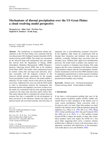
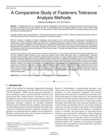
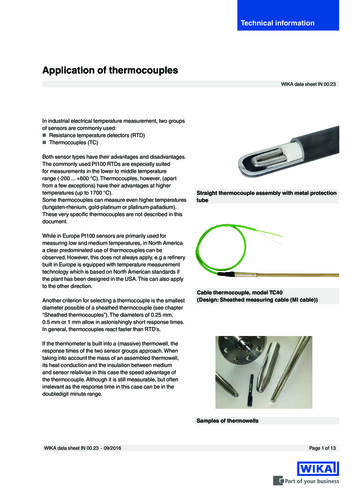
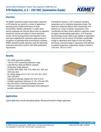
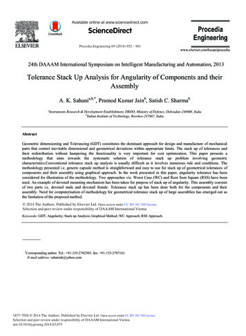
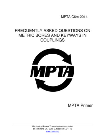
Table 1—Characteristics of Study Subjects*SubjectNo.Age, yrSex1234567891011121314151617181920Mean6 MMFMMMMMMMFMMMMMMMMMBMI,kg/m2Pao2,mm HgPaco2,mm HgVC, L (%)FEV1, L(%)FEV1/VC, 46.352.351.653.751.444.052.642.348.034.891.53 (41)2.52 (60)1.15 (55)1.85 (50)2.1 (48)2.27 (47)1.52 (39)1.8 (47)1.43 (35)2.95 (64)1.83 (68)1.96 (45)2.77 (70)1.61 (43)1.43 (34)1.04 (30)2.35 (63)2.96 (70)1.99 (52)2.56 (65)1.98 (51.30)0.57 (12.54)0.92 (32)0.84 (26)0.4 (24)0.76 (27)0.92 (27)1.04 (28)0.8 (26)1(32)0.64 (21)1.12 (33)0.72 (33)0.84 (25)1.8 (53)0.68 (24)0.6 (20)0.64 (25)1.04 (36)1.16 (36)1.32 (40)0.76 (24)0.90 (29.6)0.30 55.4726.09*BMI 5 body mass index; NA 5 not available; M 5 male; F 5 female.All patients had moderate to severe stable COPD,with a predicted mean FEV1 of 29.6 6 7.6%, apredicted mean FVC of 51.3 6 12.5%, and a predicted mean residual volume to total lung capacityratio of 155.47 6 26.09%. No subject had any clinical sign suggesting an associated sleep apnea syndrome. Mean body mass index was 23.94 6 4.17kg/m2. Mean Pao2 at rest was 54.9 6 5.54 mm Hg,and most patients had hypercapnia (meanPaco2 5 48.03 6 4.89 mm Hg). Two patients hada Pao2 value . 60 mm Hg in room air and in stablestate, but they were included in the study. The firstpatient was clinically unstable for many years, experiencing dyspnea with clinical signs, ECG, and chestradiographs indicating right heart failure. On entering the study, this patient was treated by ambulatoryliquid oxygen because of significant desaturationswith exercise. The second patient had been hypercapnic for several years and had a typical COPDpattern with frequent bronchospastic exacerbations.All subjects were current or ex-smokers with asmoking history of . 10 pack-years. In room air (N1),apnea index (AI) was 2.1 6 1.6 events/h and the apneahypopnea index (AHI) was 4.8 6 4.3 events/h. Oxygensupplementation with or without DODS did not induce any significant increase of nocturnal apneas orhypopneas (Fig 1) at N2 (AI, 2.1 6 1.8 events/h; AHI,4.9 6 4.0), N3 (AI, 2.3 6 2.0; AHI, 4.5 6 3.4), or N4(AI, 2.3 6 1.4; AHI, 5.0 6 3.9).Oxygen Saving With the DODSTo achieve a transcutaneous Sao2 of . 95%,oxygen flow rates were regulated slightly higherwhen using a DODS device than with nasal cannulasalone (respectively, 3.0 6 1.4 L/min and 2.4 6 0.9L/min). The mean oxygen consumption was 1.669 kgfor N2 and 1.379 kg for N4. Use of a DODS devicepermitted highly significant saving because oxygenconsumption was 0.573 kg for N3 (p 5 0.003 comparing N2 with N3), which represents approximately60% reduction in oxygen consumption (Fig 2).Nocturnal SaO2 Using Oxygen With or Without theDODSWith DODS 1 room air (N1), all 20 patients spenta mean of 52 6 35.2% (extremes, 0% to 100%) oftotal recording time with an Sao2 of , 90%. Whenreceiving oxygen, 18 patients spent 100% of thenight with an Sao2 level of 90%, whether theDODS was used (N3) or not (N2) (Fig 3). Twopatients (Nos. 1 and 18) who spent only 1% of thenight with an Sao2 level of , 90% when receivingoxygen (N2 and N4) had a worsened nocturnaldesaturation when given DODS 1 oxygen (N3),with 13% and 37% of the night, respectively, spentwith an Sao2 level of , 90%. Both patients werecharacterized as having rather low Pao2 levels in24Clinical InvestigationsDownloaded from chestjournal.chestpubs.org by guest on February 18, 2011 1999 American College of Chest Physicians
Figure 1. Values of AHI calculated from total recording time. No statistical differences were foundbetween the values recorded for each night.room air. However, they had no greater pulmonaryfunction alterations when compared with the otherpatients.Total recording time with an Sao2 of 95% wasslightly longer during N2 (69.4 6 32.4%) and N4(73.0 6 26.3%) than during N3 (61.5 6 31.0%), butthis difference was not considered statistically significant. The recording time with an Sao2 between 90%and 95% was 29.8 6 31.5% for N2, 26.4 6 26.2% forN4, and 35.9 6 27.3% for N3 (p 5 0.093; not significant [NS]).Sleep Characteristics When Using Oxygen With orWithout the DODSAn EEG recording for N3 was not available forone patient because of an EEG clock failure. Therefore, final polysomnographic results are presentedfor only 19 patients (Table 2). Regardless of therecording conditions, REM sleep time was belowthe 20% level usually found in non-COPD patients8: oxygen alone (N2), 12.3 6 8.7%; DODS 1oxygen (N3), 16.4 6 7.8%; and oxygen alone (N4),Figure 2. The mean amounts of oxygen consumed were identical for N2 and N4. Using a DODSdevice (N3) allowed highly significant savings (*, p 5 0.003).CHEST / 116 / 1 / JULY, 1999Downloaded from chestjournal.chestpubs.org by guest on February 18, 2011 1999 American College of Chest Physicians25
Figure 3. Nocturnal oxygen saturation values are expressed in percentage of total recording time.There were no statistical differences for any parameters between the two oxygen nights N2 and N4.Times spent at different Sao2 levels were statistically similar between N2 and N3, and between N4 andN3 (p 5 0.093).15.3 6 7.0%. The percentage of the night spent inNREM sleep was 83.7 6 7.9% for N3 comparedwith 87.7 6 8.7% for N2 and 84.7 6 7.0% for N4(p . 0.05). The percentage of stage 1/2 NREM sleepwas 45.7 6 14.2% during N3, 51.2 6 16.4% duringN2, and 49.6 6 12.5% during N4 (NS). The percentage of stage 3/4 NREM sleep was 37.8 6 14% for N3vs 36.5 6 13.1% for N2 and 35.1 6 14.1% for N4(NS) (Table 2). The patients spent slightly more timein REM and stage 3– 4 NREM sleep during N3, andthey woke up slightly less often, but the differencecompared with N2 and N4 was NS.In normal individuals, SOL is normally observedbetween 15 and 20 min. Mean values obtainedfrom our COPD patients were always . 30 minbut were not statistically different between thenights: 38.4 6 33.5 min for N2, 52.8 6 50.7 minfor N3, and 38.2 6 33.0 min for N4. REM SOL,normally found between 60 and 90 min, was151.4 6 101.6 min for N2, 109.0 6 68.8 min forN3, and 110.2 6 91.0 min for N4. The quantity ofsleep assessed by the sleep efficiency index, whichis normally 90%, was less than normal in thisstudy, but no significant difference was observedbetween N3 and N2 or N4 (Table 2). The efficiency index was 69.6 6 14.0%, with a mean number of arousals per night of 14.8 6 6.9 for N3;71.0 6 15.3%, with a mean number of arousals of16.4 6 8.3 for N2; and 68.5 6 15.0%, with a meannumber of arousals of 16.1 6 4.9 for N4. Patients1 and 18, who had less satisfactory Sao2 corrections while receiving oxygen 1 DODS whensleeping, did not have significantly different sleepparameters.DiscussionRespiratory Pattern During Sleep in COPDPatients With and Without the DODSSleep-related hypoxemia is a primary issue inmanaging severe COPD patients. Nocturnal desaturations are well related to the severity of diurnal Sao2at rest,8,9 but they cannot be reliably predicted fromarterial oxygen desaturation during maximal exercise.10 In a vast majority of COPD patients, nocturnal hypoxemia is mainly related to hypoventilationduring REM sleep because of a marked decrease ofcentral respiratory drive and hypotonia of accessoryrespiratory muscles.8 Hypoventilation also occursduring NREM sleep, secondary to a decreased basalmetabolic rate and an increased upper airways resistance.8 Additional ventilation/perfusion mismatchdoes occur during REM sleep,11 but the extent ofthis phenomenon is not well known. Sleep apneas orhypopneas are triggering factors of nocturnal hypoxemia in only a minority of COPD patients. Sleepparameters are profoundly altered in COPD patients8,12–14 with a reduction of TST, REM sleep, andstage 3– 4 NREM sleep. In contrast to reportedstudies,13,15,16 current research has demonstratedthat sleep pattern and quality are improved bynocturnal oxygen therapy.8,17–19As previously mentioned, we observed a highlysignificant oxygen saving when using the DODSdevice (approximately 60%). Despite this oxygensaving, correction of nocturnal oxygenation (definedby the conventional Sao2 threshold of 90%) was aseffective as with oxygen alone, except for two patients. EEG records showed a marked reduction of26Clinical InvestigationsDownloaded from chestjournal.chestpubs.org by guest on February 18, 2011 1999 American College of Chest Physicians
Table 2—Sleep Architecture*N2 (O2)REM sleep, %NREM sleep, %Stage 1-2, %Stage 3-4, %SOL, minREM latency, minSleep efficiency, %Arousals, No.12.3 6 8.787.7 6 8.751.2 6 16.436.5 6 13.138.4 6 33.5151.4 6 101.671 6 15.316.4 6 8.3N3(O2 1 DODS)N4 (O2)16.4 6 7.815.3 6 783.7 6 7.984.7 6 745.7 6 14.2 49.6 6 12.537.8 6 1435.1 6 14.152.8 6 50.7 38.2 6 33109 6 68.8 110.2 6 9169.6 6 1468.5 6 15.14.8 6 6.916.1 6 4.9p s given as mean 6 SD.TST and sleep efficiency (# 70%) with or withoutthe DODS. The duration of wakefulness duringsleep was higher than expected, with an average of100 min, but not statistically different when usingoxygen with or without the DODS. In addition to theunderlying respiratory disease, the higher number ofarousals and the increased REM and NREM sleeplatencies were also responsible for the poor quality ofsleep. Although none of these patients had obstructive apnea syndrome, the number of arousals washigh (at least 14 per night), with no significantdifference between nights. A higher mean value ofSOL was observed when patients usedoxygen 1 DODS (N3) than when they used oxygenalone (N2 or N4), and this was the only EEGparameter suggesting an alteration of sleep associated with DODS use; however, this difference wasnot statistically significant (p 5 0.563). The constraints related to the protocol (early bedtimes andwaking times) may have been responsible for lengthening of the sleep latencies and an artificial interruption of the morning sleep. Sleep staging revealed ahigher percentage of stage 3– 4 NREM sleep in ourpatients (37.8% with the DODS and 35.8% withoutthe DODS) as compared with healthy adults (normally 25%). This observation is unusual in COPDpatients in whom stage 3– 4 NREM and REM sleepare commonly decreased and contribute to daytimesleepiness. In healthy adults, stage 3– 4 NREM sleepis concentrated during the first half of the sleepcycle, and REM and stage 1–2 NREM sleep areobserved during the second half. Stage 3– 4 NREMsleep sometimes disappears from the last two cyclesof sleep, which generally comprises four to six cycles.In our patients, a long SOL and an early awakeningcould possibly account for this abnormal sleep distribution, with a relatively large proportion of stage3– 4 NREM sleep. Moreover, age-related modifications of the various sleep stages should be taken intoaccount20: a reduction of stage 3– 4 NREM sleep anda more homogeneous distribution of the varioussleep stages during the night are often observed withincreasing age. Compared with healthy adults, ourCOPD patients had a decreased percentage of REMsleep but without any significant difference wheneither oxygen alone or oxygen 1 DODS was used.Interest in DODS Use Among COPD PatientsOxygen-conserving devices, such as reservoir nasalcannulas, transtracheal oxygen catheters, or the demand oxygen delivery valves, are effective ways toreduce oxygen costs and increase patient autonomy.4,6,21–25 The characteristic of a demand oxygendelivery valve is to deliver the gas at the beginning ofeach inspiration. In COPD patients breathing at arespiratory rate of 20/min and an inspiratory toexpiratory ratio of 1 : 2, each respiratory cycle lasts3 s with 1 s devoted to inspiration and 2 s devoted toexpiration.26 Only the gas inhaled during the firsthalf of inspiration (representing two thirds of thetidal volume) participates in alveolar ventilation, thelast third ventilating the dead space. Because onlyoxygen inhaled during the first 0.5 s of the respiratory cycle is used for oxygen exchange, oxygentherapy might be maximized by delivering the gasonly during the first 0.5 s of inspiration.A number of studies have shown that DODSdevices ensure good quality oxygenation atrest4,6,26 –29 or during exercise,28 –32 although a recentstudy has cast some doubt on their ability to correctthe most profound desaturations during exercise.33Indeed, very few studies have confirmed that suchDODS devices do not influence quality of sleep andcan be safely prescribed in COPD patients. To ourknowledge, only the study reported by Bower et al28has addressed this issue. These authors demonstrated the efficacy of DODS devices in maintainingSao2 during sleep in six hypoxemic COPD or restrictive patients.28 However, a French study concerningtwo DODS devices (Optimox and COS5 [NellcorPuritan Bennett]) reported unsatisfactory results forquality of nocturnal oxygen therapy among patientswith chronic respiratory insufficiency.34 These results were partly attributed by the authors to theabsence of the apnea card with the Optimox valve.Furthermore, the COS5 valve equipped with thesafety system triggered by apnea did not ensuremore satisfactory Sao2. These authors reported thatthe oxygen flow rate when using the DODS wasequivalent to the flow rate delivered by continuousnasal oxygen therapy and was not previously adjustedindividually according to the transcutaneous oxygensaturation as performed in our study. Indeed, theoxygen flow rate we used with DODS was adjustedto the patient requirements and was finally higherthan the continuous oxygen flow rate (respectively,3.0 6 1.4 L/min and 2.4 6 0.9 L/min). Similar conCHEST / 116 / 1 / JULY, 1999Downloaded from chestjournal.chestpubs.org by guest on February 18, 2011 1999 American College of Chest Physicians27
ditions were applied in the study conducted byKerby et al6 among hospitalized patients.Limits and Tolerance of the DODS DeviceChallenging issues still persist concerning the tolerance of DODS devices. Patients may hear a slightclicking sound and may feel a small burst of oxygenassociated with activation of the valve during inspiration. Some studies have adopted a subjective approach to this problem, and questionnaires revealedthat patients often complain of the auditory discomfort at night.34 Because of the Medilog systemtechnology, our study may have overlooked thepresence of microarousals from a clicking sound or asudden burst of oxygen. However, such discomfortdid not influence significantly the sleep parametersof our patients. A more serious difficulty may belinked to the changing respiratory pattern associatedwith nocturnal apneas or mouth breathing duringsleep. This issue could not be addressed in our studybecause patients did not have any nocturnal apneawhen breathing either room air or oxygen. As a safetyfactor, the apnea card appears essential to avoidinterruptions of oxygen flow rate, and it may contribute to good oxygen saturation in patients with sleepapnea syndrome and COPD (overlap syndrome). Inthe study by Bower et al,28 the oxygen-saving devicesensed the negative inspiratory pressure wheneverthe patient was sleeping with open or closed mouth.Finally, use of the DODS device appears to be safe;unlike continuous oxygen therapy, pulsed oxygentherapy does not induce nasal dryness.4,35 Kerby etal6 estimated that the oxygen savings and humidifiercosts would cover the expense of a DODS devicewithin a 2-year period.ConclusionIn summary, the DODS device ensures goodquality oxygenation among a great majority of patients with moderate to severe COPD without sleepapnea syndrome and no significant alteration ofnocturnal neurophysiologic profiles, provided thatthe oxygen flow rate is adjusted individually to asatisfactory transcutaneous Sao2. However, persistence of nocturnal desaturation in very few patientsin our study still justifies providing a night of systematic ventilation polygraphy recording when prescribing a DODS device. Moreover, further studies arerequired to confirm the efficacy of this device inpatients with an abnormal AHI.ACKNOWLEDGMENT: The authors are grateful to Drs. H.Ziani-Bey, A. Verdure, and A. Portmann for their assistance inthis work and their valuable guidance in constructing the study,and to Mr. Richard Medeiros for his advice in editing themanuscript.References1 Nocturnal Oxygen Therapy Trial Group. Continuous or nocturnal oxygen therapy in hypoxemic chronic obstructive lungdisease: a clinical trial. Ann Intern Med 1980; 93:391–3982 Report of the Medical Research Council Working Party.Long term domiciliary oxygen therapy in chronic cor pulmonale complicating chronic bronchitis and emphysema. Lancet1981; 1:681– 6853 Vergeret J, Brambilla C, Mounier L. Portable oxygen therapy:use and benefit in hypoxaemic COPD patients on long termoxygen therapy. Eur Respir J 1989; 2:20 –254 Tiep BL, Lewis MI. Oxygen conservation and oxygen conserving devices in chronic lung disease: a review. Chest 1987;92:263–2725 Robert D, Leger P, Perrin F. Evaluation of an intermittentflow oxygen system. Bull Eur Physiopathol Respir 1986;22:315–3186 Kerby GR, O’Donohue WJ, Romberger DJ, et al. Clinicalefficacy and cost benefit of pulse flow oxygen in hospitalizedpatients. Chest 1990; 97:369 –3727 Rechtshaffen A, Kales A, eds. A manual of standardizedterminology techniques and scoring systems for sleep stagesof human subjects. Bethesda, MD: National Institute ofNeurological Disease and Blindness, 1968; NIH publication2048 Douglas NJ. Sleep in patients with chronic obstructive pulmonary disease. Clin Chest Med 1998; 19:115–1259 Muir JF, Rose D, Aubry P, et al. Perturbations ventilatoires etanomalies du sommeil de l’insuffisant respiratoire chroniqueobstructif grave. Presse Med 1983; 12:1701–170610 Mulloy E, McNicholas WT. Ventilation and gas exchangeduring sleep and exercise in severe COPD. Chest 1996;109:387–39411 Catterall JR, Douglas NJ, Calverley PMA, et al. Transienthypoxemia during sleep in chronic obstructive pulmonarydisease is not a sleep apnea syndrome. Am Rev Respir Dis1983; 128:24 –2912 Douglas NJ, Flenley DC. Breathing during sleep in patientswith obstructive lung disease. Am
liquid oxygen therapy has recently been developed; and, currently, portable gas cylinders may be ob-tained from a compressor linked to a concentrator. The use of these ambulatory devices raises the issue of oxygen preservation either from an economic point of view or to maximize patient autonomy. Demand oxygen delivery system (DODS) devices