Transcription
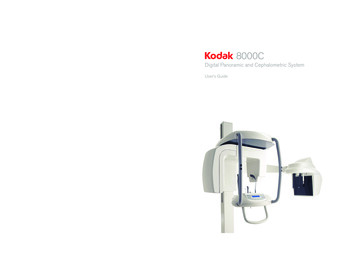
8000C8000C SystemTrophyA Subsidiary of Carestream Health, Inc.4 rue F. Pelloutier – Croissy-Beaubourg77435 Marne la Vallée Cedex 2 (France) 33 1 64 80 85 00www.kodakdental.com Carestream Health, Inc., 2010.The Kodak trademark and trade dressare used under license from Kodak.User’s GuideFor more information, visit:SM735Ed0203/2010Digital Panoramic and Cephalometric SystemUser’s Guide
NoticeCongratulations on your purchase of the KODAK 8000C Digital Panoramic and Cephalometric Extraoral ImagingSystem. The KODAK 8000C unit is the KODAK 8000 equipped with the cephalostat unit. Thank you for yourconfidence in our products and we will do all in our power to ensure your complete satisfaction.The User Guide for the KODAK 8000C Digital Panoramic and Cephalometric Extraoral Imaging System includesinformation on the cephalometric features. For the panoramic features, see the KODAK 8000 Extraoral ImagingSystem (SM722) User Guide. We recommend that you thoroughly familiarize yourself with this Guide in order tomake the most effective use of your system.WARNING: We recommend that you consult the “Safety, Regulatory and the TechnicalSpecification User Guide” before using the KODAK 8000C Extraoral Imaging Systems.The information contained in this Guide may be subject to modification without notice, justification or notificationto the persons concerned.No part of this Guide may be reproduced without the express permission of Carestream Health, Inc.The US Federal law restricts this device to sale by or on the order of a physician.This document is originally written in English.Manual Name: KODAK 8000C Digital Panoramic and Cephalometric Extraoral Imaging System User GuidePart Number: SM735Revision Number: 02Print Date: 03/2010The brand names and logos reproduced in this Guide are copyright.KODAK is a trademark of KODAK used under License.KODAK 8000C Digital Panoramic and Cephalometric Extraoral Imaging System, complies with Directive93/42/CEE relating to medical equipment.0086ManufacturerCare stream Hea lth, Inc.150 Verona StreetRoche ster NY 14 608Authorized Representative in the European CommunityEC REPTROPHY4, Rue F. Pelloutier, Croissy-Beaubourg77435 Marne la Vallée Cedex 2, France
Contents1—About This GuideConventions in this Guide. . . . . . . . . . . . . . . . . . . . . . . . . . . . . . . . . . . . . . . . . . . . . . . . . . . . . . . . . . . . . . . . . . . . . 1–12—KODAK 8000C UNIT OVERVIEWGeneral Overview . . . . . . . . . . . . . . . . . . . . . . . . . . . . . . . . . . . . . . . . . . . . . . . . . . . . . . . . . . . . . . . . . . . . . . . . . . .Mobile Components . . . . . . . . . . . . . . . . . . . . . . . . . . . . . . . . . . . . . . . . . . . . . . . . . . . . . . . . . . . . . . . . . . . . . .General Functional Components . . . . . . . . . . . . . . . . . . . . . . . . . . . . . . . . . . . . . . . . . . . . . . . . . . . . . . . . . . . .Digital Sensor Locations. . . . . . . . . . . . . . . . . . . . . . . . . . . . . . . . . . . . . . . . . . . . . . . . . . . . . . . . . . . . . . . . . . .Laser Locations . . . . . . . . . . . . . . . . . . . . . . . . . . . . . . . . . . . . . . . . . . . . . . . . . . . . . . . . . . . . . . . . . . . . . . . . . .Control Panel . . . . . . . . . . . . . . . . . . . . . . . . . . . . . . . . . . . . . . . . . . . . . . . . . . . . . . . . . . . . . . . . . . . . . . . . . . . . . . .X-Ray Remote Control Overview . . . . . . . . . . . . . . . . . . . . . . . . . . . . . . . . . . . . . . . . . . . . . . . . . . . . . . . . . . . . . . .Positioning Accessories and Replacement Parts . . . . . . . . . . . . . . . . . . . . . . . . . . . . . . . . . . . . . . . . . . . . . . . . . NG SOFTWARE OVERVIEWComputer System Requirements . . . . . . . . . . . . . . . . . . . . . . . . . . . . . . . . . . . . . . . . . . . . . . . . . . . . . . . . . . . . . . .General Software Overview . . . . . . . . . . . . . . . . . . . . . . . . . . . . . . . . . . . . . . . . . . . . . . . . . . . . . . . . . . . . . . . . . . .KODAK Dental Imaging Software . . . . . . . . . . . . . . . . . . . . . . . . . . . . . . . . . . . . . . . . . . . . . . . . . . . . . . . . . . .Cephalometric Acquisition Interface Module . . . . . . . . . . . . . . . . . . . . . . . . . . . . . . . . . . . . . . . . . . . . . . . . . .Cephalometric Acquisition Interface Module . . . . . . . . . . . . . . . . . . . . . . . . . . . . . . . . . . . . . . . . . . . . . . . . . . . . .Cephalometric Acquisition Window Overview . . . . . . . . . . . . . . . . . . . . . . . . . . . . . . . . . . . . . . . . . . . . . . . . .Cephalometric Program Pane . . . . . . . . . . . . . . . . . . . . . . . . . . . . . . . . . . . . . . . . . . . . . . . . . . . . . . . . . . .Cephalometric Patient Pane . . . . . . . . . . . . . . . . . . . . . . . . . . . . . . . . . . . . . . . . . . . . . . . . . . . . . . . . . . . .Cephalometric Parameter Pane. . . . . . . . . . . . . . . . . . . . . . . . . . . . . . . . . . . . . . . . . . . . . . . . . . . . . . . . . GETTING STARTEDSwitching on the Unit . . . . . . . . . . . . . . . . . . . . . . . . . . . . . . . . . . . . . . . . . . . . . . . . . . . . . . . . . . . . . . . . . . . . . . . .Starting the Imaging Software . . . . . . . . . . . . . . . . . . . . . . . . . . . . . . . . . . . . . . . . . . . . . . . . . . . . . . . . . . . . . . . . .Creating a Patient Record. . . . . . . . . . . . . . . . . . . . . . . . . . . . . . . . . . . . . . . . . . . . . . . . . . . . . . . . . . . . . . . . . . . . .Accessing the Cephalometric Acquisition Window . . . . . . . . . . . . . . . . . . . . . . . . . . . . . . . . . . . . . . . . . . . . . . . .4–14–24–24–35—ACQUIRING CEPHALOMETRIC IMAGESAcquiring a Lateral Image . . . . . . . . . . . . . . . . . . . . . . . . . . . . . . . . . . . . . . . . . . . . . . . . . . . . . . . . . . . . . . . . . . . . . 5–1Preparing the Unit and Setting the Acquisition Parameters . . . . . . . . . . . . . . . . . . . . . . . . . . . . . . . . . . . . . . 5–1Acquiring a Frontal AP or PA Image. . . . . . . . . . . . . . . . . . . . . . . . . . . . . . . . . . . . . . . . . . . . . . . . . . . . . . . . . . . . . 5–5Preparing the Unit and Setting the Acquisition Parameters . . . . . . . . . . . . . . . . . . . . . . . . . . . . . . . . . . . . . . 5–5Preparing and Positioning the Patient . . . . . . . . . . . . . . . . . . . . . . . . . . . . . . . . . . . . . . . . . . . . . . . . . . . . . . . . 5–6Acquiring an Oblique Image . . . . . . . . . . . . . . . . . . . . . . . . . . . . . . . . . . . . . . . . . . . . . . . . . . . . . . . . . . . . . . . . . . . 5–9Preparing the Unit and Setting the Acquisition Parameters . . . . . . . . . . . . . . . . . . . . . . . . . . . . . . . . . . . . . . 5–9Preparing and Positioning the Patient . . . . . . . . . . . . . . . . . . . . . . . . . . . . . . . . . . . . . . . . . . . . . . . . . . . . . . . 5–10Acquiring a Submento-Vertex Image . . . . . . . . . . . . . . . . . . . . . . . . . . . . . . . . . . . . . . . . . . . . . . . . . . . . . . . . . . . 5–12Preparing the Unit and Setting the Acquisition Parameters . . . . . . . . . . . . . . . . . . . . . . . . . . . . . . . . . . . . . 5–12Preparing and Positioning the Patient . . . . . . . . . . . . . . . . . . . . . . . . . . . . . . . . . . . . . . . . . . . . . . . . . . . . . . . 5–13KODAK 8000C Digital Panoramic and Cephalometric Extraoral Imaging System User Guide (SM735) Ed 02iii
ContentsAcquiring a Carpus Image . . . . . . . . . . . . . . . . . . . . . . . . . . . . . . . . . . . . . . . . . . . . . . . . . . . . . . . . . . . . . . . . . . . . 5–16Preparing the Unit and Setting the Acquisition Parameters . . . . . . . . . . . . . . . . . . . . . . . . . . . . . . . . . . . . . 5–16Preparing and Positioning the Patient . . . . . . . . . . . . . . . . . . . . . . . . . . . . . . . . . . . . . . . . . . . . . . . . . . . . . . . 5–17X-Ray Dose Emission Information . . . . . . . . . . . . . . . . . . . . . . . . . . . . . . . . . . . . . . . . . . . . . . . . . . . . . . . . . . . . . . 5–196—MAINTENANCEDaily . . . . . . . . . . . . . . . . . . . . . . . . . . . . . . . . . . . . . . . . . . . . . . . . . . . . . . . . . . . . . . . . . . . . . . . . . . . . . . . . . . . . . . . 6–1Monthly . . . . . . . . . . . . . . . . . . . . . . . . . . . . . . . . . . . . . . . . . . . . . . . . . . . . . . . . . . . . . . . . . . . . . . . . . . . . . . . . . . . . 6–1Annually . . . . . . . . . . . . . . . . . . . . . . . . . . . . . . . . . . . . . . . . . . . . . . . . . . . . . . . . . . . . . . . . . . . . . . . . . . . . . . . . . . . . 6–17—TROUBLESHOOTINGQuick Troubleshooting . . . . . . . . . . . . . . . . . . . . . . . . . . . . . . . . . . . . . . . . . . . . . . . . . . . . . . . . . . . . . . . . . . . . . . . . 7–1iv
Chapter 1About This GuideConventions in this GuideThe following special messages emphasize information or indicate potential risk topersonnel or equipment:WARNINGWarns you to avoid injury to yourself or others by following thesafety instructions precisely.CAUTIONAlerts you to a condition that might cause serious damage.IMPORTANTAlerts you to a condition that might cause problems.NOTEEmphasizes important information.TIPProvides extra information and hints.KODAK 8000C Digital Panoramic and Cephalometric Extraoral Imaging System User Guide (SM735) Ed 021–1
Conventions in this Guide1–2About This Guide
Chapter 2KODAK 8000C UNIT OVERVIEWThe KODAK 8000C digital panoramic and cephalometric unit is designed to carry out thefollowing radiological examinations: PanoramicMaxillary SinusTemporomandibular Joints (TMJ)Lateral cephalometricFrontal (PA or AP) cephalometricOblique cephalometricSubmento-vertex cephalometricCarpus cephalometricGeneral OverviewThe KODAK 8000C digital panoramic and cephalometric unit is composed of thefollowing functional components: The unit head that contains all the electronic controlThe rotative armThe fixed arm with a control panelThe panoramic digital sensorThe x-ray source assemblyThe x-ray remote controlThe chin rest and bite blockThe chin rest baseThe panoramic chin rest and bite blockThe temple supportsThe hand gripsThe cephalostat armThe cephalostat headThe head clamps and ear conesThe nasion supportThe acquisition software (see “Imaging Software Overview”)The following figures illustrate the general overview of the KODAK 8000C digitalpanoramic and cephalometric units.KODAK 8000C Digital Panoramic and Cephalometric Extraoral Imaging System User Guide (SM735) Ed 022–1
General OverviewIMPORTANTThe Cephalostat can be positioned either on the right or theleft side of the KODAK 8000 unit.Mobile ComponentsFigure 2-1 illustrates the up and down movement of the KODAK 8000C digitalpanoramic and cephalometric units mobile component and the rotation of the rotativearm.Figure 2–12–2KODAK 8000C UNIT OVERVIEWKODAK 8000 and KODAK 8000C Units Mobile Components
General OverviewGeneral Functional ComponentsFigure 2-2 illustrates the general functional components of the KODAK 8000C digitalpanoramic and cephalometric units.Figure 2–2KODAK 8000 and KODAK 8000C Units Functional Components1416115171611839127106421351ON/OFF button10Collimator selector2Chin rest and bite block11Unit rotative arm3Temple supports12X-Ray remote control4Temple supports control knob13PC hosting the imaging and the acquisitionsoftware5Hand Grips14Cephalostat arm6Control panel15Cephalostat head7Height adjustment buttons16Head clamps and ear cones8Panoramic digital sensor17Nasion support9X-Ray source assemblyKODAK 8000C Digital Panoramic and Cephalometric Extraoral Imaging System User Guide (SM735) Ed 022–3
General OverviewDigital Sensor LocationsFigure 2-3 illustrates the locations of the digital panoramic and digital cephalometricsensors of the KODAK 8000C digital panoramic and cephalometric units.Figure 2–3KODAK 8000 and KODAK 8000C Units Digital Sensor Locationssystem2–4KODAK 8000C UNIT OVERVIEW
General OverviewLaser LocationsFigure 2-4 illustrates the location of the lasers of the KODAK 8000C digital panoramicand cephalometric units.Figure 2–4KODAK 8000 and KODAK 8000C Units Laser Beam Locations43211Mid-sagittal plane positioning laser beam2Frankfort plane positioning laser beam3Canine plane positioning laser beam4Cephalometric Frankfort plane positioning laser beamKODAK 8000C Digital Panoramic and Cephalometric Extraoral Imaging System User Guide (SM735) Ed 022–5
Control PanelControl PanelThe control panel is an alphanumeric, digital soft touch console. It allows the operator tocontrol certain unit functions. It also displays the operating parameters and errormessages.Figure 2–5Unit Control Panel8Digital Panoramic and Cephalometric SystemkV12–6mAS2341X-Ray emission LED: Yellow, indicates the x-rays are being emitted.2Display Screen: Displays the current acquisition parameters and the error messages.3Reset button: Resets the unit arm to the initial position to enable the patient to enterand exit the unit.4Laser beam button: Activates the laser positioning beams to correctly position thepatient.KODAK 8000C UNIT OVERVIEW
X-Ray Remote Control OverviewX-Ray Remote Control OverviewThe x- ray remote control enables you to launch a radiological image acquisition via theexposure button from outside the x-ray room. You must press and hold the exposurebutton until the end of acquisition. Premature release of the exposure button interruptsthe acquisition.Figure 2–6X-Ray Remote Control11Exposure button: launches image acquisition.KODAK 8000C Digital Panoramic and Cephalometric Extraoral Imaging System User Guide (SM735) Ed 022–7
Positioning Accessories and Replacement PartsPositioning Accessories and Replacement PartsThe following accessories are used when positioning a patient. They are delivered withthe KODAK 8000 digital panoramic and KODAK 8000C digital panoramic andcephalometric unit.Table 2-1 and Table 2-2 list the panoramic and cephalometric positioning accessories.Table 2–1Panoramic Positioning Accessories and Replacement PartsAccessoryDescriptionPanoramic chin rest and TMJ x2Maxillary sinus chin restTMJ x2 and TMJ x4 nose restStandard bite blockBite block for edentulous patientsA set of right and left temple supportsx2Single use sheaths for bite blocks(500 pcs box)2–8KODAK 8000C UNIT OVERVIEW
Positioning Accessories and Replacement PartsTable 2–2Cephalometric Positioning Accessories and Replacement PartsAccessoryDescriptionHead clamps with ear conesx2Nasion supportKODAK 8000C Digital Panoramic and Cephalometric Extraoral Imaging System User Guide (SM735) Ed 022–9
Positioning Accessories and Replacement Parts2–10KODAK 8000C UNIT OVERVIEW
Chapter 3IMAGING SOFTWARE OVERVIEWComputer System RequirementsThis section specifies the minimum computer system requirements for KODAK 8000Cdigital panoramic and cephalometric extraoral imaging system software.IMPORTANTIt is MANDATORY to check that the computer system configuration iscompatible with the computer system requirements for the KODAK8000C software. If necessary you MUST update your computersystem configuration. KODAK 8000C MUST be connected to thecomputer via a point-to-point Ethernet link and not via a LAN. DO NOTplace the computer and the peripheral equipment connected to it inthe immediate vicinity of the patient in the unit. Leave at least 1.5 mdistance from the unit. The computer and the peripheral equipmentmust conform to the IEC 60950 standard.Table 3–1Minimum Computer System RequirementsItemViewingAcquisitionCPU2 GHz Intel Duo Core2 GHz Intel Duo CoreRAM2 GB2GB Hard disk drive Graphic board1.2 GB for softwareinstallation Operating system (32 bits) RAM has a major impact onsystem performance.1.2 GB for software installation80 GB free space to use thesoftwareNvidia / ATI based board supportingOpen GL 1.2 with 256 MB of videoRAM on AGP x8 video bus (forexample: Nvidia GeForce 6800 GT)MonitorComments1 monitor17" or larger1024 x 768 minimum screenresolution - 32 bits colormodeNvidia / ATI based boardThe video RAM has majorsupporting Open Glide 1.2 withimpact on system performance.256 MB of video RAM on AGP x8video bus (for example: NvidiaGeForce 6800 GT) Windows 2000 SP4 Windows XP Home / Proedition SP2 1 monitor17"1280 x 880 minimumscreen resolutionYour monitor is a vitalcomponent in displaying qualityimages. Low-quality screens willprevent you from properdiagnoses and treatment.Windows XP Home / Proedition SP2Windows VistaWindows VistaKODAK 8000C Digital Panoramic and Cephalometric Extraoral Imaging System User Guide (SM735) Ed 023–1
General Software OverviewTable 3–1Minimum Computer System Requirements (Continued)ItemViewingEthernet interface1 Ethernet interfaceCommentsAcquisition1 Ethernet interfaces (100Mbits)If there is a LAN connection asecond Ethernet interface isrequired.CD/DVD driveA DVD-BURNER drive is required.A DVD-BURNER drive is required.Backup MediaRemovable/portable, external harddisk driveRemovable/portable, external hard We strongly recommend a dailydisk drive.backup of x-ray images andpatient records.General Software OverviewThe KODAK 8000C digital panoramic and cephalometric extraoral imaging systemoperates with the following software: KODAK dental imaging softwareCephalometric acquisition interface moduleKODAK Dental Imaging SoftwareThe KODAK dental imaging software is a user-friendly working interface that wasdesigned and developed specifically for radiological diagnosis. It is the common imagingplatform for all our digital systems for dentistry.The KODAK dental imaging software has the following features: Patient record management using Patient Window featuresExtraoral and intraoral image management using Imaging Window featuresNOTEFor a complete information on the KODAK Dental ImagingSoftware, see the “KODAK Dental Imaging Software, Quick StartGuide”.Cephalometric Acquisition Interface ModuleThe cephalometric acquisition interface module is a user-friendly working interface thatwas designed and developed specifically for KODAK 8000C digital panoramic andcepahlometric extraoral imaging system.3–2IMAGING SOFTWARE OVERVIEW
Cephalometric Acquisition Interface ModuleCephalometric Acquisition Interface ModuleCephalometric Acquisition Window OverviewThe Cephalometric Acquisition Window is the main cephalometric interface with theKODAK 8000C cephalometric extraoral imaging system that provides you with imagingacquisition functions.Figure 3–11Cephalometric Acquisition WindowInformation button: About: Identifies the Software and the Firmware versions Reset of the Values: Resets to the manufacturing parameter settingsMemorize settings: Memorizes the user preference settings for each patient type (kV,mA and seconds)2Preview Screen: Displays the acquired image in real time.3Selected Parameter Display: Displays the current acquisition parameter settings.4System Status Screen: Displays various alert or warning messages originating from the unit.5X-Rays ON / OFF button: Turns off the x-ray emissions to demonstrate the acquisition processfor the patient.6Generator Cooling indicator: Indicates the automatic cooling time (mm:ss) required for thegenerator to reach 0 for a new acquisition.KODAK 8000C Digital Panoramic and Cephalometric Extraoral Imaging System User Guide (SM735) Ed 023–3
Cephalometric Acquisition Interface Module7Positioning laser button: Activates the laser positionning beams to correctly position thepatient.8Reset button: Resets the rotative arm in the start position.9Ready Indicator LED Green indicates the unit is ready to start acquisition. Black indicates the unit is not ready to start acquisition.10Exit button: Closes the Acquisition Window.11X-Ray Emission indicator: Yellow, indicates the x-ray emission status.12Selector Button: Selects different acquisition setting options. Click Program to select examination type options. Click Patient to select patient type parameters.Click Parameters to select exposure parameter options.The Selector button enables you to access the following 3 panes: 3–4Program pane: Examination type optionsPatient pane: Patient type parameter optionsParameters pane: Exposure parameter optionsIMAGING SOFTWARE OVERVIEW
Cephalometric Acquisition Interface ModuleCephalometric Program PaneThe cephalometric Program pane enables you to choose different radiological exams aswell as different acquisition formats.Figure 3–2Cephalometric Program PaneRadiological exam options:Clickfor a lateral exam.Clickorexam (AP and PA).Clickfor a frontalClickClickfor a submento-vertex exam.for a carpus exam.for an oblique exam.NOTEThe above list of exam types are only a sample of exam options ofthe Program pane.KODAK 8000C Digital Panoramic and Cephalometric Extraoral Imaging System User Guide (SM735) Ed 023–5
Cephalometric Acquisition Interface ModuleCephalometric Patient PaneThe cephalometric Patient pane enables you to choose different patient parameters. Theselection of the patient parameters influences the quality of the image. The selectedparameters must be based on the patient age and morphology.Figure 3–3123–6Cephalometric Patient PanePatient type parameters:Clickif the patient is a child.Clickif the patient is adult.Patient size parameters:Clickif the patient is small.Clickif the patient is medium.Clickif the patient is large.IMAGING SOFTWARE OVERVIEW
Cephalometric Acquisition Interface ModuleCephalometric Parameter PaneThe cephalometric Parameter pane enables you to choose exposure parameters for theradiological image acquisition. If the default parameter setting is not adapted to yourpatient type, you can manually adapt the parameter settings to the patient type and savethis setting as the default setting.Figure 3–41Cephalometric Parameter PaneRadiation dose options:kV: KilovoltmA: MilliampereS: Second2Fine-tuning buttons:Click3to fine-tune the kV, mA and the second.Saving parameter button:Clickto save the selected parameters.KODAK 8000C Digital Panoramic and Cephalometric Extraoral Imaging System User Guide (SM735) Ed 023–7
Cephalometric Acquisition Interface Module3–8IMAGING SOFTWARE OVERVIEW
Chapter 4GETTING STARTEDSwitching on the UnitBefore switching on the unit, check that: The installation of the unit is complete.The PC is switched on.IMPORTANTYou must switch on the PC and wait for it to be ready for theconnection before switching on the unit.To switch on the unit, follow these steps:1. On the unit column, press the ON button.2. You must wait for a minute for the connection between the unit and the PC to beestablished. If you start the imaging software before the connection is established anerror message is displayed. Click OK, close the imaging software and wait for theconnection to be established.3. You can now proceed to start the imaging software.IMPORTANTTo increase the operating life of the x-ray tube, if the unit has not beenused for a month, you must follow the following procedures beforeuse.1. In the Panoramic Acquisition Window, select the Parameter pane.2. Select the following series of parameter settings: 70 kV - 6.3 mA80 kV - 10 mA85 kV - 10 mA3. Leave the x-ray room and close the door. For each parameter setting, from the x-rayremote control, press and hold the button to launch the x-rayThe unit is now ready to be used for acquisition.KODAK 8000C Digital Panoramic and Cephalometric Extraoral Imaging System User Guide (SM735) Ed 024–1
Starting the Imaging SoftwareStarting the Imaging SoftwareTo start the imaging software, follow these steps:1. On your desktop, double-click.ORFrom your PC, click Start All Programs Kodak Kodak Dental Software.A blank Patient Window is displayed.2. Create or open an existing patient record.Creating a Patient RecordTo create a patient record, follow these steps:1. In the Patient Window, from the toolbar, clickORFrom the menu bar, select Patient New.2. Enter the required patient information. The Last name, the First name and the Dateof birth fields are required.3. From the menu bar, select Picture Insert Picture to add a *.tif or *.bmp picture ofthe patient to the record. Select the picture from your directory and click Open.4. Click OK to save. The patient record is automatically assigned a 7-digit numberstarting with a letter (for example, M0000001).5. Clickto access the Imaging Window.6. Select an image acquisition.4–2GETTING STARTED
Accessing the Cephalometric Acquisition WindowAccessing the Cephalometric Acquisition WindowTo access the Acquisition Windows, follow these steps:1. In the Imaging Window, from the toolbar, clickAcquisition Window.to access the Cephalometric2. Prepare the acquisition parameters and launch an acquisition.KODAK 8000C Digital Panoramic and Cephalometric Extraoral Imaging System User Guide (SM735) Ed 024–3
Accessing the Cephalometric Acquisition Window4–4GETTING STARTED
Chapter 5ACQUIRING CEPHALOMETRIC IMAGESAcquiring a Lateral ImageBefore acquiring a lateral image, check that you have: Reset the unit rotative arm to the start position for patient to enter the unit.Selected the patient record.Accessed the Imaging Window.Accessed the Cephalometric Acquisition Window.Preparing the Unit and Setting the Acquisition ParametersTo set the acquisition parameters, follow these steps:1. Raise and block the sensor manually.2. On the x-ray source assembly, set the collimator selector to LA.3. Position the head clamps manually for the lateral exam.IMPORTANTYou must position the head clamps manually because theyare not positioned automatically from the Program pane examtype selection. In this case, the relevant exam type selectionicon becomes active.4. In the Cephalometric Acquisition Window, click the Program button to access theProgram pane. In the Program pane thefor a lateral exam is active.5. Click the Patient button to access the Patient pane. Select the patient: TypeSize6. If the default parameter setting is not adapted to your patient type, click theParameter button and in the Parameter pane select the appropriate parameters. Tosave the new parameter settings as the default settings, clickMemorize settings.fand select7. Preparing and Positioning the PatientTo prepare and position the patient, follow these steps:KODAK 8000C Digital Panoramic and Cephalometric Extraoral Imaging System User Guide (SM735) Ed 025–1
Acquiring a Lateral Image1. Ask the patient to remove all metal objects.2. Ask the patient to wear a lead apron. Ensure that the apron lays flat across thepatient’s shoulders.3. Press and hold the height adjustment buttons to raise the cephalostat head.4. Open the head clamps and ask the patient to stand up straight, in front of thecephalometric unit, in the appropriate position.5. Press and hold the height adjustment buttons to level the ear cones to the patient’sauditory canals.6. Insert gently one cone in the auditory canal of the patient. Turn gently the button toclose the arms. Insert gently the second cone in the auditory canal of the patient.7. On the control panel, clickto turn ON the Frankfort laser positioning beam.Align the patient with the Frankfort laser beam.5–2ACQUIRING CEPHALOMETRIC IMAGES
Acquiring a Lateral Image8. Lower the nasion support to a vertical position.KODAK 8000C Digital Panoramic and Cephalometric Extraoral Imaging System User Guide (SM735) Ed 025–3
Acquiring a Lateral ImageLaunching the X-rayTo launch the x-ray, follow these steps:1. Leave the x-ray room and close the door. You must keep visual contact with thepatient during acquisition.IMPORTANTTo stop the acquisition, if any problem, release the exposurebutton of the remote control or press the red emergency stopbutton.2. Launch the x-ray with the remote control. Press and hold the exposure button until theend of acquisition. Theturns yellow, indicating x-ray emission. The image appearson the Preview Screen of the Cephalometric Acquisition Window. When theacquisition ends, the Cephalometric Acquisition Window disappears and theacquired image is transferred automatically to the Imaging Window.3. Check the image quality, if satisfactory, remove the ear cones and the nasion supportand release the patient.5–4ACQUIRING CEPHALOMETRIC IMAGES
Acquiring a Frontal AP or PA ImageAcquiring a Frontal AP or PA ImageBefore acquiring a frontal AP or PA image, check that you have: Reset the unit rotative arm to the start position for patient to enter the unit.Selected the patient record.Accessed the Imaging Window.Accessed the Cephalometric Acquisition Window.Preparing the Unit and Setting the Acquisition ParametersTo acquire a frontal AP or PA image, follow these steps:1. Raise and block the sensor manually.2. On the x-ray source assembly, set the collimator selector to AP/PA.3. Position the head clamps manually for the frontal AP or PA exam.IMPORTANTYou must position the head clamps manually because theyare not positioned automatically from the Program pane examtype selection. In this case, the relevant exam type selectionicon becomes active.4. In the Cephalometric Acquisition Window, click the Program button to access theProgram pane. In the Program pane, clickfor a frontal PA exam.5. Click the Patient button to access the Patient pane. Select the patient: TypeSize6. If the default parameter setting is not adapted to your patient type, click theParameter button and in the Parameter pane select the appropriate parameters. Tosave the new parameter settings as the default settings, clickMemorize settings.fand selectKODAK 8000C Digital Panoramic and Cephalometric Extraoral Imaging System User Guide (SM735) Ed 025–5
Acquiring a Frontal AP or PA ImagePreparing and Positioning the PatientTo prepare and position the patient, follow these steps:1. Ask the patient to remove all metal objects.2. Ask the patient to wear a lead apron. Ensure that the apron lays flat across thepatient’s
General Software Overview The KODAK 8000C digital panoramic and cephalometric extraoral imaging system operates with the following software: KODAK dental imaging software Cephalometric acquisition interface module KODAK Dental Imaging Software The KODAK dental imaging software is a user-friendly working interface that was Guide".