Transcription
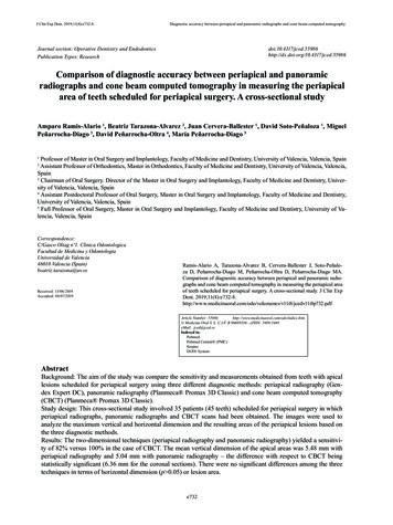
J Clin Exp Dent. 2019;11(8):e732-8.Diagnostic accuracy between periapical and panoramic radiographs and cone beam computed tomographyJournal section: Operative Dentistry and EndodonticsPublication Types: .4317/jced.55986Comparison of diagnostic accuracy between periapical and panoramicradiographs and cone beam computed tomography in measuring the periapicalarea of teeth scheduled for periapical surgery. A cross-sectional studyAmparo Ramis-Alario 1, Beatriz Tarazona-Alvarez 2, Juan Cervera-Ballester 1, David Soto-Peñaloza 1, MiguelPeñarrocha-Diago 3, David Peñarrocha-Oltra 4, María Peñarrocha-Diago 5Professor of Master in Oral Surgery and Implantology, Faculty of Medicine and Dentistry, University of Valencia, Valencia, SpainAssistant Professor of Orthodontics, Master in Orthodontics, Faculty of Medicine and Dentistry, University of Valencia, Valencia,Spain3Chairman of Oral Surgery. Director of the Master in Oral Surgery and Implantology, Faculty of Medicine and Dentistry, University of Valencia, Valencia, Spain4Assistant Postdoctoral Professor of Oral Surgery, Master in Oral Surgery and Implantology, Faculty of Medicine and Dentistry,University of Valencia, Valencia, Spain5Full Professor of Oral Surgery, Master in Oral Surgery and Implantology, Faculty of Medicine and Dentistry, University of Valencia, Valencia, Spain12Correspondence:C/Gasco Oliag nº1. Clinica OdontologicaFacultad de Medicina y OdontologiaUniversidad de Valencia46010 Valencia (Spain)beatriz.tarazona@uv.esReceived: 13/06/2019Accepted: 04/07/2019Ramis-Alario A, Tarazona-Alvarez B, Cervera-Ballester J, Soto-Peñaloza D, Peñarrocha-Diago M, Peñarrocha-Oltra D, Peñarrocha-Diago MA.Comparison of diagnostic accuracy between periapical and panoramic radiographs and cone beam computed tomography in measuring the periapical areaof teeth scheduled for periapical surgery. A cross-sectional study. J Clin ExpDent. volumenes/v11i8/jcedv11i8p732.pdfArticle Number: 55986http://www.medicinaoral.com/odo/indice.htm Medicina Oral S. L. C.I.F. B 96689336 - eISSN: 1989-5488eMail: jced@jced.esIndexed in:PubmedPubmed Central (PMC)ScopusDOI SystemAbstractBackground: The aim of the study was compare the sensitivity and measurements obtained from teeth with apicallesions scheduled for periapical surgery using three different diagnostic methods: periapical radiography (Gendex Expert DC), panoramic radiography (Planmeca Promax 3D Classic) and cone beam computed tomography(CBCT) (Planmeca Promax 3D Classic).Study design: This cross-sectional study involved 35 patients (45 teeth) scheduled for periapical surgery in whichperiapical radiographs, panoramic radiographs and CBCT scans had been obtained. The images were used toanalyze the maximum vertical and horizontal dimension and the resulting areas of the periapical lesions based onthe three diagnostic methods.Results: The two-dimensional techniques (periapical radiography and panoramic radiography) yielded a sensitivity of 82% versus 100% in the case of CBCT. The mean vertical dimension of the apical areas was 5.48 mm withperiapical radiography and 5.04 mm with panoramic radiography – the difference with respect to CBCT beingstatistically significant (6.36 mm for the coronal sections). There were no significant differences among the threetechniques in terms of horizontal dimension (p 0.05) or lesion area.e732
J Clin Exp Dent. 2019;11(8):e732-8.Diagnostic accuracy between periapical and panoramic radiographs and cone beam computed tomographyConclusions: The sensitivity of periapical radiolucencies detected using CBCT was significantly greater than with thetwo-dimensional imaging techniques. Significant differences between the latter and CBCT were only observed in thecase of the vertical measurements.Key words: Periapical lesion, apicoectomy, CBCT, periapical radiography, panoramic radiography.Introductionis that there is no difference neither in the sensibility norin none of the measurements between the three methods.Apical periodontitis is defined as radiolucency associated to the most apical portion of the root of the tooth andmeasuring at least twice as wide as the normal periodontal width (1). Such lesions are usually accompanied bybone reabsorption, producing a radiographically identifiable periapical area (2).Apical periodontitis is monitored based on the clinicaland radiographic findings. Radiological assessment isessential for the successful and timely diagnosis, sincethe condition may be asymptomatic (3,4). The prevalence of apical periodontitis is greater in teeth that havebeen subjected to endodontic treatment (5), being observed in 36% of all endodontically treated teeth, depending on the population studied (6).In relation to the radiological study of periapical lesions,a number of authors have shown cone beam computedtomography (CBCT) to offer greater diagnostic sensitivity than periapical radiography (4,7). The fact of beingable to see the images in three dimensions improves andadvances the diagnosis (8). However, the few studiesthat have compared the dimensions of the periapicalradiolucency using two-dimensional radiography andCBCT have reported no significant differences betweenthe two techniques (9,10).On the other hand, despite the widespread use of panoramic radiography in dental clinics, no studies have established quantitative comparisons of the areas obtainedwith two-dimensional radiography and CBCT. Furthermore, both bidimensional radiographs, have importantlimitations, as they are overlays of certain anatomicalstructures, the fact that lesions limited to cancellousbone can go unnoticed, until the cortical bone is erodedor neither these lesions will be detected until the bonemineral loss reaches 30-50% (1).Studying the previous size of the periapical lesion is important because it is strongly related to the prognosis ofthe apical surgery. Higher rates of successful are guaranteed with lesions minors than 5 mm, versus higherlesions, hence the importance of studying the size beforeapical surgery (11,12).In keeping with the above-mentioned observations, thepresent study compares: (i) the sensitivity to detect apical lesions in teeth scheduled for periapical surgery and(ii) the size of these lesions, using three different radiographic diagnostic techniques: periapical radiography,panoramic radiography and CBCT. The null hypothesisMaterial and Methods-Study design and participantsThis study was conformed to STROBE guidelines forcross-sectional human observational study (13). Thestudy was carried out in our Dental School (ValenciaUniversity, Valencia, Spain) between March 2015 andMarch 2017. All patients scheduled for periapical surgerywere included. Written informed consent was obtainedfrom all participants, and the study was approved by thelocal Ethics Committee (Reference: H1490891777899).-Selection of casesPatients with teeth scheduled for periapical surgery, withthe obtainment of periapical radiographs, panoramic radiographs and CBCT scans. The exclusion criteria were:images of deficient quality (incomplete visualization ofthe roots or cortical components) and images with thethree techniques obtained in a period of time superior totwo months.-Radiographic examinationThe digital periapical radiographs were obtained withthe Gendex Expert DC system (Gendex Dental Systems,Hatfield, USA), using the Vista Scan radiological sensor(Dürr Dental AG, Bietigheim-Bissingen, Germany) (4 x3 cm) with the following exposure parameters: 65 kV, 7mA, and times from 0.08 to 0.18, depending on the toothevaluated. All the periapical radiographs were obtainedusing a parallel technique (Super-Bite, Kerr). The digitalimages were processed and filed using DBSWIN software (Dürr Dental). The panoramic radiographs wereobtained with a Planmeca system (Promax 3D Classic, Helsinki, Finland), using the Planmeca Romexisprogram for image processing. The exposure parameterswere: 68 kV, 10 mA, 19 seconds.The CBCT scans were obtained with a Planmeca system (Promax 3D Classic, Helsinki, Finland), using thefollowing operating parameters: 90 kV, 10 mA, 15 seconds for 180º rotation, and a voxel size of 0.15 mm.The field of view (FOV) of the detector panel was 40 x40 mm. Planmeca Romexis Viewer software was usedto process the CBCT images. Slice thickness was 0.20mm. For each tooth the sections were modified to alignthe root axis with the vertical plane in the coronal andsagittal views.The CBCT scans had been requested to precisely evae733
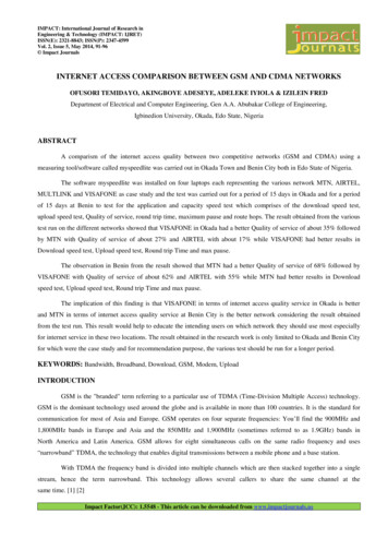
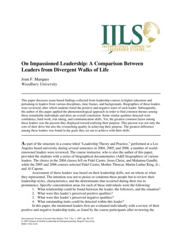
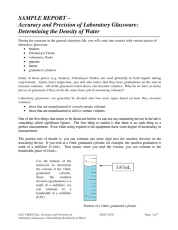
J Clin Exp Dent. 2019;11(8):e732-8.Diagnostic accuracy between periapical and panoramic radiographs and cone beam computed tomographyluate the anatomical structures affected and the endodontic conditions of the teeth, with a view to performingperiapical surgery, and were not obtained as a routinestudy – thereby complying with the ALARA (As LowAs Reasonably Achievable) criterion. The images wereevaluated by two blinded examiners (both certified oralsurgeons, J.C. and A.R.) not related to the treatment andfollow-up of the patients.-Calibration of examinersCone beam computed tomography images were used forcalibration and evaluation of inter-examiner accuracy,measuring the distance from the apex of the mesial rootof the first mandibular molar to the upper margin of themandibular canal (14). Ten random measurements wereobtained by both examiners, yielding a difference between them of 0.14 mm per image (range 0.0-0.28 mm).The level of agreement between reviewers is obtainedthrough de intraclass correlation coeficient (ICC) andinterpreted according the Landhis and Koch scale (15) .-Evaluation of imagesIn the global study, each measurement was made by thetwo examiners, with calculation of the average of bothmeasurements. This was the value subsequently used inthe data analysis, except in those cases where the twomeasurements differed by more than 0.2 mm. In such cases a third measurement was made jointly by both examiners (14). The measurements were made in three sessions spaced one week apart. In each session one-thirdof the images obtained with the different radiographictechniques were evaluated on a random basis (7).-Linear and area measurementsThe two-dimensional (2D) radiographs and CBCT scanswere displayed on a Full HD monitor with a resolutionof 1920 x 1080 pixels in a dimly lit room. The periapical lesions were measured as follows: Vertical measurements: the maximum vertical dimension (in mm) of thearea was recorded. Horizontal measurements: the maximum dimension (in mm) perpendicular to the previouslyobtained vertical measurement was recorded.The same measurement protocol was used for periapical radiography (Fig. 1), panoramic radiography (Fig.2) and CBCT (Fig. 3). In the latter technique the measurements were obtained from the sagittal and coronalsections. The corresponding area was calculated as arectangle (vertical measurement multiplied by horizontal measurement), according to a previous study (9). Theexaminers were able to modify size, brightness and contrast of the images of all three techniques.-Statistical analysisInter-examiner agreement was assessed by contrastingthe normal distribution of the differences in measurements using the Kolmogorov-Smirnov test, with calculation of the coefficient of variation (CV) and theintraclass correlation coefficient (ICC). The Studentt-test for dependent samples was used to compare theFig. 1: Vertical and horizontal measurements obtained by periapicalradiography.Fig. 2: Panoramic radiograph with the measurements made on theradiolucent area of tooth 15.Fig. 3: Sagittal (A) and coronal (B) sections used to obtain the measurements of the periapical area of tooth 15.diagnostic accuracy of the three radiological techniques.The Mann-Whitney U-test was used to assess the impactof patient gender upon lesion size, while the Spearmannonlinear correlation coefficient was used to evaluatethe influence of age. The level of statistical significancewas defined as 5% (α 0.05). The statistical analysis wasconducted using statistical program R (Version R 3.5.1).Results-Sample characteristics and descriptive dataThe initial sample size consisted of 82 patients, of which47 were excluded after application of the inclusion ande734
J Clin Exp Dent. 2019;11(8):e732-8.Diagnostic accuracy between periapical and panoramic radiographs and cone beam computed tomographyexclusion criteria. Patients excluded from the study, withreasons were:1. Patients with periapical or panoramic radiographs, orCBCT scans, not allowing full visualization of the rootsand cortical component of the affected teeth (n 29).2. Poor image quality (n 4).3. Time intervals among the three radiographic studies 2 months (n 6).4. Images taken after the apicectomy (n 8).The final study sample therefore consisted of 35 patients(45 teeth): 18 males and 17 females, with a mean ageof 47 years (range 19-74). The upper incisors were theteeth most frequently affected by periapical disease. Thedistribution of the teeth is shown in Table 1.periapical radiographs because the distal root was notavailable on the image, so the sample size is different inthis method with regard to the other two. There were nosignificant differences between the two-dimensional techniques (periapical radiography and panoramic radiography), though significant differences were observed inthe vertical measurements between the two-dimensionaltechniques and CBCT, with higher values when the latter technique was used (p 0.05) (Table 2).-Horizontal measurementsWith regard to the horizontal measurements, the meanvalues were 4.80 mm in the case of the periapical radiographs (n 44, SD 3.43) and 5.01 in the case of thepanoramic radiographs (n 45, SD 3.91). In the case ofTable 1: Description of the study sample.Study variableDescriptive statisticsSample size35 patients (45 periapical areas)Gender18 males (51.4%) and 17 females (48.6%)Age47 years (19-74)Affected teeth:Upper incisors15 (33.3%)Lower incisors4 (8.8%)Upper canines2 (4.4%)Lower canines1 (2.2%)Upper premolars8 (17.8%)Lower premolars3 (6.6%)Upper molars5 (11%)Lower molars7 (15.5%)-Detection of periapical areasThe conventional two-dimensional techniques (periapical radiography and panoramic radiography) bothshowed a sensitivity of 82%. Of the 45 periapical areas,8 were not detected by either technique: two corresponded to the anterior sector (incisors and canines) and 6to the posterior sector (premolars and molars). In percentage terms, the two-dimensional techniques failed toregister 4.5% of the anterior lesions and 13.5% of theposterior lesions. In contrast, CBCT registered all 45radiolucent areas. Inter-examiner agreement was almostperfect, with CV 3% and ICC 0.99 in all cases.-Vertical measurements of periapical areasThe mean vertical measurement of the apical area wasfound to be 5.48 mm with periapical radiography (n 44,SD 3.93) and 5.04 mm with panoramic radiography(n 45, SD 3.61 mm). The results in the case of CBCTwere 6.36 mm in the coronal sections (n 45, SD 3.65)and 6.38 mm in the sagittal sections (n 45, SD 3.45).There was a lower molar which wasn t measured onCBCT the values were 5.63 mm in the coronal sections(n 45, SD 2.95) and 5.10 mm in the sagittal sections(n 44, SD 2.26). No significant differences were observed on comparing the different radiographic techniques:all three were therefore considered to offer similar results in relation to the horizontal measurements (p 0.05)(Table 3).-Area of periapical bone defectsWith regard to the calculation of lesion area from thevertical and horizontal measurements, the resultswere 37.33 mm2 for the periapical radiographs (n 44,SD 44.49) and 36.37 mm2 for the panoramic radiographs (n 45, SD 47.63). In the case of CBCT the area was44.76 mm2 in the coronal sections (n 45, SD 49.32) and36.59 mm2 in the sagittal sections (n 44, SD 29.41).Likewise in this case there were no significant differences among the different radiographic techniques: allthree were therefore considered to offer similar results inrelation to the area measurements (Table 4). The samplesize differs from one unit between the different methods,e735
J Clin Exp Dent. 2019;11(8):e732-8.Diagnostic accuracy between periapical and panoramic radiographs and cone beam computed tomographyTable 2: Difference in measurements (mean and standard deviation [SD]), 95% confidence interval (95%CI), t-test for dependent samples (p-value) referred to the vertical dimension (height) among the different radiographic techniques.Difference betweenmeasurements95%CI for thedifferenceP-valueMeanSDLowerlimitUpperlimitRx PERIAPICAL – Rx PANORAMIC0.472.31-0.231.180.182Rx PERIAPICAL – CBCT coronal-0.891.97-1.49-0.290.005*Rx PANORAMIC – CBCT coronal-1.312.75-2.20-0.520.003*Rx PERIAPICAL – CBCT sagittal-0.901.91-1.48-0.320.003*Rx PANORAMIC – CBCT sagittal-1.342.66-2.19-0.560.002*Table 3: Difference in measurements (mean and standard deviation [SD]), 95% confidence interval (95%CI), t-test for dependent samples (pvalue) referred to the horizontal dimension (width) among the different radiographic techniques.Difference betweenmeasurements95%CI for the differenceMeanSDLower limitUpper limitRx PERIAPICAL – Rx PANORAMIC-0.233.36-1.250.79P-value0.654Rx PERIAPICAL – CBCT coronal-0.832.37-1.55-0.110.024Rx PANORAMIC – CBCT coronal-0.623.37-1.630.390.224Rx PERIAPICAL – CBCT sagittal-0.402.78-1.260.460.351Rx PANORAMIC – CBCT sagittal-0.363.58-1.450.730.507Table 4: Difference in measurements (mean and standard deviation [SD]), 95% confidence interval (95%CI), t-test for dependent samples (pvalue) referred to lesion area among the different radiographic techniques.Difference betweenmeasurements95%CI for the differenceP-valueMeanSDLower limitUpper limit0.7630.15-8.409.93Rx PERIAPICAL – CBCT coronal-7.7119.57-13.7-1.770.012Rx PANORAMIC – CBCT coronal-8.3931.51-17.81.080.081Rx PERIAPICAL – CBCT sagittal-1.8728.47-10.636.890.669Rx PANORAMIC – CBCT sagittal-4.8233.98-15.25.510.352Rx PERIAPICAL – Rx PANORAMICbecause there was an area, due to its size, in periapicalradiography and in sagittal sections it couldn t be measured, as its margin was not observed completely so vertical and/or horizontal measure couldn t be registered.Lastly, no significant associations were found betweenlesion area and patient age or gender.0.868rizontal measurements and areas obtained with the threeimaging techniques, and assessed their diagnostic validity and the possible influence of other associated factorssuch as patient gender and age.-Sensitivity of three methodsIs well known that CBCT sensitivity for the prevalenceof periapical radiolucencies was 100% (4,16). The findings of the present study are in agreement with the above-mentioned trend. However, the prevalence decreasedto 82% when using the two-dimensional radiographictechniques (periapical radiography and panoramic radiography). The failure rate in identifying periapical lesionsDiscussionThe present study compared the three radiodiagnostic techniques currently used for the measurement and analysis of radiolucencies produced by periapical infection.Specifically, we compared the different vertical and hoe736
J Clin Exp Dent. 2019;11(8):e732-8.Diagnostic accuracy between periapical and panoramic radiographs and cone beam computed tomographywith these techniques in posterior and anterior teeth was13.5% and 4.5%, respectively. Liang et al. (17) recordeda similar percentage of 5% in anterior teeth with periapical radiography, thus coincidingwith our own observation that anterior sector lesions aremore easily visualized than posterior lesions. No significant associations were found between lesion area andpatient age or gender.The literature offers similar data (14,16), with a higherperiapical lesion non-visualization rate associated totwo-dimensional imaging techniques. Lesions close tothe maxillary sinus are very likely to go undetected withtwo-dimensional methods, due to overlapping –
Results: The two-dimensional techniques (periapical radiography and panoramic radiography) yielded a sensitivi-ty of 82% versus 100% in the case of CBCT. The mean vertical dimension of the apical areas was 5.48 mm with periapical radiography and 5.04 mm with panoramic radiogr