Transcription
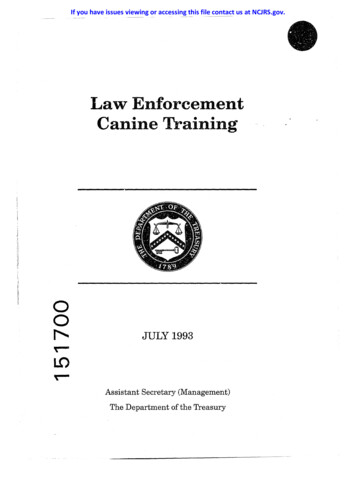
JDO 57 CASE REPORTCanine Substitution Treatment of Class IIIMalocclusion, Crossbite with a CongenitallyMissing Upper Incisor and a Peg Lateral IncisorHistory: Upper right lateral incisor (UR2) is congenitally missing, and upper left lateral incisor (UL2) is peg-shaped.Diagnosis: A 30-year-old male presented with increased facial height (58.5%), and a markedly increased mandibular plane (SN-MP49 ), but a normal facial profile (13 ). Intraoral examination revealed an asymmetric Class III malocclusion, lingual crossbite of theupper right first molar (UR6), anterior crossbite from canine to canine (UR3-UL3), missing UR2, peg-shaped UL2, and upper midlinedeviation 4mm to the left. The ABO Discrepancy Index (DI) was 50 points.Treatment: The peg-shaped UL2, and both lower first premolars (LR4, LL4) were extracted. A full fixed passive self-ligation (PSL)Damon Q appliance was bonded on all permanent teeth. Four bite turbos were bonded on lower arch: LR6, LR3, LR1, and LL6. Theanterior crossbite was corrected with Class III elastics, and the maxillary anterior spaces were closed in the upper arch to achievebilateral canine substitution. Torque control of the U3s was accomplished with specific bracket selection and torquing auxiliarysprings. Increasing the lower facial height to correct the anterior crossbite increased the facial convexity, but the patient maintainedlip competence.Outcome: This very difficult malocclusion (DI 50) was treated in 34 months to an acceptable result: ABO Cast-Radiograph Evaluation(CRE) 29 points, and Pink & White Esthetic Score 4. (J Digital Orthod 2020;57:52-67)Key words:Canine substitution, missing lateral incisor, crossbite, bite turbos, early light short elastics (ELSE), torquing auxiliary spring, peg lateralincisorDiagnosis and EtiologyA 30-year-old male presented for orthodontic consultation to evaluate his “protrusive chin,” but the problemappeared to be a protrusive lower lip. There were no contributing medical history nor known habits. Facialevaluation showed a long convex face (Fig. 1), and the occlusion was Class III with an anterior crossbite (Fig. 2).Radiographic evaluation documented a very steep mandibular plane, and impacted lower third molars (Fig. 3).In the frontal plane, facial structures were relatively symmetric, but the occlusal plane was canted inferiorlyon the right side. There was asymmetric condylar translation (Fig. 4), but were no signs nor symptoms oftemporomandibular joint dysfunction. Intraoral examination revealed an asymmetric Class III malocclusion(more severe on the left) with a maxillary midline that was deviated 4mm to the left. The right maxillary lateralincisor (UR2) was peg-shaped, and the contralateral lateral incisor (UL2) was congenitally missing. In addition,crossbites were noted for the upper right first molar and the entire maxillary anterior segment (Fig. 1).52
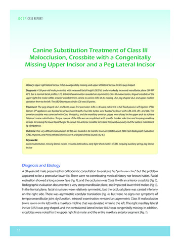
Class III Malocclusion, Crossbite with a Missing Upper Incisor and a Peg Lateral Incisor JDO 57Dr. Claire JY Chen,Lecturer, Beethoven Orthodontic Course (Left)Dr. Angle Lee,Editor, Journal of Digital Orthodontics (Center left)Dr. Chris H. Chang,President, Beethoven Orthodontic CenterPublisher, Journal of Digital Orthodontics (Center right)Dr. W. Eugene Roberts,Editor-in-chief, Journal of Digital Orthodontics (Right)Cephalometric analysis (Table 1) revealed excessive facial height (Na-ANS-Gn 58.5%) with a normal facialprofile, but lower lip protrusion was excessive to the E-Line (5mm). Bimaxillary retrusion (SNA 79 , SNB 75 ),high mandibular plane angle (SN-MP 49 , FMA 41 ), and retrusive maxillary incisors (97 ) were associated witha skeletally retrusive mandible (ANB 4 ). The panoramic radiograph showed that the mandibular 3rd molarswere impacted (Fig. 3). The American Board of Orthodontics (ABO) Discrepancy Index (DI) was 50 points, asshown in the supplementary Discrepancy Index (Worksheet 1). Fig. 1: Pre-treatment facial and intraoral photographs53
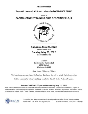
JDO 57 CASE REPORT Fig. 2: Pre-treatment dental models (casts)Fig. 4:Radiographs of the mandibular condyles in the closed positionare shown bilaterally in the left and right images, respectively.The corresponding open mouth positions are shown in thecenter left and center right images, respectively. Although theexcursions are asymmetric, there were no signs or symptoms oftemporomandibular disorder.CEPHALOMETRIC SUMMARYSKELETAL ANALYSISSNA (82º)SNB (80º)ANB (2º)SN-MP (32º)FMA (25º)PRE-TxPOST-TxDIFF.79 75 4 49 41 79 74 5 51 43 0 1 1 2 2 97 11 mm87 0 mm95.5 6 mm82 2 mm1.5 5 mm5 -1 mm5 mm0 mm1 mm1 mm4 mm13 16 3 58.5%58%0.5%DENTAL ANALYSISU1 To NA mm (4 mm) 2 mmU1 To SN (110º)L1 To NB mm (4 mm)L1 To MP (90º)FACIAL ANALYSISE-LINE UL (2-3 mm)E-LINE LL (1-2 mm)Convexity: G-Sn-Pg’(13º)%FH: Na-ANS-Gn(53%) 54Fig. 3: Pre-treatment panoramic and cephalometric radiographs Table 1: Cephalometric summary
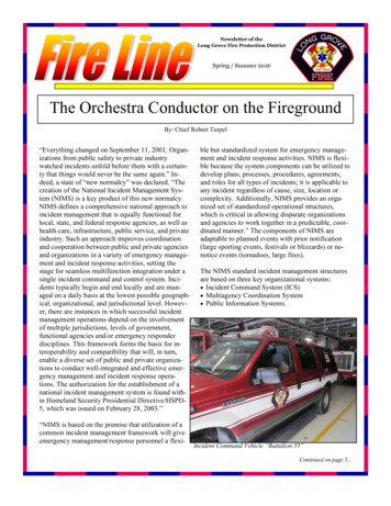
Class III Malocclusion, Crossbite with a Missing Upper Incisor and a Peg Lateral Incisor JDO 57Specific Objectives of Treatment1. Retract the lower dentition to correct the anteriorcrossbite.2. Extract the UR2 and close edentulous spaces toachieve bilateral canine substitution.3. Achieve ideal overjet and overbite relationships.Plan A 4. Correct intermaxillary sagittal and frontaldiscrepancies.Fig. 5: Treatment Plan AExtract the peg-shaped lateral incisor, correct the anterior crossbite,open space for implants, and restore both maxillary lateral incisorswith implant-supported prostheses. Restore the fractured lower leftcentral incisor with composite resin.5. Finish with a cast radiograph score of no morethan 30 points.Treatment PlanPlan A: (Fig. 5) Optimize upper lateral incisor spaces withpreprosthetic orthodontics. Extract the upper left peg-shaped lateral incisor. Restore both upper lateral incisors with implantsupported prostheses. Restore the lower left central incisor withcomposite resin.Plan B: (Fig. 6) Extract the upper right peg lateral incisor andboth lower first premolars. Reshape upper canines to simulate lateralincisors. Restore the lower left central incisor withcomposite resin.Plan B Fig. 6: Treatment Plan BExtract upper right peg lateral incisor, extract both lower firstpremolars, and substitute the upper canines for the lateral incisors.Restore lower left central incisor with composite resin. See text fordetails.After carefully considering both options, the patientchose canine substitution instead of implantsupported prostheses.Appliances and Treatment ProgressAfter the peg lateral (UR2) and lower first premolars( LR4, LR5 ) were extracted, a 0.022-in slot, passiveself-ligating ( P SL ) Damon Q bracket system(Ormco, Glendora, CA) was installed on both arches.Standard torque brackets were used except for hightorque brackets on the lower incisors. The archwire55
JDO 57 CASE REPORTsequence for both the upper and lower archwiresfor the UR3, UR2 and UR1 ( Fig. 8 ). Brackets werewas: 0.014-in CuNiTi, 0.014x0.025-in CuNiTi,rebonded accordingly.0.017x0.025-in TMA, and 0.016x0.025-in SS.In the 9 th month, an expanded 0.017x0.025-inFuji II Type II glass Ionomer cement (GC America,TMA archwire was placed on the upper arch and aAlsip, IL) was used to build bite turbos on the lingual0.014x0.025-in CuNiTi was inserted in the lower arch.surfaces of the LR3 and LR1, as well as the occlusalPower chains and power tubes were used for closingsurfaces of both lower first molars (Fig. 7) to facilitateanterior crossbite correction. A tongue depressorwas provided with instructions to apply light andsteady pressure in a labial direction to move theupper central incisors out of crossbite. Early lightshort elastics (Parrot 5/16” 2-oz.) were applied bilaterallyfrom the upper first molars to the lower canines tocorrect the Class III molar relationship (Fig. 7).In the 6th month of treatment, a progress panoramicradiograph revealed axial inclination problems Fig. 8:A progress panoramic film revealed second order axial inclinationproblems: UR4, UR3, and UR1. Brackets were repositioned. See textfor details.1M 56Fig. 7:Crossbite correction - resin bite turbos (blue ovals) were bonded on the lingual surface of the LR3 and LR1 and on the occlusal surfaces of theLR6 and LL6 (lower right image). Early light short elastics (yellow) were applied bilaterally from upper first molars to lower canines. See text fordetails.
Class III Malocclusion, Crossbite with a Missing Upper Incisor and a Peg Lateral Incisor JDO 57the upper anterior spaces. In the 10th month, thewere removed and teeth in the maxillary anteriorClass III elastics were stopped once the overjet wassegment were restored with composite resin tocorrected.optimize dental esthetics.In the 12th month, the lower archwire was changedto 0.016x0.022-in SS to stabilize the arch-form as theRetentionpremolar extraction spaces were closed. A figure-After the fixed appliances were removed, uppereight ligature maintained firm contact betweenand lower clear overlay retainers were deliveredthe six lower anterior teeth. The bracket on thewith directions specifying full-time wear for thelower right second molar was rebonded. Seventeenfirst six months, and then nights only afterwards.months ( 17M ) into treatment, torquing springsInstructions on home care and maintenance of thewere applied to the substituted canines to deliverretainers were also provided.lingual root torque (Fig. 9). They were removed at18-27 months when adequate axial inclinationswere achieved. From 23-28 months, detailing wasTreatment Resultsperformed with progressive wire-bending. BracketsFacial esthetics were substantially improved bywere repositioned on the maxillary central incisors.correction of the lower lip protrusion, and theBoth substituted canines were reshaped to simulatesmile line was optimized by improving the cant oflateral incisors.the occlusal plane (Fig. 10 ). ABO Cast-RadiographEvaluation (CRE) score was 29 points. The major CREAfter 34 months of active treatment, all appliances12M discrepancy were marginal ridges, buccal-lingual17MFig. 9:At twelve months (12M) into treatment, a torquing spring was placed on canines bilaterally to decrease root prominence (yellow dotted ovalon the left). Note the spring was incorrectly positioned (blue oval). At seventeen months (17) into treatment, note the corrected position of thetorquing spring with the arm under the main archwire (red oval). After five months of torque application, root prominence is improved (yellowdotted oval on the right). See text for details.57
JDO 57 CASE REPORT Fig. 10: Post-treatment facial and intraoral photographsmolar relationships, and overjet (Worksheet 2). Bothanterior and posterior crossbites were resolved,and the molar relationships were Class I (Fig. 11 ).The post-treatment panoramic and cephalometricradiographs are shown in Fig. 12. Lip balance wasimproved, and lip competence was maintaineddespite a 3 increase in facial convexity (Fig. 13, Table 1).The post-treatment cephalometric analysis wasconsistent with a Class II skeletal pattern (ANB 5 ),high mandibular plane angle (SN-MP 51 , FMA 44 ),and increased lower facial height (58% ) ( Table 1 ). 58Fig. 11: Post-treatment study models (casts)Superimposed cephalometric tracings showedretraction of the upper and lower incisors, as well
Class III Malocclusion, Crossbite with a Missing Upper Incisor and a Peg Lateral Incisor JDO 57as anterior movement and extrusion of the lowermolars. The mandible was rotated clockwise(posteriorly), and the lower lip was retracted (Fig. 13).The patient was quite satisfied with the result.DiscussionAccording to epidemiological studies, 1 maxillarylateral incisors show the highest genetic variancein the dentition. The most common anomaly is aunilateral undersized (often peg-shaped ) maxillarylateral incisor. Less commonly the condition isbilateral and may be associated with a contralateralcongenitally absent lateral.2 Agenesis prevalence formaxillary laterals is 2-9% over a variety of differentethnic groups; the data is similar for the mandibularsecond premolars ( 3%). However, the congenitalabsence of third molars is much more prevalent (25-35%).3 Fig. 12: Post-treatment panoramic and cephalometric radiographs Fig. 13:Pre-treatment (black) and post-treatment (red) cephalometric tracings are superimposed on the anterior cranial base (left) to showan improved facial profile. Maxillary superimposition (upper right) documents protraction and extrusion of upper molars. Mandibularsuperimposition (lower right) shows incisor retraction and molar protraction.59
JDO 57 CASE REPORTCanine substitution was deemed the best andTreatment of Peg Lateral Incisorscomprehensive treatment option for the patient.2If the root is well formed, a peg lateral incisor canbe restored with a porcelain crown or sometimes aCanine Substitutionveneer. Porcelain restorations are the most commontreatment for peg lateral incisors because theyWhen restoring the esthetic zone (maxillary anteriorrequire little or no restorative preparation of thesegment when smiling),3-5 it is important to considertooth. If interproximal space is adequate, a porcelainthe type of malocclusion, crowding/spacing,restoration is bonded over a tapered peg lateral tointermaxillary tooth size relationships, canine4restore normal form and function.positions, shape/color of canines, and maxillarylip length.4 Furthermore, a detailed assessment oftooth form and the supporting gingiva is indicated:Treatment of Missing Lateral Incisorsworn incisal edges, shape of individual teeth, incisalIn achieving optimal esthetics and function,contact relationship, contours of gingival margins,a coordinated, interdisciplinary approach isand probability of black triangles when the dentition5often necessary. Treatment may involve canineis ideally aligned. The correction of all applicablesubstitution, a tooth-supported restoration orfactors should be simulated with a wax or digital2-6The presentset-up prior to initiating orthodontic treatment.malocclusion with maxillary lateral incisorThe decision to reshape teeth and/or to add toothdeficiencies (Fig. 14) was complicated by an anteriorstructure should be carefully evaluated in relationcrossbite and Class III molar relationship (Fig. 2 ).to the ideal width-to-length ratios of the Goldenan implant-supported prosthesis.Proportion.51. Indications for Canine Substitutiona. Malocclusion:Two malocclusion patterns are particularly amenableto canine substitution: 1. Angle Class II with nocrowding in the mandibular arch which can befinished in a Class II occlusion; and 2. Angle Class Iwith crowding in the mandibular arch that requires 60Fig. 14:A periapical radiograph shows the peg lateral (UR2) and the missingUL2. There was inadequate space for the prosthetic restoration ofthe maxillary lateral incisors. See text for details.extraction of premolars (Fig. 15). For these situations,canine-protected occlusion is not usually a priority,so anterior group function in all excursions is a good
Class III Malocclusion, Crossbite with a Missing Upper Incisor and a Peg Lateral Incisor JDO 57convex profile is also acceptable. The protrusiveprofile and less prominent nose that is typical ofAsians are favorable factors for canine substitutionbecause retraction results in a more ideal lipprotrusion. 8 On the other hand, a convex profile,retrusive mandible, and deficient chin prominenceare unfavorable characteristics for caninesubstitution (Fig. 3). Correcting the anterior crossbiteby opening the bite with bite turbos is usually aArisky procedure for patients with a long face, butfortunately the patient was able to maintain lipcompetence despite a 3 increase in facial convexity(Figs. 12 and 13, Table 1).c. Canine size and shape:In comparison to the adjacent canine, the lateralincisor has a flat labial surface, and narrowerdimensions at the cementoenamel junction ( CEJ )for both mesio-distal and bucco-lingual width. 7BTo simulate a maxillary lateral incisor, the outlineform of the crown must be reduced at the cusp tip,as well as in mesio-distal width and facio-lingual Fig. 15:(A) Angle Class II malocclusion with no crowding in the mandibulararch(B) Angle Class I malocclusion with crowding in the lower arch thatrequires extractions.depth. Furthermore, resin restoration of the outlineextensions is required for the mesio-incisal and distoincisal line angles (Fig. 16). According to Thordarsonand Zachrisson,6 tooth sensitivity may persist for 1-3option. Nordquist and McNeill7 found no differencedays after crown reduction, but there is no long-termin occlusal function or periodontal status betweensensitivity if the high-speed reduction is performedcanine-protected and group function occlusion.with copious water spray.9,10b. Profile:d. Soft issue (lip level):Kokich5 feels a straight profile is the most favorablePatients with a high lip line when smiling arefor canine substitution in Caucasians, but a mildlychallenging for effective management of the61
JDO 57 CASE REPORT(B)(A)MesialLingualDistalLabialIncisal Fig. 16:The suggested outline form for a canine substituting for a lateralincisors is shown by the purple dotted line in the frontal (A) andprofile (B) views. See text for details. Fig. 17:Bracket position adjustments are shown to assist in achievingthe preferred high-low-high gingival margin profile that mimicsnatural soft tissue margins. See text for details.maxillary anterior esthetic zone. The periodontallength is decreased, while the buccolingual surfacessupport is especially important: control of gingivitis,are recontoured with crown reshaping, and thesoft tissue contours, and root prominence. Theline angles are restored with composite resin ( Fig.present patient (Fig. 10) has the opposite problem:16 ). In addition, the adjacent premolar is maskeda relatively low lip line. Thus, detailed coordinationto simulate a canine by intrusion to improve theof upper to lower arch alignment is a high priority,gingival emersion profile. An additional importantbecause inadequate maxillary incisor exposure isstep is to lengthen the labial surface with compositean increasingly important issue for aging adults,resin to achieve group function.11particularly men.3. Bracket/Torque Selection2. Bonding PositionA high torque bracket ( Table 2 ) is recommended62Restoring the natural contours of the gingivalfor lingual root torque to simulate the natural labialmargins is particularly important for maxillary canineinclination of a lateral incisor. 9 A torquing springsubstitution (Fig. 16 ). Optimal esthetics requires ais also helpful if additional lingual root torque ismore incisive gingival margin compared to theneeded to decrease the root prominence of theadjacent central incisor and first premolar. The incisorcanine. In contrast, buccal root torque is neededto canine high-low-high gingival margin principleto increase the first premolar root prominence.7is best achieved with coordinated orthodontics andHowever, these mechanics must be appliedrestorative procedures. The canine can be intrudedjudiciously to avoid alveolar dehiscence andby a more incisal bracket position (Fig. 17). Crowngingival recession. For mesially substituted first
Class III Malocclusion, Crossbite with a Missing Upper Incisor and a Peg Lateral Incisor JDO 57 le 2: Torque value of Damon Q brackets.premolars, the preferred buccal crown torque isrelatively perpendicular. A standard first premolarbracket is preferred because it has more negativetorque (-11 ). Furthermore, the buccal crown torquecompensates for the intrusion of 1st premolars (Table2).8 If additional torque compensation is required fora specific tooth, it is best achieved with a torquingauxiliary (Fig. 9).4. Interdisciplinary TreatmentWhen the canine is darker in comparison to theadjacent central incisor and first premolar, aselective tooth whitening procedure is indicated.If that conservative approach is inadequate, it maybe necessary to place porcelain veneer or crown(1) Straight or mildly convex facial profile forCaucasians, or a protrusive profile in Asians.(2) Angle Class I or II malocclusion with crowding inthe lower arch.(3) Mimic natural esthetics with careful attention todental and soft tissue morphology.(4) Torquing spring auxiliaries are helpful forcorrection of labial contour and/or rootprominence.(5) Veneer prostheses may be necessary to achieveoptimal results for demanding patients.restorations to achieve harmonious esthetics.AcknowledgmentConclusionsSpecial thanks to Mr. Paul Head for proofreading thisCanine substitution is an effective long-term solutionarticle.for selected patients. The initial examination andthe follow-up diagnosis are critical for the successof this comprehensive interdisciplinary treatment.Important considerations include:63
JDO 57 CASE REPORTReferences1. Medina AC. Radiographic study of prevalence and distributionof hypodontia in a pediatric orthodontic population inVenezuela. Pediatr Dent 2012;34(2):113-6.2. Park JH. Orthodontic treatment of a congenitally missingmaxillary lateral incisor. J Esthet Restor Dent 2010;22:297-313.3. Kokich VO Jr. Early management of congenitally missing teeth.Semin Orthod 2005;11(3):146-151.4. Zachrisson BU. Improving orthodontic results in cases withmaxillary incisors missing. Am J Orthod 1978;73:274-89.5. Kokich VO Jr, Kinzer GA. Managing congenitally missinglateral incisors, part 1: Canine substitution. J Esthet RestorDent 2005;17:1-6.6. Thordarson A, Zachrisson BU, Mjör IA. Remodeling ofcanines to the shape of lateral incisors by grinding: a long-termclinical and radiographic evaluation. Am J Orthod DentofacialOrthop 1991;100:123-32.7. Nordquist GG, McNeill RW. Orthodontic vs. restorativetreatment of the congenitally absent lateral incisor - longterm periodontal and occlusal evaluation. J Periodontol1975;46(3):139-43.8. Huang TK. Chang CH, Roberts WE. Missing maxillary centralincisor treated with medial substitution of the lateral incisor,canine and first premolar. Int J Orthod Implantol 2015;38:78-89.9. Schwaninger B, Shaye R. Management of cases with upper incisormissing. Am J Orthod Dentofacial Orthod 1977;71:396-405.10. Zachrisson B, Rosa M, Toreskog S. Congenitally missingmaxillary lateral incisors: Canine substitution. Am J OrthodDentofacial Orthop 2011;139:434-45.11. Drummond S, Capelli J Jr. Incisor display during speechand smile: Age and gender correlations. Angle Orthod2016;86(4):631-7.64
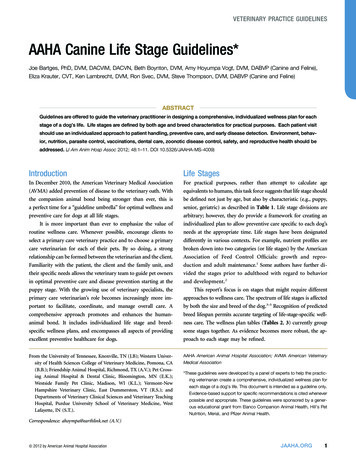
DISCREPANCY INDEX WORKSHEETEXAM YEAR2009ClassIII Malocclusion,Crossbitewith a MissingUpperIncisorand a Peg Lateral IncisorABOID#96112P(Rev.ATIENTCHAO-YUENCHIUCASE #19/22/08)TOTAL D.I. SCORE25OVERJETLINGUAL POSTERIOR X-BITE0 mm. (edge-to-edge) 1 pt.1 Ð 3 mm. 0 pts.3.1 Ð 5 mm. 2WORKSHEETpts.DISCREPANCYINDEX5.1 Ð 7 mm. 3 pts.P(Rev.ATIENTCHIUC7.1ASEÐ 9# mm. 1 9/22/08) CHAO-YUEN4 pts.T OTAL25 5 pts.9 mm.D.I. SCORE 50Discrepancy Index WorksheetNegative OJ (x-bite) 1 pt. per mm. per tooth OVERJET 1 pt. per tooth2 pts. per toothTotalTotal SN-MPCEPHALOMETRICS 38¡ANB degree6¡ or -2¡Each38¡5516Each degree 26¡Each degree 6¡1 to MP 99¡SN-MP 38¡Eachdegree 99¡Each degree 38¡500Class I to end onEnd on Class II or IIIFull Class II or IIIBeyond Class II or III(See Instructions) 26¡ 2 pts. 4 pts.22x 2 pts.114x 1 pt. 1 pt.x 1 pt. x 1 pt. 4 1 pt. 2x 1 pt. 2 pts.2x 2 pts. Total 1 pt.24Each degree 26¡4x 1 pt. OTHER (See Instructions)41 toMP 99¡ teethSupernumerary x 1 pt.1 pt. Ankylosisofperm.teethx2pts.Each degree 99¡2x 1 pt. 2 Anomalous morphologyx 2 pts. Impaction (except 3rd molars)x 2 pts. 0Midline discrepancy ( 3mm)Total @ 2 pts.8 Missing teeth (except 3rd molars)x 1 pts. Missing teeth, congenital1 x 2 pts. OTHER(SeeInstructions)Spacing (4 ormore,per arch)x 2 pts. (Mx cent. diastema 2mm)SpacingSupernumerary teethx 1@pt.2 pts. Toothtranspositionx2pts.Ankylosis of perm. teethx 2 pts. Skeletal asymmetry(nonsurgical tx)3 pts.Anomalousmorphologyx 2@pts. 2 Addl.treatmentcomplexitiesx2pts.1rdImpaction (except 3 molars)x 2 pts. 1 pt.2 pts.4 pts.7 pts.010 x 1 pt. 20Each degree26¡ -2¡CROWDING (only one arch)1 OCCLUSIONÐ 3 mm. 1 pt.3.1Ð 5 Imm. 2 0pts.Classto end onpts.5.1Ð 7onmm.4 2pts.EndClass II or III pts. per side Full7 mm. 7 4pts.Class II or IIIpts. per sideBeyond Class II or III 1 pt. per mm.Total 1 additional1TotalOCCLUSION02 2 pts.perdegreetooth 6¡ TotalEachLATERAL OPEN BITEANTERIOR OPEN BITE2 pts. per mm. per tooth0 mm. (edge-to-edge), 1 pt. per tooththen 1 pt. per additional full mm. per toothTotal 0Total 00CROWDING (only one arch)1 Ð 3 mm. OPEN BITE LATERAL3.1 Ð 5 mm. 2 5.1pts.Ðpermm. per tooth 7 mm. 7 mm. TotalBUCCALPOSTERIORX-BITEEach degree -2¡x 1 pt. 0 ANTERIORÐ 3 mm. 0 pts.OPEN BITE3.1 Ð 5 mm. 2 pts.5.1Ð 7 mm.pts.0 mm.(edge-to-edge), 1 pt. per3toothImpinging(100%) 5then 1 pt. per additional full mm.pts.per tooth 10 EXAM YEAR2009BUCCALPOSTERIORABO ID#96112 X-BITEOVERBITETotalTotalTotalLINGUAL POSTERIOR X-BITECEPHALOMETRICS(See Instructions)01 pt. per toothTotal ANB 6¡ or -2¡0 4 pts.0 mm. (edge-to-edge) 1 pt.1 Ð 3 mm.Total 0 pts. 53.1 Ð 5 mm. 2 pts.OVERBITE5.1Ð 7 mm. 3 pts.7.1 Ð 9 mm. 4 pts.0 Ð 3 mm. 0 pts. 9 mm. 5 pts.3.1 Ð 5 mm. 2 pts.5.1 Ð 7 mm. mm. per3 pts.tooth NegativeOJ (x-bite) 1 pt. perImpinging (100%) 5 pts.TotalTotalJDO 57pts.pts.pts.022222Midline discrepancy ( 3mm)@ 2 pts. Identify: Anomalousmorphologyx URMissing teeth(except 3rd molars)1 pts.peg lateralMissing teeth, congenitalx 2 pts. 42Spacing (4 or more, per arch)Total x 2 pts. 62Spacing (Mx cent. diastema 2mm)@ 2 pts. Tooth transpositionx 2 pts. Skeletal asymmetry (nonsurgical tx)@ 3 pts. Addl. treatment complexitiesx 2 pts. Identify: 0 pts.2 pts. per side4 pts. per side1 pt. per mm.2pts.pts.Total 4pts.additionalTotal 0265
JDO 57 CASE REPORTOcclusal ContactsCast-Radiograph Evaluation1ABO Cast-Radiograph EvaluationCaseTotal#29CRE ScorePatientTotal C-R Eval Score:161Alignment/Rotations41111Marginal RidgesOcclusal Relationships614112111 11Interproximal Contacts0Buccolingual Inclination6111111Root AngulationOverjet2161 111111INSTRUCTIONS: Place score beside each deficient tooth and enter total score for each parameterin the white box. Mark extracted teeth with ÒXÓ. Second molars should be in occlusion.66
Class III Malocclusion, Crossbite with a Missing Upper Incisor and a Peg Lateral Incisor JDO 57IBOI Pink & White Esthetic Score4Total Score: Total 1. Pink Esthetic Score6266222225553533553334414411414111. M & D Papillae0 1 22. Keratinized Gingiva0 1 23. Curvature of Gingival Margin0 1 24. Level of Gingival Margin0 1 25. Root Convexity ( Torque )0 1 26. Scar Formation0 1 21. M & D Papilla0 1 22. Keratinized Gingiva0 1 23. Curvature of Gingival Margin0 1 24. Level of Gingival Margin0 1 25. Root Convexity ( Torque )0 1 26. Scar Formation0 1 22. White Esthetic Score ( for Micro-esthetics )4443334443331111112225552226661Total 31. Midline0 1 22. Incisor Curve0 1 23. Axial Inclination (5 , 8 , 10 )0 1 24. Contact Area (50%, 40%, 30%)0 1 25. Tooth Proportion (1:0.8)0 1 26. Tooth to Tooth Proportion0 1 21. Midline0 1 22. Incisor Curve0 1 23. Axial Inclination (5 , 8 , 10 )0 1 24. Contact Area (50%, 40%, 30%)0 15. Tooth Proportion (1:0.8)0 1 26. Tooth to Tooth Proportion0 1 2267
President, Beethoven Orthodontic Center Publisher, Journal of Digital Orthodontics (Center right) Dr. W. Eugene Roberts, Editor-in-chief, Journal of Digital Orthodontics (Right) Cephalometric analysis (Table 1) revealed excessive facial height (Na-ANS-Gn 58.5%) with a normal facial profile, but lower lip protrusion was excessive to the E-Line .