Transcription
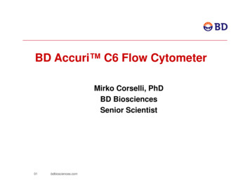
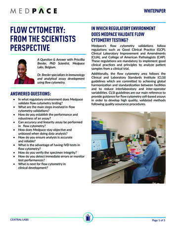
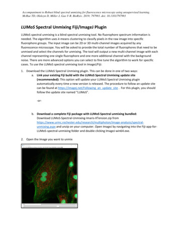
Spectral Flow Cytometry DefinitionNolan and Condello (2013) Current Protocols in CytometrySpectra of dyes excited by blue (488 nm) laser
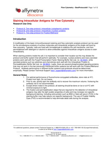
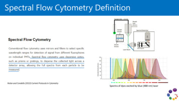
Cytek Aurora’s Optical DesignUnique Optical Design High Sensitivity Collection Optics Lasers are spatially separated. Eachexcitation laser has an associated solid statemulti channel semiconductor array detectormoduleFull Spectrum Analysis Entire emission spectrum is captured acrossthe different modules and then stitchedtogether to create a spectral signature thatcombines emission information from allthree excitation wavelengthsSpectral Unmixing Spectral unmixing algorithms calculate thecontribution of each known fluorophore’sspectra to the total collected emission signal
5 Laser Aurora: Optical Design
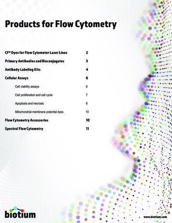
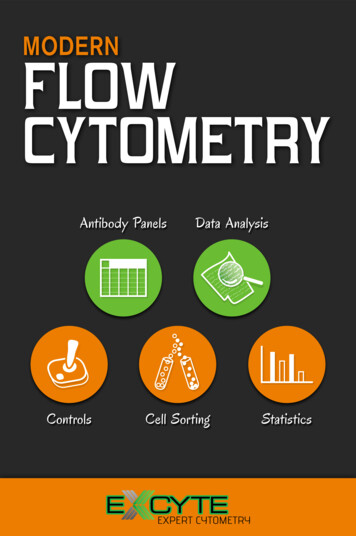
Full Spectrum SignaturesFull spectrum capture enables theuse of novel unmixing algorithmfor data analysis.PEEmission spectra excited by the UV,Violet, Blue, Yellow-Green and Redlasers are measured from the laserline to the infrared region.APCThe entire emission spectra offluorescent dyes excited by theonboard lasers is measuredBV785UVVioletBlueYellow GreenRed
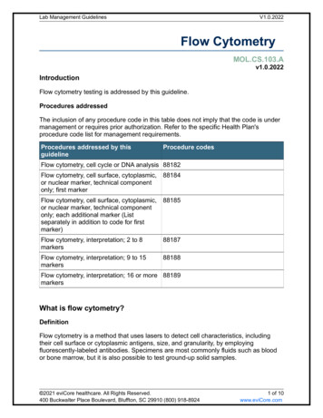
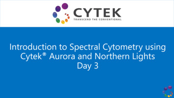
Full Spectrum Enables Use of Highly Overlapping DyesAPCAlexa Fluor 647Plot gated on singlet lymphocytesAPCAlexa Fluor 647Markers that are co expressed CANeffectively be used in combination
5 Laser Aurora: Detector Arrays
Ultraviolet Laser Unique SignaturesBUV395BUV661Live/Dead BlueBUV737BUV496BUV805BUV563
Violet Laser Unique SignaturesBV421Alexa Fluor 405, Super Bright 436,Zombie VioleteFluor 450, VioBlue, Pacific Blue,Live/Dead VioletBV480eFluor 506BV510, VioGreen, ZombieAqua, Live/Dead AquaBV570Pacific Orange,Live/Dead YellowSuper Bright 600, BV605Zombie YellowQdot605BV650, Super Bright 645Qdot655Super Bright 702, BV711Qdot705BV750BV785Qdot800
Blue Laser Unique SignaturesBB515, Vio 515, sVio 515Alexa Fluor 488, FITC, VioBright FITC, Zombie Green,Live/Dead GreenAlexa Fluor 532PerCPPerCP-Cy5.5PerCP-Vio700, PerCP-eFluor710
Yellow Green Unique SignaturesPEPE-Cy5PE-eFluor 610, PE/Dazzle 594, PE-TxRed, PE-CF594,Live/Dead RedPE-Cy5.5PE-Alexa Fluor 610Zombie RedPE-Alexa Fluor 700PE-Cy7, PE-Vio 770
Red Laser Unique SignaturesAPCZombie NIRAlexa Fluor 647, Vio 667, sVio 667, eFluor 660,Live/Dead Far RedAPC-Alexa 750, APC/Fire 750, APC-Cy7, APC-Vio 770,APC-eFluor 780, APC-H7APC-Cy5.5Live/Dead NIRAlexa Fluor 700, APC-R700
Compensation vs. Spectral UnmixingConventional Cytometer - CompensationSpectral Analyzer - UnmixingFITCFITCPEPEFITC into PE spillover Each fluorochrome is associated with a primary detector.For an n color assay, n detectors are needed Each fluorochrome is detected in multiple channels. In the 5 laserAurora analyzer, there are 64 fluorescent channels. Using single stained controls, spillover is mathematicallyremoved by subtracting out the % photons of lightcontribution from the non-primary color into the primarydetector, a mathematical process called compensation The number of detectors has to be higher than the number offluorochromes. A compensation matrix is calculated: it is a square matrix,nxn Single stained controls are used to establish the signatures of eachfluorochromes Unmixing is used to determine which combination of referencecontrols best fits the multicolor spectral signature of a multicolorsample An unmixing matrix is calculated: it is an nx64 rectangular matrix
Unmixing Workflow in Aurora1. Run UNSTAINED control2. Run individual dye spectra controls (Reference Controls)3. Unmix (equivalent to Compensation step in conventional cytometer)Reference Spectra from Single Stain ControlsRaw WorksheetUnmixed WorksheetUnmixingAlgorithm
Raw vs. Unmixed DataRAW DATAUNMIXED DATA Parameters are the instrument channels(V1, V2, etc) Parameters are the fluorochromesincluded in the assay Visualized in raw worksheet Visualized in unmixed worksheet Large fcs file size: up to 64 parameters FSC and SSC Smaller fcs file size: number of fluors FSC and SSC Can be unmixed as many times asdesired Can not be used to unmix
Requirements for Optimal Reference ControlsFluor BNeed to calculate spillover (slope) between fluorochromesX( )X(-)Fluor AHow to get an accurate calculation? The more separate the two data points are, the better the calculation Bright particles are necessary for this Both particles need to have IDENTICAL autofluorescence characteristics If negative particles are beads, then the positive particle need to be the exact same beads (same lot) There is need to have enough events for both data points Stopping rules need to be adjusted according to the sample type and marker used The fluorescence spectrum of the positive data points needs to be IDENTICAL to the one in the multicolor sample Special considerations when using tandem dyes The spectrum of the reagent binding to beads may be different to the spectrum of the same reagent whenbound to cells!
Unstained Control vs Negative Population in Reference Control In addition to the Reference Controls, an Unstained Control is mandatory for SpectralUnmixing This control is NOT needed for spillover calculation This control is used for measurement of autofluorescence ALWAYS needed for unmixing even without autofluorescence extraction This control needs to exactly match the particle type and sample prep procedure usedin the multicolor samples If Reference Controls do not have a negative population: New software 2.1 allows for additional unstained controls Negative cells MUST match the cells used as reference controls for spillover calculation Negative beads MUST match beads used as reference controls for spillover calculation
Reference Controls QC Examples (1)Unstained control troubleshooting, human PBMCs ExpectedXProvided by User
Reference Controls QC Examples (2)Qdot 605 control troubleshooting ExpectedXProvided by User
Reference Controls: Making Good ChoicesShould I use beads or cells as controls? Beads are easy to use and it is very likely that they will have a bright positive signal. It’s also easy tocollect enough events. HOWEVER, users need to assess whether the signature of the reagents used to stain the beadsmatches the one when stained on cells If possible, compare unmixing results using beads vs cells as reference controls Users also need to assess how forgiving a specific assay is if there are errors in the calculationsI want to use cells, but my marker is rare or very dim. What can I do? If a fluorochrome is NOT a tandem, replace with a marker highly expressed in a distinct population(CD3, CD4, CD8, B220 etc). Example: instead of using CD25 PE, use CD4 PE. If fluorochrome is a tandem, only option is to use beads stained with exactly same reagent (samelot)
Rules for Using Beads as Controls Fluorochrome spectrum signature needs to be IDENTICAL to be onewhen antibody is bound to cells Beads should be treated as the cells in order to ensure fluorochromes havebeen in the same “environment” (exposure to same buffers, for same amountof time, etc) Intensity does matter: beads need to be equally bright or brighterthan cells to be an adequate control for a given fluor Each of these requirements are equally important
Panel Design: Gathering Information1. STARTING POINT: BIOLOGY!!!a)Antigen Classification: primary, secondary and tertiaryb)Antigen co-expression2. What fluorochromes should I use for my assay?a)How many antigens I want to detect?b)What are the best X number of fluors that I can use?3. What antibodies are commercially available?Make a table, antigens vs. fluor
Antigen Classification Primary: high density, on and offexpressionSecondary: relatively high density,continuous expressionTertiary: Uncharacterized orexpressed at low levels.Y. Mahnke and M. Roederer. Clin Lab Med:2007. 27:469Primarye.g. CD4Secondarye.g. CCR7Tertiarye.g. PD-1
Antigen RIgDCD27CD38CD1cCD95CD8CD57Level of Antigen ExpressionPD-1CD25TCR g/dCD1cCD123CD11c
PEPE-Vio770PE-eFluor PE-Cy7BV421BB700eFluor 660APCAPC-Vio 770APC-eFluor 780Super Bright 436Alexa Fluor 647Vio667APC-Cy7BB515BUV661BV785BV711PE-Alexa Fluor 700BV480VioBright FITCBV750BUV737PE-Texas RedPerCP-eFluor 710APC-Fire 750VioBlueBV605Alexa Fluor 488Super Bright 600BUV805Super Bright 702BV650APC-H7Super Bright 645BUV563VioGreenPacific BlueAPC-Alexa Fluor 750FITCeFluor 450APC-Cy5.5Alexa Fluor 700PerCP-Vio700PE-Alexa Fluor 610PerCP-Cy5.5BV570BV510Qdot 655BUV496Alexa Fluor 405BUV395eFluor 506PerCPQdot 605Qdot 800Qdot 705Alexa Fluor 532Pacific OrangeFluorochrome Brightness Ranking400350300250200150100500
Assessing Antigen ResolutionMULTICOLOR SCENARIOMarker A Fluor XSINGLE STAINEDCountsMarker A Fluor XCountsSINGLE COLOR SCENARIOMFI Fluor XMarker A Fluor XMULTICOLOR TUBECountsMarker A Fluor YMFI Fluor YMain Contributors for Resolution Reduction Instrument Performance Instrument Setup Fluorochrome BrightnessCountsMFI Fluor XMFI Fluor XMain Contributors for Resolution Reduction SPREAD!!! Antibody titer
Quantification of Impact of Spread in ResolutionMFI Fluor YMarker A Fluor XStain IndexMarker A Fluor XFluor X DOES NOTspread into Fluor YFluor Y DOES NOTspread into Fluor XMFI Fluor YMULTICOLOR SCENARIOSINGLE COLOR SCENARIOStain IndexMarker A Fluor YMFI Fluor XMFI Fluor XFluor X DOESSPREAD into Fluor YFluor Y DOES NOTspread into Fluor XMFI Fluor YMarker A Fluor YMFI Fluor YMFI Fluor XStain Indices UnchangedStain Index Marker A Fluor YDECREASES in presence of fluor XMFI Fluor XConsiderations CO EXPRESSION ANTIGEN LEVEL OF EXPRESSION Data used for calculations has to be unmixed using a certain combination of fluorochromes
Panel Design and Highly Overlapping Dyes (1)Co-expression and antigen classification are needed for correct fluorochrome choice.FMOantigen and overlapping fluorcco-expressed
Panel Design: Fluor AssignmentSame rules apply as conventional cytometry!1. Fluorochrome assignment for tertiary antigensa) Assess reagent availability (often not too many options available)b) Assign brightest fluor available (use fluor brightness ranking)2. Fluorochrome assignment for secondary antigensa) Based on CO-EXPRESSION of antigens expressed at intermediate levelsb) If no co-expression, use any bright dye still availablec) If co-expression: if available, use a bright dye that does not spread into selected fluor for tertiary antigens If only available dyes have spread, use a dim dye to minimize spread impact3. Fluorochrome assignment for primary antigensa) Often available in many colorsb) Try to assign to dyes that are dim and that have minimal spread in other dyes (examples: FITC, Pacific Blue, BV510,Alexa 532, APC H7)
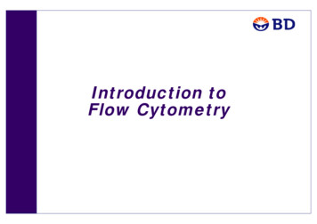
Cross Stain Index Matrix for 30 V421SupereFluor 450Bright exa Fluor Alexa FluorPerCPPerCP-Cy5.5488532eFluor 710PEPEDazzle594PE-Cy5PE-Cy7APCAlexa 05BV421Super Bright 436eFluor exa Fluor 488Alexa Fluor 532PerCP-Cy5.5PerCP-eFluor 710PEPE-Dazzle594PE-Cy5PE-Cy7APCAlexa Fluor 647APC-R700APC-Fire 750How to read this table: the fluor in the row impacts the one in the column. Red means the fluor in that row hassignificant spread into the dye in the column (for example PE into BV570). Areas in bright pink and red is wheremore attention to panel design is needed.APC-Fire750
Cytek Assay Settings1. Strongly suggested settings to use as a starting point for any application2.What are Cytek assay settings?a) Settings established using biological samplesb) Ensure optimal resolution for each detectorc) Leave enough room to accommodate bright markersd) Ensure unique spectrum with accurate emission peak for all currently tested dyese) Spread minimized as much as possible (remember. there will always be spread!)3.When to modify these settings?a) ONLY if signals are off scaleb) Increasing the gains will not result in more resolution and in contrast can result in increased spread!
Instrument Setting Adjustment: Example 1Off scaleIn scaleFluorochromeCD8 BV421CD4 Super Bright 436CD4 eFluor450BV421Super Bright 436eFluor450Issue: BV421 signal in V1-V3 is off scale1.Decrease V1 (primary channel of BV421)gain until V1 is on scale2.change V2 and V3 gains proportionally tomaintain the minor differences in thespectrum of BV421, Super Bright 436,eFluor 450Only three channels gain needs to be changedso that we don’t sacrifice other dyes resolutionwhile keeping reasonable spectrums for alldyes.BV421Super Bright 436eFluor450
Same rules apply as conventional cytometry! 1. Fluorochrome assignment for tertiary antigens. a) Assess reagent availability (often not too many options available) b) Assign brightest fluor available (use fluor brightness ranking) 2. Fluorochrome assignment for secondary antigens. a) Based on CO-EXPRESSION of antigens expressed at intermediate .