Transcription
Introduction to Spectral Cytometry using Cytek Aurora and Northern LightsDay 3
What happens when I have too much caffeine!
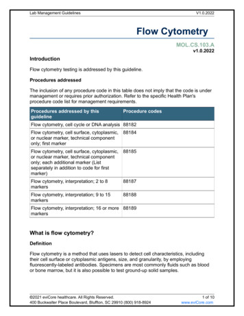
A B cell panel for human PBMCsMarkerFluorLevel of Expression /Cell FrequencyControl: Beadsor BV785BV605Pacific BlueBV650B711High/ intermediateHigh/ intermediateIntermediate/ highHigh/ highHigh/intermediateHigh/ intermediateLow/ lowCellsCellsCellsCellsCellsCellsBeadsPBMCsControl Stained WithAny bright BV510Any bright BV421CD27 BV785, same lotCD62L BV605, same lotAny bright Pacific BlueIgM BV650, same lotIgG BV711, same lot
Intracellular Cytokine Staining/Proliferation Assay,cultured PBMCsMarkerFluorCFSECD3PE CF594CD4APCCD8APC R700IFNgefluor 450IL-4PE Cy7IL-17PerCP e710viability Aqua Amine DyeunstainedLevel of Expression /Cell FrequencyControl: Beadsor CellsControl Stained WithHigh/highLow/ highLow/highIntermediate/highIntermediate/ int.High/lowLow/lowhigh/lowUnstim cellsUnstim cellsUnstim cellsCellsStim cellsStim cellsBeadsCellsPBMCs culturedCFSE well titrated*CD3 PE-CF594, same lotAny bright APC, but CD4 is goodCD8 APC-R700, same lotAny bright eFluor 450IL-4 PE-Cy7, same lot, higher stopping gate for enough eventsIL-17 PerCPeFluor710, same lotAqua Amine DyeUnstim and stimulatedSeparate controls*Switch CFSE to Cell Trace Violet – much easier to titrate into range
You are planning to perform an assay for detection of FOXP3 and to follow the protocol below. Please write downthe protocol you will follow (step by step) to stain beads to be used as controls for surface antigens AND forintracellular antigensCells1. Add surface Abs2.Incubate/wash3.Fix Perm4.Wash5.AF647 Ab6.Incubate/wash7.Run!Beads for Surface AgsBeads for i.c. Ags
Reference Control QCPE control:GoodPositive peak SpectrumNegative peak Spectrum
Reference Control QCPE control:neg peak has pos signal – ALWAYS WASH YOUR BEADS/CELLS
Reference Control QCSuper Bright 780 Control: not highest peak gated, highest peak has incorrect signature (has SB436 sig) tandemdegradation, pos peak in bottom plot is the donor, the acceptor is no longer fluorescing
Reference Control QCBB700 Control: Pos gate too wide, too heterogeneous, plot 2 and 3 show different spectra, lower peak looks like carryover
Reference Control QCPE-Cy5 Control: a lot of tailing on pos peak – usually when beads get stuck on the side of the tube and encountervariable amounts of Ab, this is also a problem with old beads that don’t bind uniformly
Knowledge Review Analyze full spectrum signatures Understand how full spectrum signatures are generated Identify unique signatures Normalize spectrum signatures QC and troubleshoot signatures Understand the fundamentals of spectral unmixing How it works How it compares to compensation Understand the workflow in Spectroflo Understand the requirements for optimal reference controls How to QC reference controls
ObjectivesBy the end of Day 3, you should be able to: Understand how to plan and design a multicolor panel Understand the fundamentals of panel design Understand the tools available for fluorochrome selection and successful paneldesign Panel performance evaluation and optimization strategies
Know the BiologyPlanning a Multicolor Experiment
Panel Design: Gathering InformationIdentify Antigens Level of expression: primary, secondary, tertiary Antigen co-expressionSelect Fluorochromes Unique signature Brightness Spread considerationsCheck Availability Are the reagents available in the antibody/fluor combination
Antigen ClassificationPrimaryHigh densityon or off expressionSecondaryCD3CD4CD8Intermediate densitycontinuous expressionCD45RACCR7CD127HLA-DRTertiaryLow densityunknown expressionPD-1CD25TCR g/dFlurochrome BrightnessLevel of Antigen ExpressionCD4CD127CD25Mahnke, Y. and Roederer, M. Clin Lab Med. 2007 September ; 27(3): 469
Know your co-expression
Selecting Fluorochromes with Unique SignaturesSeveral tools available tohelp:1. Fluorochrome selectionguidelines2. Online spectrum viewer3. Similarity & complexityindices
Laser UniqueLook at andBlueIdentifyUniqueSignaturesdyes to use
Fluorochrome selectionStain index
Assessing Brightness: Stain Index Ranking of 65 DyesStain IndexDimMidBrightVery Bright
Example: 35 Fluorochrome SelectionApprox. EmissionWavelength 30750780800UVVioletBlueYellow GreenRedBUV395BUV496BV421Super Bright 436eFluor 450BV480BV510BUV563Pacific OrangeBV570BUV661BV605BV650Live/Dead CP-Cy5.5PerCP-eFluor710PE-Cy5APCAlexa Fluor 647APC-R700BUV737BUV805BV750BV785Qdot 800PE-Cy7APC-Fire 750
Fluorochrome selectionUnderstanding Spread
Assessing Antigen ResolutionMULTICOLOR SCENARIOMarker A Fluor XSINGLE STAINEDCountsMarker A Fluor XCountsSINGLE COLOR SCENARIOMFI Fluor XMarker A Fluor XMULTICOLOR TUBECountsMarker A Fluor YMFI Fluor YMain Contributors for Resolution Reduction Instrument Performance Instrument Setup Fluorochrome BrightnessCountsMFI Fluor XMFI Fluor XMain Contributors for Resolution Reduction Spread Antibody titer
Quantification of Impact of Spread in ResolutionMFI Fluor YMarker A Fluor XStain IndexMarker A Fluor XFluor X DOES NOTspread into Fluor YFluor Y DOES NOTspread into Fluor XMFI Fluor YMULTICOLOR SCENARIOSINGLE COLOR SCENARIOStain IndexMarker A Fluor YMFI Fluor XMFI Fluor XFluor X DOESSPREAD into Fluor YFluor Y DOES NOTspread into Fluor XMFI Fluor YMarker A Fluor YMFI Fluor YMFI Fluor XStain Indices UnchangedStain Index Marker A Fluor YDECREASES in presence of fluor XMFI Fluor XConsiderations CO EXPRESSION ANTIGEN LEVEL OF EXPRESSION Data used for calculations has to be unmixed using a certain combination of fluorochromes
Fluor X DOESSPREAD into Fluor YFluor Y DOES NOTspread into Fluor XMFI Fluor YCross Stain Index Calculation1Stain Index Marker A Fluor Y2 DECREASES in presence of fluor XMFI Fluor XBy unmixing CD4 stained cellswith multiple fluors we canassess how much the positivepopulation spread into otherdyes (rSD) .Spread % ReductionWe use that spread to calculate how much thesingle stained Stain Index (1) is reduced by thespread introduced by another dye (2)25
Cross Stain Index Matrix: 35 Color PanelBUV395LIVE DEAD BlueBUV496BUV563BUV661BUV737BUV805BV421Super Bright 436eFluor 450BV480BV510Pacific OrangeBV570BV605BV650BV711BV750BV785Qdot CF568PE-Dazzle594PE-Cy5PE-Cy7APCAlexa Fluor 647APC-R700APC-eFluor 780Dye in row impacts dye in columnNoImpactHighImpactUnmixed CD4 PE-Cy7BUV805Unmixed CD4 APCBUV395BUV395LIVE DEAD BlueBUV496BUV563BUV661BUV737BUV805BV421Super Bright 436eFluor 450BV480BV510Pacific OrangeBV570BV605BV650BV711BV750BV785Qdot 800BB515FITCCF514PerCPPerCP-Cy5.5PerCP-eFluor 710PECF568PE-Dazzle594PE-Cy5PE-Cy7APCAlexa Fluor 647APC-R700APC-eFluor 780APCPE-Cy7
35-Color PanelApprox. EmissionWavelength 30750780800UVVioletBlueYellow GreenRedCD45RA BUV395CD16 BUV496PD-1 BV421CD123 SB436CD161 eFluor 450IgD BV480CD3 BV510CD14 BUV563CD20 Pacific OrangeHLA DR BV570CD11c BUV661CD28 BV605CXCR3 BV650Live/Dead BlueCCR6 BV711CD86 BB515CD57 FITCCD19 CF514CD335 PECD4 CF568CD24 PE Dazzle594CD45 PerCPCD95 PE-Cy5CD11b PerCP Cy5.5TCR γδ PerCP eFluor710CD27 APCCD33 Alexa647CD127 APC-R700CD56 BUV737CD45RO BUV805CXCR5 BV750CCR7 BV785CD8 Qdot 800PrimaryCD25 PE Cy7SecondaryTertiaryCD38 APC-eFluor 780
Panel Design and Highly Overlapping Dyes (1)TCRgd BV421-ACo-expression and antigen classification are needed for correct fluorochrome choice.No SB436 SB436NOT co-expressed SB436Co-expressed
Panel Design and Highly Overlapping Dyes (2)No SB436 oreFluor450 eFluor450Co-expressed SB436NOT co-expressed SB436Co-expressed
Panel EvaluationMaking sure your panel is optimal
Single Stained ControlsSingle Stained Controls allow the full evaluation of: Antibody performance/titer/staining conditions Adequacy of reference/compensation controlsMarker A Fluor XSINGLE STAINEDCounts Quality of panel design TAKE THE TIME TO ANALYZE FOR EACH MARKERMarker A Fluor XMULTICOLOR TUBECountsMFI Fluor XMFI Fluor X THE SINGLE STAINED TUBE vs. THE MULTICOLORTUBE IS THE FOUNDATION FOR MC PANELOPTIMIZATION
Evaluation of Single Stained Controls vs MulticolorSingle ColorCD4 CF568CD38 APCeF780CD57 FITCCD45RA BUV395Multicolor32
Evaluation of Single Stained Controls vs MulticolorSingle ColorCD95 PE-Cy5TCRγδ PerCPeF710CD28 BV605CD56 BUV737Multicolor33
Exercise 3Aims: Demonstrate your panel design Kung Fu! You don’t need to know NK cell biology As a general guide anything with a CD# higherthan 200 is a tertiary marker, the rest of the Agsare fairly explanatory
Exercise 3 – Panel Design Steps1.2.3.4.Classify the antigens Go to vendor websites, check staining examples Gating strategyConsider co-expression Gating strategy – know your biologyCharacterize spread of available fluors – use stain index and spread matrix Bright fluors that receive low spread Dim fluors that contribute low spreadAssign Fluors Tertiary – very bright/bright fluors that receive low spread Secondarya)b)bright/mid fluors that receive low spreadmid fluors that contribute low spread Primary – dim fluors that contribute low spread
Exercise 3 NK Cell Panel DesignNK Cell PanelSpecificityAlt 7LFA-2FCgRIIINCAMPurposeNK cellsnon-NK cellsKIR2DL1KIR2DL2NKG2DNKp46NKp30NK receptors
Exercise 3NK Cell Panel DesignCD4Gating StrategyCD3 CD4-CD3-CD3NKT cellsCD56CD8CD16NKT cellsCD56CD56NK cellsCD56CD16NK 14CD335CD337NK Cell PanelClassify/Fluor 335CD337Alt NameLFA-2FCgRIIINCAMPurposeNK cellsnon-NK cellsKIR2DL1KIR2DL2NKG2DNKp46NKp30NK receptors
12534
Cross Stain Index for 24 FluorophoresFluors in the rows spread into fluors in thecolumnsFluors in the columns receive spread from fluorsin the rows
* Reagent availableDimMidBright*************Very Bright****************** *************
80615660680700720750780780810NK Panel Design Exercise 3Violet 405nmAntigen(1) Dim(2) Mid(3) Bright(4) Very BrightBlue 488 nmFluorAntigenYellow-Green 561 nmFluorAntigenFluorRed 640 nmAntigenFluorBV421 (4)eF450 (2)BV480 (3)BV510 (2)BB515 (3)FITC (2)AF532 (1)BV570 (2)BV605 (2)BV650 (2)PE (4)PE-CF594(4)APC (4)PE-Cy5 (4)BV711 (3)BV750 (3)BV785 (3)PerCP-Cy5.5 (1)PerCP-eF710 (3)AF700 (3)PE-Cy7 (4)APC-F750 (2)ZombieNIR
Questions?
Introduction to Spectral Cytometry using . As a general guide anything with a CD# higher than 200 is a tertiary marker, the rest of the Ags are fairly explanatory. Exercise 3 -Panel Design Steps 1. Classify the antigens . Flow Cytometry Experiment Planning Steps Author: Maria Jaimes