Transcription
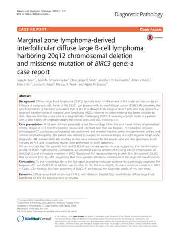
Hatem et al. Diagnostic Pathology (2016) 11:137DOI 10.1186/s13000-016-0588-xCASE REPORTOpen AccessMarginal zone lymphoma-derivedinterfollicular diffuse large B-cell lymphomaharboring 20q12 chromosomal deletionand missense mutation of BIRC3 gene: acase reportJoseph Hatem1, April M. Schrank-Hacker1, Christopher D. Watt1, Jennifer J. D. Morrissette1, Adam I. Rubin2,Ellen J. Kim2, Sunita D. Nasta3, Mariusz A. Wasik1 and Agata M. Bogusz1*AbstractBackground: Diffuse large B-cell lymphoma (DLBCL) typically leads to effacement of the nodal architecture by aninfiltrate of malignant cells. Rarely ( 1%), DLBCL can present with an interfollicular pattern (DLBCL-IF) preserving thelymphoid follicles. It has been postulated that DLBCL-IF is derived from marginal zone B cells and may represent alarge-cell transformation of marginal zone lymphoma (MZL), however no direct evidence has been provided todate. Here we describe a rare case of a diagnostically challenging DLBCL-IF involving a lymph node in a patientwith a prior history of lymphadenopathy for several years and MZL involving skin.Case presentation: A 53-year old man presented to our Dermatology Clinic due to a 1-year history of generalizeditching, fatigue of 2–3 month’s duration, nausea and mid back rash that was biopsied. PET (positron emissiontomography)/CT (computed tomography) was performed and revealed inguinal, pelvic, retroperitoneal, axillary, andcervical lymphadenopathy. The patient was referred to surgery for excisional biopsy of a right inguinal lymph node.Diagnostic H&E stained slides and ancillary studies were reviewed for the lymph node and skin specimens. B-cellclonality by PCR and sequencing studies were performed on both specimens.We demonstrate that this patient’s MZL and DLBCL-IF are clonally related, strongly suggesting that transformationof MZL to DLBCL had occurred. Furthermore, we identified a novel deletion of the long arm of chromosome 20(del(20q12)) and a missense mutation in BIRC3 (Baculoviral IAP repeat-containing protein 3) in this patient’s DLBCLthat are absent from his MZL, suggesting that these genetic alterations contributed to the large cell transformation.Conclusions: To our knowledge, this is the first report providing molecular evidence for a previously suspected linkbetween MZL and DLBCL-IF. In addition, we describe for the first time del(20q12) and a missense mutation in BIRC3in DLBCL. Our findings also raise awareness of DLBCL-IF and discuss the diagnostic pitfalls of this rare entity.Keywords: Diffuse large B-cell lymphoma (DLBCL) with deletion 20q(del(20q)), Interfollicular diffuse large B celllymphoma (DLBCL-IF), Marginal zone lymphoma* Correspondence: Agata.Bogusz@uphs.upenn.edu1Department of Pathology and Laboratory Medicine, Hospital of theUniversity of Pennsylvania, 7 E Gates Pavilion, 3400 Spruce Street,Philadelphia 19104-4283, PA, USAFull list of author information is available at the end of the article The Author(s). 2016 Open Access This article is distributed under the terms of the Creative Commons Attribution 4.0International License (http://creativecommons.org/licenses/by/4.0/), which permits unrestricted use, distribution, andreproduction in any medium, provided you give appropriate credit to the original author(s) and the source, provide a link tothe Creative Commons license, and indicate if changes were made. The Creative Commons Public Domain Dedication o/1.0/) applies to the data made available in this article, unless otherwise stated.
Hatem et al. Diagnostic Pathology (2016) 11:137BackgroundDiffuse large B-cell lymphoma (DLBCL), the most common type of non-Hodgkin lymphoma, is defined as aneoplasm of large B lymphocytes that display a diffusegrowth pattern [1]. DLBCL is a quite diverse group ofmalignancies with respect to cell morphology, pathogenesis, clinical presentation, and therapeutic response [1–3]. Gene expression profiling studies identified twomajor subgroups of nodal DLBCL based on the “cell oforigin”: the germinal center (GC) B-cell-like DLBCL (GCBDLBCL) and the prognostically less favorable activatedB-cell-like DLBCL (ABC DLBCL) [4]. Immunohistochemical signatures were developed to translate the molecularsignatures and distinguish between the GCB DLBCL andnon-GCB DLBCL [5].The World Health Organization (WHO) classificationdistinguishes numerous subtypes of DLBCL [1]. In themajority of the cases, DLBCL leads to effacement of thenodal architecture by a diffuse infiltrate of malignantcells. Rarely, however, DLCBL can show an interfollicular pattern (DLBCL-IF) of proliferation, preserving thelymphoid follicles [6]. These cases constitute only about1% of all DLBCL, frequently display a polymorphous appearance microscopically due to the admixture of nonneoplastic inflammatory cells, and often present a diagnostic challenge. Previous reports indicate that DLBCLIF are predominantly of non-GCB type [6, 7]. Interestingly, the interfollicular large B cells in normal lymphnode also show a non-GCB phenotype and share someimmunophenotypic characteristics with monocytoid Bcells [8]. It has been postulated that DLBCL-IF is derivedfrom marginal zone B cells and may represent a largecell transformation of an underlying MZL [7], however,no direct evidence has been provided to date. The overall survival rate and prognosis of the DLBCL-IF seemsto be better than that of a non-IF DLBCL as controlgroup (DLBCL-CG) as the majority of cases present instage 1 or 2, show significantly lower International Prognostic Index (IPI) scores than the DLBCL-CG [6]. Consequently, it has been previously suggested that DLBCLIF is a distinct clinicopathologic entity [7].In the case described here, we provide a direct evidence genetically linking DLBCL-IF and MZL. Furthermore, we identify a novel del(20q12) and a BIRC3missense mutation in DLBCL-IF, but not the patient’spreceding MZL involving skin, strongly suggesting thatthese genomic alterations are at least in part responsiblefor the large cell transformation.Materials and methodsHistology and immunohistochemistryFormalin-fixed paraffin-embedded tissue sections werestained with hematoxylin and eosin (H&E). Immunohistochemical stains were performed on 4 μm tissuePage 2 of 10sections using an Autostainer (Leica BOND platform,Buffalo Grove, IL) according to manufacturer’s instructions. Sections were deparaffinized in xylene and gradedalcohols. Detection of the antibodies was performedusing a chromogenic substrate, diaminobenzene(DAKO). The following antibodies were used: CD1a,CD3, CD5, CD10, CD20, CD23, CD30, CD79a, BCL6,Oct-2, BOB.1 (all from Leica), CD2, CD8, BCL2, CD45,CD68, CD138, CD163, MUM-1/IRF4, ALK, Ki67, p53(all from Dako), BCL1 (Fisher), c-MYC (Epitomics),PAX5 and CD15 (both BD Bioscience), CD4 (Biocare),CD56 (Zymed/Invitrogen), CD57 (Thermofisher), PD-1(Abcam), TIA1 (Immunotech), and panCK (Biogenex),Molecular analysis for clonalityDNA was extracted from either fresh tissue (i.e., skin) orparaffin embedded (i.e., lymph node) tissue. PCR amplification was subsequently performed using two sets offluorescently-labeled primers (InVivoScribe Technologies) that hybridize to a conserved V-framework (i.e.,FR2 or FR3) region and the conserved J-region of immunoglobulin heavy chain (IGH) gene. The PCR products were size separated by capillary electrophoresis on a3500xL Genetic Analyzer (Life Technologies). Data wereanalyzed (GeneMapper v5.0 software) and then reviewedfor peak patterns consistent with a clonal expansion.Fluorescence in situ hybridization (FISH) analysisFISH was performed on 3 μm formalin-fixed paraffinembedded sections using the Vysis LSI D20S108 (20q12)SpectrumOrange probe and the Vysis CEP 8 (D8Z2,8p11.1-8q11.1 alpha satellite) SpectrumGreen (AbbottMolecular Laboratories, Abbott Park, IL), according tothe manufacturers’ instructions. In brief, slides weredeparaffinized using xylene incubation ( 3), followed byethanol wash steps (100, 70%). Prior to hybridization theslides were treated with Dako Pre-Treatment solution(Dako, Inc K5799) followed by digestion with pepsin(37 C, 15 min). Slides were dehydrated in ethanol (70,85, 100%), dried and the FISH probes were added andincubated overnight. The following morning the slideswere washed, counterstained with DAPI and manuallyvisualized and scored.Gene mutation analysisDetection of single nucleotide variants (SNVs) and insertions/deletions (indels) analysis of paraffin-embeddedskin and lymph node tissue samples was performed bythe University of Pennsylvania clinical genomics laboratory, the Center for Personalized Diagnostics. The genessequenced were part of a custom, targeted nextgeneration sequencing amplicon panel for 68hematologic malignancy-associated genes (TruSeq Custom Amplicon, Illumina Inc.) based on previously
Hatem et al. Diagnostic Pathology (2016) 11:137described analyses [9, 10]. Briefly, individual librarypreparations were pooled and concurrently sequencedon the Illumina MiSeq (Illumina, Inc.). To assure a minimum read depth of 250 , the mean depth of coverageacross the entire panel was 2000 . Detection of SNVswas validated to a 4% allele frequency and large indels toa 1% allele frequency. A separate assay was performed inparallel to sequence the CEBPA gene using long rangePCR to isolate the gene and library preparation was performed using the tagmentation-based Nextera librarypreparation kit (Illumina, Inc.). The CEBPA gene was sequenced in tandem with the hematologic nextgeneration sequencing panel and reported together withthe custom heme-NGS panel.A custom bioinformatics pipeline was utilized to detect alterations [11]. Manual review of the data including visualization of variants was performed on allsamples following bioinformatics processing variants,with variants compared with our knowledgebase andon-line databases for further curation, using humanreference sequence UCSC build hg 19 (NCBI build37.1) for comparison. Single nucleotide polymorphisms (SNPs) with a minor allele frequency (MAF) 0.1% were considered benign and were not reported;these calls were based on the Exome Variant Server(http://evs.gs.washington.edu/EVS), the ExAC browser(http://exac.broadinstitute.org) and dbSNP. Reportedvariants used nomenclature based on the HumanGenome Variation Society nomenclature guidelines(http://www.hgvs.org/mutnomen) and internally categorized into five different categories (benign, likelybenign, variant of uncertain significance, likelypathogenic, pathogenic); the categories likely benign,variant of uncertain significance and likely pathogenicwere reported as variants of uncertain significance inthe electronic health record.Flow cytometryFlow cytometry was performed on representative tissuefrom the inguinal lymph node using the FACSCantoIIflow cytometer from BD Immunosystems. Samples wereprepared by aliquoting 50–75 μL of prepared cell suspension into reagent tubes. Twenty microliter of rabbitblocking reagent was added to the B cell clonality tubeand allowed to incubate at 37 C for 20 min and 20 μLof rabbit serum was added to the remainder of the tubes.Laboratory prepared and premixed antibody cocktailwas added in 20 μL aliquots to each designated tube andallowed to incubate in the dark for 20 to 30 min. Thecells were then washed with 3 to 5 ml of wash bufferthat was added to each tube, followed by centrifuging at1450 rpm for 5 min at 10 C. The cell pellet was resuspended in 0.5 ml of wash buffer and 10 μL of DAPIPage 3 of 10working solution was added to each tube. Data is thenacquired on the flow cytometer.Case presentationClinical findingsA 53-year-old man was evaluated in our DermatologyClinic due to a 1-year history of generalized itching, fatigue of 2–3 month’s duration, nausea over the pastmonth, and a new left mid back rash. A physical examwas essentially unremarkable except for a 1.2 cm red/purple plaque on the left mid back that was biopsied.Basic blood work was within normal limits. PET/CT wasperformed and revealed inguinal, pelvic, retroperitoneal,axillary, and cervical lymphadenopathy. The patient wasreferred to surgery for excisional biopsy of a right inguinal lymph node. Of note, a CT scan performed 4 yearsearlier had demonstrated inguinal lymphadenopathy(3.8 cm lymph node). No additional workup was done atthat time. A follow-up CT scan performed 2 years latershowed mildly prominent bilateral axillary lymphadenopathy. One year prior to the current evaluation, the patient noted swelling in the right and left axilla.Pathologic findingsSkin biopsyThe H&E-stained sections demonstrated a dense nodular lymphoid infiltrate in the superficial and deeper dermis without involvement of the epidermis (Fig. 1a). Theinfiltrate was composed of predominantly small lymphocytes with scattered larger transformed cells with immunoblast morphology (Fig. 1b). Immunohistochemicalstains demonstrated nodules of CD20 positive B cellsaround and within follicles, with scattered larger interfollicular B cells also present (Fig. 1c). CD3 stainhighlighted abundant interfollicular T lymphocytes (notshown). Essentially all neoplastic B cells as well as manyof the interfollicular T cells were positive for BCL2(Fig. 1d). BCL6 was negative in the tumor cells. Thefindings were consistent with cutaneous involvement bymarginal zone lymphoma.Excisional biopsy of the right inguinal lymph nodeThe H&E stained sections showed a lymph node with anoverall well-preserved nodal architecture including reactive lymphoid follicles with well-demarcated mantlezones (Fig. 2a). The interfollicular zones were, however,expanded by numerous large atypical mononuclear cellsadmixed with abundant eosinophils, small lymphocytes,plasma cells and histiocytes (Fig. 2a and b). The largercells had oval to slightly irregular nuclear contours, vesicular chromatin, prominent nucleoli, and scant to moderate amount of clear to amphophilic cytoplasm (Fig. 2cand d). Immunohistochemical stains showed that thelarge cells were positive for B-cell markers CD20
Hatem et al. Diagnostic Pathology (2016) 11:137Page 4 of 10Fig 1 Histological findings of the skin biopsy from initial presentation. a Low magnification (25 ) demonstrating a nodular infiltrate in thesuperficial and deeper dermis (H&E). b At higher power (400 ) the majority of the cells are small and have irregular nuclei and inconspicuousnucleoli. There are scattered large transformed immunoblasts (H&E). c CD20 stain highlights the nodules of B lymphocytes (CD20immunohistochemical stain, 50 ) d The B lymphocytes in the nodules are positive for BCL2 (BCL2 immunohistochemical stain, 50 )(Fig. 2e), PAX5 (Fig. 2f ), and CD79a, as well as BCL6(subset), CD30 (Fig. 2g), MUM1(Fig. 2h), CD23, c-MYC(40–50%), p53 (dim in 40% of the large cells), BOB.1(variable) and Oct-2 (variable). The Ki-67 proliferationindex in the interfollicular areas was high, exceeding70%. Key negative stains included BCL2, CD10, andCD15. CD138 highlighted plasma cells with a kappato lambda ratio of 3:1. An in-situ hybridization studyfor Epstein-Barr virus (EBER) was negative. CD20,PAX5 and CD79a stains also marked the abundantreactive follicles with CD10 , BCL6 and BCL2- germinal centers and BCL2 mantle zones. The proliferation index within the germinal centers was high asexpected ( 90%) and highlighted the polarized lightand dark zones. The infiltrating small T lymphocyteswere positive for CD2, CD3, CD5 and CD7 (majorsubset). The CD4 to CD8 ratio was increased ( 5:1).CD57 and PD-1 were positive in a subset of germinalcenter T cells whereas TIA1, perforin, and granzymeB were positive in scattered small interfollicular Tcells with a pattern similar to CD8 staining. CD56and ALK-1 stains were negative. CD68, CD163, andCD1a stained scattered histiocytes and accessory cellsrespectively.Ancillary studies on the lymph node specimenFlow cytometry studies were performed on representative tissue from the right inguinal lymph node. Asshown in Fig. 3a–c, there was a large population ofCD19 B lymphocytes (51% of all lymphoid cells)with a strong lambda bias bordering on overt restriction (kappa: lambda ratio of 0.33 and 0.27 for theCD19 and CD20 cells, respectively). Minor subsetsof all B lymphocytes were CD5 (Fig. 3a) or CD10 (Fig. 3d); these cells expressed predominantly kappaand, hence, were not part of the apparent lambdaclone.Molecular studies for IGH gene rearrangement performed on the skin biopsy showed a 281 base pair (bp)peak in framework 2, as well as 94 bp and 115 bp peaksin framework 3 (Fig. 4). Examination of tissue obtainedfrom the right inguinal lymph node demonstrated thesame 281 bp peak in framework 2 and the same 115 bppeak in framework 3 whereas the 94 bp peak seen in theskin specimen was not seen in the lymph node (Fig. 4b).The overall results indicate that the lymphoproliferativedisorders involving lymph node and skin are clonally related. The significance of the 94 bp peak is unclear, butit may represent a separate (sub-)clonal expansion. No
Hatem et al. Diagnostic Pathology (2016) 11:137Page 5 of 10Fig. 2 Histological findings of the inguinal lymph node excision 2 weeks after initial presentation. a Low-power view (25 ) shows a lymph node with apreserved architecture with intact capsule, patent sinuses and polarized germinal centers with well-demarcated mantle zones. The interfollicular areas areexpanded (H&E). b At higher magnification (100 ) the interfollicular infiltrate is composed of numerous large transformed immunoblasts in the backgroundof small lymphocytes, abundant eosinophils, scattered plasma cells and histiocytes (H&E). c At 200 magnification (H&E) and (D) at 400 magnification thelarge atypical cells have large oval to slightly irregular nuclei with vesicular chromatin, prominent nucleoli and scant to moderate amounts of clear toamphophilic cytoplasm (H&E). e The large atypical cells are immunoreactive for CD20 (CD20 immunostain, 50 ) as well as for f PAX5 (PAX5 immunostain,200 ), g CD30 (CD30 immunostain, 200 ) and h MUM1 (MUM1 immunostain, 200 )clonal T-cell receptor (TCR) rearrangement was detected in the lymph node specimen.FISH studies for rearrangements of MYC, BCL2, andBCL6 performed on the lymph node were also negative.Previous report described del(20q) in MZL with aggressive features [12]. Since this patient’s DLBCL wasthought be derived from a MZL, additional FISH analysis for a 20q12 deletion was performed on both skinand lymph node material to determine whether thisabnormality may be present. The analysis revealed that38/150 (25.3%) of cells in the involved lymph node wereharboring del(20q12) (Fig. 5), whereas the skin lesionwas negative for this deletion (data not shown).Targeted DNA sequencing studies using ourhematologic malignancy panel on the lymph node tissuedid not reveal any known disease-associated mutations,however it did identify two variants of uncertain significance (Table 1): a missense variant in BIRC3 (Baculoviral
Hatem et al. Diagnostic Pathology (2016) 11:137Page 6 of 10Fig. 3 Flow cytometric analysis of representative tissue from the inguinal lymph node. a The CD19/CD5 plot demonstrates and admixture ofCD19-positive B cells and CD5-positive T cells. b Gating on the CD19 positive B cells demonstrates a clear lambda bias. c the lambda bias of isalso seen when gating on CD20 positive B cells. d The majority of the CD19 positive B cells were found to be CD20 positive and CD10 negative.The presence of a subset of CD10 positive B cells and subset positivity for kappa light chain staining is compatible with germinal center B cellsthat are admixed with the malignant B cellsIAP repeat-containing protein 3) at amino acid 572 converting the wild type residue, cysteine, to tyrosine inBIRC3 (p.C572Y c.1715G A) in 491 reads out of atotal 9155 sequence reads for an allele frequency of5.36% and a missense variant in EZH2 gene (Enhancerof Zeste Homolog 2) at amino acid 322 converting thewild type residue, Asparagine, to Serine (p.N322S c.965A G) in 818 reads out of a total 1561 sequencereads for an allele frequency of 52.40% . Subsequent sequencing studies of the skin revealed the same missensevariant in EZH2 as in the lymph node in 1097 reads outof a total 2165 sequence reads for an allele frequency ofFig. 4 IGH PCR analysis of both specimens revealed a shared pattern of peaks. a Primers for FR2 region identified a clonal peak at approximately281 bp in the skin and the lymph node. b Similarly, a reproducible peak at approximately 115 bp was identified with primers targeting the FR3region of IGH. An additional 94 bp peak was identified in a polyclonal background in the skin but not lymph node specimen
Hatem et al. Diagnostic Pathology (2016) 11:137Page 7 of 10Fig. 5 FISH analysis of the lymph node specimen. Representative images of FISH analysis of the lymph node specimen using probes specific forchromosome 20 (20q12, red) and centromere of chromosome 8 (green). a Cell positive for del(20q) and b cell negative for del(20q)50.67% but no other alterations, including the BIRC3mutation, were identified.DiscussionWe report a rare case of a DLBCL with an interfollicularpattern of involvement (DLBCL-IF) with sparing of thenodal architecture including lymphoid follicles. This pattern is common in peripheral T-cell lymphoma, nototherwise specified (PTCL, NOS) that shows proliferation of malignant cells in the paracortex [1]. Our caseshowed that the large malignant cells were admixed withan inflammatory infiltrate including abundant eosinophils. It has been reported that approximately 60% ofDLBCL-IF cases contain reactive inflammatory cells thatare admixed with the large cells, a feature that is frequently seen also in PTCL, NOS [1, 6, 7]. Thus, DLBCLIF can mimic histological picture of PTCL, NOS and itis of utmost importance to be aware of this entity, asPTCL-NOS carries a worse prognosis than DLBCL-IFand calls for different therapeutic approaches. Therefore,immunohistochemistry studies are required to differentiate between these two entities. It is important to keep inmind that both these types of lymphoma can be immunoreactive for CD30. Furthermore, it is critical to alwaysconsider DLBCL-IF in the differential diagnosis of atypical interfollicular proliferations. Since distinguishing itfrom a reactive interfollicular hyperplasia may be sometimes difficult, given the abundant background of inflammatory cells often seen in this type of lymphoma,additional ancillary testing including IGH gene rearrangement studies and flow cytometry may prove particularly useful to establish the diagnosis.It has been postulated that DLBCL-IF may be derivedfrom marginal zone B cells and may represent a large-cell transformation of a MZL [7, 8]. To date, however,there was no direct evidence to confirm this hypothesis.Here we show for the first time a direct link between aMZL and DLBCL-IF by demonstrating the presence ofthe same clonal IGH rearrangement in the patient’s skinand nodal biopsies. Furthermore we identifieddel(20q12) in the DLBCL-IF. This genetic abnormalityhas not been reported to date in a DLBCL of any kind.Deletion of chromosome 20q is a common cytogeneticabnormality in various myeloid neoplasms, most notablymyelodysplastic syndrome [1, 13, 14] but its associationwith lymphoid malignancies is rare and limited to a fewreports. This includes a case of lymphoplasmacyticlymphoma/Waldenström macroglobulinemia [15] andtwo cases of marginal zone lymphoma [12, 16]. Anotherstudy of 64 patients with chronic lymphocytic leukemia(CLL) revealed that del(20q) appears to be a therapyrelated abnormality in CLL involving both myeloid andlymphoid cells and may represent disease progression[17, 18]. Given that del(20q12) in this patient was identified in the lymph node and not skin lesion and that thepatient has not received any prior chemotherapy at thattime, this abnormality is likely related to transformationof MZL to DLBCL and implicates a loss of importantgene(s) likely with tumor suppressor function. Potentialcandidate genes that map to this region include MYBL2[19], Dido [20], L3MBTL1 [21] and SAMHD1 [22].Next-generation sequencing studies using a custompanel of 68 hematologic malignancy-associated genes revealed the same missense variant in EZH2 in both theskin and lymph node specimen and a missense variantin BIRC3 that was present only in the lymph node specimen. The EZH2 variant was listed in all three databasessearched at very low allele frequencies (5 cases inTable 1 Summary of identified missense variants in the lymph node and skin biopsy specimensGene alterationProtein changeNucleotide changeFrequency in lymph nodeFrequency in skinBIRC3C572Yc.1715G A5.36%0%EZH2N322Sc.965A G52.40%50.67%Abbreviations: A adenine, C cysteine, c. coding, G guanine, N asparagine, p. protein, S serine, Y tyrosine
Hatem et al. Diagnostic Pathology (2016) 11:137COSMIC, in 4/13002 alleles in the exome variant server,and 37/121,370 alleles in the ExAC database).The missense variant in BIRC3 (p.C572Y c.1715G A), not reported in any of the three databases searched(see methods section), was detected only in the lymphnode but not skin specimen at an allele frequency of5.36%. Morphologic examination revealed that thetumor comprises about 10-15% of the lymph node specimen. When these two observations are taken together,the data imply that the BIRC3 mutation may be presentin a heterozygous state within the neoplasm (i.e., atumor burden of approximately 10% with a heterozygousmutation would be expected to generate an allelic burden of approximately 5%). So, even though this varianthas not been previously described, the circumstantialevidence implies this is an oncogenic mutation that occurred in the malignant DLBCL-IF cells.The fact that this mutation was only seen in thelymph node and not in the skin biopsy strongly suggests that it may have contributed to transformationof this patient’s MZL to DLBCL. BIRC mutations arenot among the recurrent mutations in the “standard”DLBCL according to a recent report [23]. BIRC3 genelocated on chromosome 11 at 11q22.2 encodes cIAP2(cellular inhibitor of apoptosis 2), that acts as an E3ubiquitin ligase that regulates NF-kappa-B signalingand affects a variety of cell functions including apoptosis, proliferation and immune modulation [24, 25].Of note, proteins from the IAP family are potentialtherapeutic targets in hematological malignancies [26].BIRC3 mutations have been identified in various smallB cell lymphomas including chronic lymphocyticleukemia (CLL) and splenic marginal zone lymphoma(SMZL) [27, 28]. More recent studies demonstrate therole of the ubiquitin ligases, both cIAP2 and a closelystructurally related cIAP1, encoded by the BIRC2gene, in NF-κB signaling triggered by the B cell receptor (BCR) in DLBCL cells [29].The alteration in EZH2 gene (p.N322S c.965A G)was observed with an allele frequency of approximately50% in both specimens and this most likely represents abenign/low-oncogeneic potential polymorphism ratherthan a highly oncogenic mutation. EZH2 gene on 7q36encodes the histone methyltransferase which initiatesepigenetic silencing of genes [30] and plays a role in normal B cell development [31]. EZH2 is mutated in a variety of tumors including lymphomas and targeting ofEZH2 protein with specific inhibitors show promisingresults in preclinical studies and early clinical trials [32–34]. Interestingly, it has been reported that EZH2 polymorphisms may contribute to susceptibility of tumor development and predict overall survival in some cancers[35–38] and may have also contributed to progression ofthe disease in this patient.Page 8 of 10ConclusionsIn summary, we present a rare case of diagnosticallychallenging DLBCL with an interfollicular pattern andprovide data that support the hypothesis that DLBCL-IFcan arise from a MZL. To our knowledge, this represents the first reported case where DLBCL-IF and MZLare shown to be clonally related. We identify a noveldel20q and a BIRC3 missense mutation in this patient’sDLBCL-IF that have not been previously reported inDLBCL. Given the absence of both these genetic abnormalities in the patient’s MZL, they both may have contributed to the large cell transformation.AbbreviationsABC DLBCL: Activated B-cell-like DLBCL; BCR: B-cell receptor; BIRC3: Baculoviral IAPrepeat-containing protein 3; cIAP2: Cellular inhibitor of apoptosis 2; CLL: Chroniclymphocytic leukemia; CT: Computed tomography; del(20q): Deletion 20q;DLBCL: Diffuse large B-cell lymphoma; DLBCL-IF: Interfollicular diffuse large B-celllymphoma; EZH2: Enhancer of zeste homolog 2; FISH: Fluorescence in situhybridization; GCB DLBCL: Germinal center (GC) B-cell-like DLBCL; IGH: Immunoglobulin heavy chain; IPI: International prognostic index; MZL: Marginal zonelymphoma; PET: Positron emission tomography; PTCL, NOS: Peripheral T-celllymphoma, not otherwise specified; WHO: The World Health OrganizationAcknowledgementsNot applicable.FundingThe author(s) received no financial support for this publication.Availability of data and materialsThe histological specimens and the corresponding data belong to theDepartment of Pathology and Dermatology of the University of Pennsylvaniaand are available upon request for review by the Editor-in-Chief.Authors’ contributionsJH and AMB drafted the manuscript; CDW, JJDM, AIR, EJK, SDN and MAWrevised the manuscript. AMB finalized the manuscript. ASH performed FISHstudies (Fig. 5). CDW analyzed the molecular data (Fig. 4). JJDM analyzedgenetic data (Table 1). All authors read and approved the final manuscript.Competing interestsThe authors declare that they have no competing interests.Consent for publicationWritten informed consent for publication of their clinical details and/orclinical images was obtained from the patient. A copy of the consent form isavailable for review by the Editor of this journal.Ethics approval and consent to participateNot applicable.Author details1Department of Patholog
sequenced were part of a custom, targeted next- generation sequencing amplicon panel for 68 hematologic malignancy-associated genes (TruSeq Cus-tom Amplicon, Illumina Inc.) based on previously Hatem et al. Diagnostic Pathology (2016) 11:137 Page 2 of 10. described analyses [9, 10]. Briefly, individual library preparations were pooled and concurrently sequenced on the Illumina MiSeq (Illumina .