Transcription
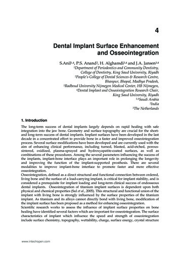
4Dental Implant Surface Enhancementand OsseointegrationS.Anil1,4, P.S. Anand2, H. Alghamdi3,4 and J.A. Jansen3,41Departmentof Periodontics and Community Dentistry,College of Dentistry, King Saud University, Riyadh2People’s College of Dental Sciences & Research Centre,Bhanpur, Bhopal, Madhya Pradesh,3Radboud University Nijmegen Medical Center, HB Nijmegen,4Dental Implant and Osseointegration Research Chair,King Saud University, Riyadh1,4Saudi Arabia2India3The Netherlands1. IntroductionThe long-term success of dental implants largely depends on rapid healing with safeintegration into the jaw bone. Geometry and surface topography are crucial for the shortand long-term success of dental implants. Implant surfaces have been developed in the lastdecade in a concentrated effort to provide bone in a faster and improved osseointegrationprocess. Several surface modifications have been developed and are currently used with theaim of enhancing clinical performance, including turned, blasted, acid-etched, poroussintered, oxidized, plasma-sprayed and hydroxyapatite-coated surfaces, as well ascombinations of these procedures. Among the several parameters influencing the success ofthe implants, implant-bone interface plays an important role in prolonging the longevityand improving the function of the implant-supported prosthesis. There are severalmodalities to improve implant-bone interface to promote faster and more effectiveosseointegration.Osseointegration, defined as a direct structural and functional connection between ordered,living bone and the surface of a load-carrying implant, is critical for implant stability, and isconsidered a prerequisite for implant loading and long-term clinical success of endosseousdental implants. Osseointegration of titanium implant surfaces is dependent upon bothphysical and chemical properties (Sul et al., 2005). This structural and functional union of theimplant with living bone is strongly influenced by the surface properties of the titaniumimplant. As titanium and its alloys cannot directly bond with living bone, modification ofthe implant surface has been proposed as a method for enhancing osseointegration.Scientific research works to assess the influence of implant surface properties on bonehealing have identified several factors which are important for osseointegration. The surfacecharacteristics of implant which influence the speed and strength of osseointegrationinclude surface chemistry, topography, wettability, charge, surface energy, crystal structurewww.intechopen.com
84Implant Dentistry A Rapidly Evolving Practiceand crystallinity, roughness, chemical potential, strain hardening, the presence of impurities, thickness of titanium oxide layer, and the presence of metal and non-metalcomposites. Among these, wettability and free surface energy of an implant surface areconsidered to be very crucial. The implant surface, including topography, chemistry, surfacecharge, and wettability, has been described as an important factor to influenceosseointegration. The influence of physical properties such as surface topography androughness on osseointegration have translated to shorter healing times from implantplacement to restoration (Cochran et al., 2002). The biologic basis underlying these clinicalimprovements continues to be explored (Kim et al., 2005, Lossdorfer et al., 2004). Albrektssonet al. (1981) suggested six factors that are particluarly important for the establishment ofreliable osseointegration: implant material, implant design, surface conditions, status of thebone, surgical technique, and implant loading conditions.2. Biology of wound healing following implant placementWound healing involves a highly orchestrated sequence of events which is triggered bytissue injury involving soluble mediators, blood cells, extracellular matrix and parenchymalcells. Ultimately, it culminates in either partial or complete regeneration or repair. Fracturehealing in bone occurs in four phases which include inflammation, soft and hard callusformation, and remodeling. Following a fracture, blood coagulation and hematomaformation takes place. This is followed by inflammation. Various chemical mediators suchas thrombin and growth factors released by activated leukocytes and platelets in thehematoma serve as chemotactic signals to many cell types which play an important role inbone healing. Unlike soft tissue healing, bone healing does not lead to scarring. Instead itleads to restoration of the bony tissue. During successful implantation, insertion of metalimplants into cortical bone eventually leads to complete healing. Following implantplacement, unlike in fracture healing, implants extend into and persist in the marrow spacesand this may have a bearing on the healing process. Although implant healing must to someextent adjust to the presence of the implant, ultimately, sound bony tissues will becompletely restored during wound healing. This adjustment involves imbedding theimplant surface in a layer of bone, continuous with the original bone.Wound healing around a dental implant placed into a prepared osteotomy follows threestages of repair- Initial formation of a blood clot occurs through a biochemical activationfollowed by a cellular activation and finally a cellular response(Stanford and Schneider,2004). During surgery, dental implant surfaces interact with blood components fromruptured blood vessels. Within a short period of time, various plasma proteins such as fibringet adsorbed on the material surface. Fibrinogen is converted to fibrin and the complementand kinin systems become activated. As in fracture healing, the migration of bone cells inperi-implant healing will occur through the fibrin of a blood clot. Since fibrin has thepotential to adhere to almost all surfaces, it can be anticipated that the migration ofosteogenic cell populations towards the implant surface will occur. However, as themigration of cells through fibrin will cause retraction of the fibrin scaffold, the ability of animplant surface to retain this fibrin scaffold during the phase of wound contraction is criticalin determining whether the migrating cells will reach the implant surface. Activation ofplatelets occurs as a result of interaction of platelets with the implant surface as well as thefibrin scaffold and this leads to thrombus formation and blood clotting.www.intechopen.com
Dental Implant Surface Enhancement and Osseointegration85Fig. 1. The implant healing process - The surface composition, roughness and topographyare interrelated surface characteristics that influence the biological response to an implant.Moreover, platelets are a rich source of many growth and differentiation factors which playa key role in the wound healing process by acting as signaling molecules for recruitmentand differentiation of the undifferentiated mesechymal stem cells at the implant surface.Plasma also contains dissolved substances such as glucose, amino acids, various ions,cholesterols, and hormones which are needed for the viability of cells and tissues. Bloodinteractions with implants lead to protein adsorption, which is dependent on the surfaceproperties of the material. As hydrophilic surfaces are better for blood coagulation thanhydrophobic surfaces, dental implants have been developed with high hydrophilic andrough implant surfaces which exhibit better osseointegration than conventional ones.Adsorption of proteins such as fibronectin and vitronectin on the surface of dental implantscould promote cell adhesion and osseointegration. During the initial remodeling, a numberof immune cells mediate early tissue response followed by migration of phagocytemacrophages. These cells initially remove the necrotic debris created by the drilling processand then undergo physiological changes which lead to expression of cell surface proteinsand production of cytokines and pro-inflammatory mediators. This cytokine-regulatedcellular recruitment, migration, proliferation and formation of an extracellular matrix on theimplant surface can be influenced by the macrophages. These cells express growth factorssuch as fibroblast growth factor (FGF-1, FGF-2, FGF-4), transforming growth factors,epithelial growth factor as well as bone morphogenetic proteins (BMPs). The end result ofthis complex cascade is promotion of a wound healing process that includes angiogenesis.3. Influence of implant surface topography on osseointegrationDental implant quality depends on the chemical, physical, mechanical, and topographiccharacteristics of the surface (Grassi et al., 2006). These different properties interact anddetermine the activity of the attached cells that are close to the dental implant surface.www.intechopen.com
86Implant Dentistry A Rapidly Evolving PracticeDental implants have been designed to provide textures and shapes that may enhancecellular activity and direct bone apposition (Huang et al., 2005). Osteogenesis at the implantsurface is influenced by several mechanisms. A series of coordinated events, including cellproliferation, transformation of osteoblasts and bone tissue formation might be affected bydifferent surface topographies (Shibli et al., 2007). Amount of bone-to-implant contact (BIC)is an important determinant in long-term success of dental implants. Consequently,maximizing the BIC and osseointegration has become a goal of treatment, which isenhanced by implant surface roughness (Soskolne et al., 2002).Albrektsson et al (1981) recognized that among the factors influencing BIC such astopography, chemistry, wettability and surface energy the most important is wettability.Surface wettability is largely dependent on surface energy and influences the degree ofcontact with the physiological environment (Kilpadi and Lemons, 1994, Zhao et al., 2005).Several evaluations have demonstrated that implants with rough surfaces show better boneapposition and BIC than implants with smooth surfaces (Buser et al., 1999, Cochran et al.,2002).Surface roughness also has a positive influence on cell migration and proliferation, which inturn leads to better BIC results, suggesting that the microstructure of the implant influencesbiomaterial–tissue interaction (Matsuo et al., 1999, Novaes et al., 2002). Implant surfaceproperties are likely to be of particular relevance to the chemical and biological interfaceprocesses in the early healing stages after implantation. It is generally accepted that these earlystages are likely to have an effect on the host response to the implant and, therefore, the longterm outcome and success of the treatment. Surface chemistry has the potential to alter ionicinteractions, protein adsorption, and cellular activity at the implant surface (Schliephake et al.,2005). These modifications may subsequently influence conformational changes in thestructures and interactive natures of adsorbed proteins and cells. Furthermore, within thecomplexities of an in vivo environment containing multiple protein and cellular interactions,these alterations may differentially regulate biologic events. Modifications to the implantsurface chemistry may lead to alterations in the structure of adsorbed proteins and havecascading effects that may ultimately be evident at the clinical level.In vivo evidence has supported the use of alterations in surface chemistry to modifyosseointegration events. Specifically, an investigation utilizing sandblasted, large-grit, acidetched (SLA) surfaces that were chemically different but had the same physical propertieswas conducted to assess BIC as a measure of osseointegration. The chemically enhancedSLA surface demonstrated significantly enhanced BIC during the first 4 weeks of bonehealing, with 60% more bone than the standard SLA surface after 2 weeks (Buser et al.,2004). The chemical modifications for the test SLA surface resulted in increased wettability(ie, in a hydrophilic surface rather than a hydrophobic one). Water contact angles of zerodegrees were seen with the chemically enhanced surface compared to 139.9 degrees for astandard SLA surface, and the hydrophilicity was maintained after drying. The chemicalcomposition of the surface was also altered, including a 50% reduction in carbonconcentration compared with the control implant surface (Rupp et al., 2006).4. Interaction between cells and the surface of the dental implantsSince surface properties of biomaterials are important parameters influencing cellular reactions towards artificial materials, the properties of dental implant surfaces are extremelyimportant in influencing the healing process leading to osseointegration and ultimatewww.intechopen.com
Dental Implant Surface Enhancement and Osseointegration87clinical success of the implant. Surface morphology modulates the response of cells to adental implant, and surfaces with defined microstructures may be useful for enhancementof the stable anchorage (Elias and Meirelles, 2010). Surface chemistry involves adhesion ofproteins, bacteria, and cells on implants. Wettability and surface energy influence theadsorption of proteins, and increase adhesion of osteoblasts on the implant surface. The cellbehavior on a hydrophilic surface is completely different from that on a hydrophobic one. Ahydrophilic surface is better for blood coagulation than a hydrophobic surface. Theexpressions of bone-specific differentiation factors for osteoblasts are higher on hydrophilicsurfaces. Consequently, dental implants manufacturers have developed high hydrophilicand rough implant surfaces which in turn exhibited better osseointegration than implantswith smooth surfaces.Fig. 2. Illustration showing the cellular phenomena at the implant bone interface duringhealing of implant5. Implant surface topographyImplant surface topography refers to macroscopic and microscopic features of the implantsurface. Although commercially pure titanium is the prime material of dental implants, thesuccess rates of different commercially available implant systems vary. The exact reason forthis is not clear. Several implant-related factors such as implant surface topography,chemical composition and surface roughness that influence osseointegration have beenstudied. It has been shown that titanium implants with adequate roughness may influencethe primary stability of implants, enhance bone-to-implant contact, and may increaseremoval torque force (Wennerberg and Albrektsson, 2009).The surface roughness of the implants can significantly alter the process of osseointegrationbecause the cells react differently to smooth and rough surfaces. Fibroblasts and epithelial cellsadhere more strongly to smooth surfaces, whereas osteoblastic proliferation and collagensynthesis are increased on rough surfaces (Boyan et al., 2001). Investigators have demonstratedthat while the adhesion of fibroblasts is lesser on rough surfaces, the adhesion andwww.intechopen.com
88Implant Dentistry A Rapidly Evolving Practicedifferentiation of osteoblastic cells are enhanced (Wennerberg and Albrektsson, 2000). It is notclear whether the height of surface irregularities is more important than the distance betweenthem, and which combination of these factors could improve osseointegration. Although theincrease in surface roughness promotes greater mechanical anchorage, the implant–boneinterface strength will not increase with the continuous increase of surface roughness.Fig. 3. The machined and nano etched implant surface6. Surface roughnessSurface topography plays an important role in the osseointegration of titanium implants (LeGuehennec et al., 2007). In vitro and in vivo studies have shown that titanium surfaceroughness influences a number of events in the behavior of cells in the osteoblastic lineage,including spreading and proliferation, differentiation, and protein synthesis (Sammons et al.,2005, Zhao et al., 2006). Implant surface roughness is divided, depending on the dimensionof the measured surface features into macro, micro, and nano-roughness.Macro roughness comprises features in the range of millimeters to tens of microns. Thisscale directly relates to implant geometry, with threaded screw and macro porous surfacetreatments. The primary implant fixation and long-term mechanical stability can beimproved by an appropriate macro roughness. This will enhance the mechanicalinterlocking between the macro rough features of the implant surface and the surroundingbone (Wennerberg et al., 1996, Shalabi et al., 2006).Micro roughness is defined as being in the range of 1–10 µm. This range of roughnessmaximizes the interlocking between mineralized bone and implant surface. Studiessupported by some clinical evidence suggest that the micron-level surface topographywww.intechopen.com
Dental Implant Surface Enhancement and Osseointegration89results in greater accrual of bone at the implant surface (Junker et al., 2009, Shalabi et al., 2006).The use of surfaces provided with nanoscale topographies are widely used in recent years.Nanotechnology involves materials that have a nano-sized topography or are composed ofnano-sized materials with a size range between 1 and 100 nm. Nanometer roughness playsan important role in the adsorption of proteins, adhesion of osteoblastic cells and thus therate of osseointegration (Brett et al., 2004).6.1 NanotopographySurface properties play a key role in biological interactions between the implant surfacesand the host bone. Modifying surface roughness has been shown to enhance BIC andimprove the clinical performance of implants. The nanometer-sized roughness and thechemistry have a key role in the interactions of surfaces with proteins and cells. Thesemicromechanical features influence the process of secondary integration (bone growth,turnover and remodeling). At the nanoscale, a more textured surface topography increasesthe surface energy which in turn increases the wettability of the surface to blood, adhesionof cells to the surface, and facilitates binding of fibrin, matrix proteins, growth anddifferentiation factors. Nanotopography, by modulating cell behavior, can influence theprocess of cell migration, proliferation, and differentiation. These surfaces thus enhance theprocess of osseointegration by hastening the wound healing following implant placement(Dohan Ehrenfest et al., 2010).Various surface modification treatments create a nanometer-scale topography that allowsthe bone to grow into and maintain the implant surface under elevated shear forces. Gritblasting, anodisation, and acid etching, are the commonly used methods for modifyingsurface roughness of metal implants. Topographical features in the nanometer ranges maybe helpful in the healing process as related to protein adsorption and cell adhesion assurface properties control the steps of adhesion, proliferation, and differentiation ofmesenchymal stem cells and, thus, condition tissue integration.Fig. 4. Showing bone healing at the nanorough surfaceNanotopography modifications are commonly described in the literature both asnanoroughness and nanofeatures. Overall surface roughness will be modified when featuresare added to the surface, that is, by adding nanofeatures the surface roughness will also bewww.intechopen.com
90Implant Dentistry A Rapidly Evolving Practicemodified. However, the modifications commonly used to produce the so-called nanoroughmaterials do not intentionally produce such nanofeatures. Reproducible surface roughnessin the nanometer range is difficult to obtain with chemical treatments. Although, all surfacesmay show nanotopography, not all of them will have significant nanostructures. Ananostructure is an object of intermediate size between molecular and micrometre-sizedstructures, and often defined between 1 and 100 nm (Dohan Ehrenfest et al., 2010).Nanofabricated samples have well-defined dimensions that aim to modulate cell activity,such as migration, attachment, proliferation and differentiation.Several investigators have revealed that nanoscale topography also influences cell adhesionand osteoblastic differentiation (Dalby et al., 2008, Webster et al., 1999). These findingsreiterate observations demonstrating that nanotopography may directly influence adherentcell behavior (Webster et al., 2000). Nanotechnology can alter the implant surface at anatomic level (Oh et al., 2005) and may influence the chemical composition of these surfaces.Nanorough titanium and nanostructured titanium can enhance osteoblast adhesion anddifferentiation compared to their nanosmooth control. Surfaces with micro- and nanoporeshave also been shown to greatly enhance osseointegration. The micro- and nanoscale surfaceproperties of metal implant, including chemistry, roughness, and wettability, could affectbone formation. It has been shown that grit-blasting with biphasic calcium phosphate (BCP)ceramic particles gave a high average surface roughness and particle-free surfaces after acidetching of titanium implants. Studies have shown that BCP grit-blasted surfaces promotedan early osteoblast differentiation and bone apposition as compared to mirror-polishedtitanium. By the process of anodic oxidation, nanoscale oxides may be deposited on surfacesof titanium implants. The nanoscale properties can be controlled by adjusting theparameters for anodization such as voltage, time, and shaking. Osseointegration of dentalimplants can be improved by the application of calcium phosphate (CaP) coating by plasmaspraying, biomimetic and electrophoretic deposition. While plasma-sprayed hydroxyapatite(HA)-coated dental implants have disadvantages related to coating delimitation andheterogeneous dissolution rate of deposited phases, an electrochemical process consisting ofdepositing CaP crystals from supersaturated solutions releases calcium and phosphate ionsfrom these coatings. This process helps in the precipitation of biological apatite nanocrystalswith the incorporation of various proteins, which in turn, promotes cell adhesion,differentiation into osteoblast, and the synthesis of mineralized collagen, the extracellularmatrix of bone tissue (Sandrine et al., 2010).Osteoclast cells are also able to resorb the CaP coatings and activate osteoblast cells toproduce bone tissue. Thus, these CaP coatings promote a direct bone-implant contactwithout an intervening connective tissue layer leading to a proper biomechanical fixation ofdental implants. Currently, titanium is the standard material for dental implants because ofits excellent biocompatibility and osseointegration properties. On account of the influence ofsurface modifications of the titanium implants on osseointegration, such modifications havebeen successfully exploited to influence bone integration and long-term stability of theimplant.7. Methods of surface modifications of implantsThe methods employed for surface modifications of implants can be broadly classified into 3types-mechanical; chemical; and physical. These different methods can be employed tochange the implant surface chemistry, morphology, and structure. The main objective ofwww.intechopen.com
Dental Implant Surface Enhancement and Osseointegration91these techniques is to improve the bio-mechanical properties of the implant such asstimulation of bone formation to enhance osseointegration, removal of surfacecontaminants, and improvement of wear and corrosion resistance.7.1 Mechanical methodsThe mechanical methods include grinding, blasting, machining, and polishing. Theseprocedures involving physical treatment generally result in rough or smooth surfaces whichcan enhance the adhesion, proliferation, and differentiation of cells.7.2 Chemical methodsMethods of surface modification of titanium and its alloys by chemical treatment arebased on chemical reactions occurring at the interface between titanium and a solution.The chemical methods of implant surface modifications include chemical treatment withacids or alkali, hydrogen peroxide treatment, sol-gel, chemical vapor deposition, andanodization. Chemical surface modification of titanium has been widely applied to altersurface roughness and composition and enhance wettability/surface energy (Bagno andDi Bello, 2004).The process of acid treatment serves to remove the surface oxide and contamination whichleads to a clean and homogenous surface. The acids commonly used include hydrochloricacid, sulfuric acid, hydrofluoric acid, and nitric acid. Acid treatment of the surfaces oftitanium implants results in uniform roughness with micro pits ranging in size from 0.5-2µm, increase in surface area, and an improvement in bioadhesion. Acid treatment ofimplants enhances osseointegration as these implants can facilitate migration and retentionof osteogenic cells at the implant surface (Takeuchi et al., 2003).Alkali treatment involves immersion of the implants in either sodium or potassiumhydroxide followed by heat treatment by rinsing in distilled water. This results in thegrowth of a bioactive, nanostructured sodium titanate layer on the implant surface. Thesurface acts as a site for the subsequent in vitro nucleation of calcium phosphates whenimmersed in simulated body fluids (SBF). This involves an initial formation of Ti-OH byrelease of sodium ions from the sodium titanate layer by the process of ion exchange. This isfollowed by formation of calcium titanate as a result of reaction with the calcium ions fromthe fluid. Being negatively charged, Ti-OH groups react selectively with the positivelycharged calcium ions in the SBF to form calcium titanate. Phosphate and calcium ions getincorporated into this calcium titanate and get transformed into apatite which can providefavorable conditions for bone marrow cell differentiation.Chemical treatment of implant surfaces with hydrogen peroxide results in chemicaldissolution and oxidation of the titanium surface. When titanium surfaces react withhydrogen peroxide, Ti-peroxy gels are formed. The thickness of titania layer formed can becontrolled by adjusting the treatment time and it has been demonstrated that, whenimmersed in SBF, thicker layers of titania gel are more favorable for the deposition of apatite(Tavares et al., 2007).Anodization is a process by which oxide films are deposited on the surface of the titaniumimplants by means of an electrochemical reaction. In this process, titanium surface to beoxidized serves as the anode in an electrochemical cell with diluted solution of acids servingas the electrolyte. The thickness of the oxide layer can be altered by altering the parameterswww.intechopen.com
92Implant Dentistry A Rapidly Evolving Practiceof the electrochemical process and it has been shown that these anodized surfacesdemonstrate improved adhesion and bonding.The sol-gel process used to deposit ceramic coatings can be employed to deposit HAcoatings on the implant surface. This results in thin layers of less than 10 µm thickness. Thisprocess improves the biological activity of the titanium implants and contributes toenhanced bone formation and osseointegration. Materials such as TiO2, CaP, TiO2-CaPcomposite, and silica-based coatings can be deposited on the titanium surface by thistechnique. Chemical vapor deposition involves chemical reactions between chemicals in thegas phase and the surface of the substrate which results in the deposition of a non-volatilecompound on the substrate.7.3 Physical methodsThe physical methods of implant surface modification include plasma spraying, sputtering,and ion deposition.Plasma spraying includes atmospheric plasma spraying and vacuum plasma spraying. Thisis used for creating titanium and CaP coatings on the surfaces of titanium implants. Onemajor concern in the use of plasma sprayed coatings is the resorption and degradability ofHA in the case of HA (PSHA) coated implants and loosening of the titanium particles in thecase of titanium plasma sprayed (TPS) implants. This can affect the stability of the implantsas well as pose a health hazard.Sputtering, a method employed to deposit thin films, has been used to deposit thin films onimplant surfaces to improve their biocompatibility, biological activity, and mechanicalproperties such as wear resistance and corrosion resistance.8. Surface treatment of titanium implants8.1 Turned surface (machined dental implants)The first generation of dental implants, termed the turned implants, had a relativelysmooth surface. After being manufactured, these implants are submitted to cleaning,decontamination and sterilization procedures. Scanning electron microscopy analysisshowed that the surfaces of machined implants have grooves, ridges and marks of thetools used for their manufacturing. These surface defects provide mechanical resistancethrough bone interlocking. The disadvantage regarding the morphology of non-treatedimplants (machined) is the fact that osteoblastic cells are rugophilic – that is, they areprone to grow along the grooves existing on the surface. This characteristic requires alonger waiting time between surgery and implant loading. The use of these implantsfollows a protocol suggested by Brånemark i.e., 3-6-month healing or waiting time priorto loading.These are the best documented implants with several reports suggesting good long-termclinical outcomes on all indications when used in sites with good bone quality using a twostage procedure. The success rates of turned implants in challenging situations such as lowbone density has been reported to be lesser than when placed in areas with good bone quality.Studies on animal models, clinical studies, and systematic reviews have suggested a positivecorrelation between surface roughness and BIC (Wennerberg and Albrektsson, 2010, Junker etal., 2009). With experimental studies clearly indicating that significantly greater amount of newbone is formed around HA coated, or oxidized implants, it has been suggested that theseimplants should be preferred over turned implants in sites with poor bone quality. Owing towww.intecho
3. Influence of implant surface topography on osseointegration Dental implant quality depends on the chemical, physical, mechanical, and topographic characteristics of the surface (Grassi et al., 2006). These different properties interact and determine the activity of the attached cells that are close to the dental implant surface. www .