Transcription
Staedt et al. International Journal of Implant w(2020) 6:81International Journal ofImplant DentistryRESEARCHOpen AccessPotential risk factors for early and latedental implant failure: a retrospectiveclinical study on 9080 implantsHenning Staedt1, Martin Rossa2, Karl Martin Lehmann3, Bilal Al-Nawas4, Peer W. Kämmerer4 and Diana Heimes4*AbstractBackground: The aim of this study was to analyze potential risk factors for early and late dental implant failure(DIF) in a clinical cohort trial.In a private practice, 9080 implants were inserted during a period of 10 years. In case of DIF, data were classifiedinto early and late DIF and compared to each other in regard of gender, age, site of implantation, implantgeometry, and patients’ systemic diseases.Results: Three hundred fifty-one implants failed within the observation period (survival rate: 96.13%). Early DIFoccurred in 293 implants (83.48%) compared to late DIF in 58 implants (16.52%). Significant earlier DIF was seen inthe mandible (OR 3.729, p 0.001)—especially in the posterior area—and in younger patients (p 0.017),whereas an increased likelihood of late DIF was associated with maxillary implants (OR 3.729, p 0.001) and olderpatients.Conclusions: Early DIF is about twice as common as late DIF. Main risk factors for early DIF are implant location inthe (posterior) mandible as well as younger age. On contrary, late DIF is rather associated with older patients,cancellous bone quality, and longer implants.Keywords: Dental implants, Early dental implant failure, Late dental implant failure, Human, Cohort study, RiskfactorBackgroundThe insertion of osseointegrated dental implants is a reliable treatment option for rehabilitating fully or partiallyedentulous patients. Despite high success rates, the individual optimization of treatment protocols is crucial forprognosis and patients’ satisfaction and analysis of potential risk factors for dental implant failure is an issue of increasing interest. Over an observation period of 10 years, asurvival rate of 85–95% can be estimated [1]. In 5%, theabsence of primary implant integration results in implantfailure [2] and an intra-individual accumulation of implantlosses might imply the existence of specific risk factors for* Correspondence: dianaheimes@web.de4Department of Oral and Maxillofacial Surgery, University Medical CenterMainz, Augustusplatz 2, 55131 Mainz, GermanyFull list of author information is available at the end of the articledental implant failure (DIF) [3, 4]. DIF can be divided intoearly and late events. Early DIF is associated with impairedbone healing. In case of insufficient bone-implant contact,fibrous scar formation leads to a loosening of the boneimplant interface [2, 5–10]. After a latency of 6 months,late DIF occurs [5, 6, 11–14].The respective risk factors can be subdivided into iatrogenic, material-associated, and patient-related factors [15].Side effects during surgery include heat-induced necrosis,poor primary stability, and incorrect positioning [9, 16,17]. The implants’ geometry—including the implant’s dimensions and its macro-design—as well as the type ofprosthetic treatment does affect loading distribution andin consequence the dental implants’ survival rate [18–20].Local risk factors include significant plaque accumulation,gingivitis, tight implant-tooth contact [9], bone quality The Author(s). 2020 Open Access This article is licensed under a Creative Commons Attribution 4.0 International License,which permits use, sharing, adaptation, distribution and reproduction in any medium or format, as long as you giveappropriate credit to the original author(s) and the source, provide a link to the Creative Commons licence, and indicate ifchanges were made. The images or other third party material in this article are included in the article's Creative Commonslicence, unless indicated otherwise in a credit line to the material. If material is not included in the article's Creative Commonslicence and your intended use is not permitted by statutory regulation or exceeds the permitted use, you will need to obtainpermission directly from the copyright holder. To view a copy of this licence, visit http://creativecommons.org/licenses/by/4.0/.
Staedt et al. International Journal of Implant Dentistry(2020) 6:81Page 2 of 10and quantity [21–24], poor oral hygiene, periodontal disorders, and chronic occlusal trauma [25]. Also, systemicfactors like xerostomia, osteoporosis, cardiovascular diseases, and diabetes mellitus are reported to influence thepatients’ wound-healing capability [5, 15, 20, 26–29].The purpose of this study was to evaluate potentialrisk factors (age, gender, site of implant placement, implant geometry, systemic disease) for early and late DIFin a retrospective cohort analysis.implant failure as well. The data was pseudonymized—therefore, no approval by the ethics commission wasneeded.MethodsStudy designPatients, who received dental implants for different reasons within a defined period of 10 years (2002-2012) participated in the present study. The patients included area subsample of all patients treated within the dentalpractice. Two hundred sixty-six patients, with a total of351 implants that failed within the observation period,were analyzed retrospectively regarding the implants’survival time and conditions. The patients’ medical records were checked for potential risk factors and, in caseof further questions, the patients or their family doctorswere consulted. A total of 9080 cases fitted the inclusioncriteria: Homogeneity was ensured by using the sameimplant system (helical HiTec Tapered Self Thread implant, Hi-Tec Implants, Herzlia, Israel) inserted by thesame dentists (S.H. and R.M.) in one practice. HiTec Tapered Self Thread implants are titanium implants designed with a tapered body and v-shaped threads. Theapex is dome shaped and contains grooves. A hexagonalinternal connection is placed within the straighthead [30].Special interest was directed toward the analysis of implant failures. Implant failure was defined in case of highimplant mobility and/or pain or infection (including periimplant radiolucency). Implants showing those signs wereremoved. Besides, a lost implant was defined to be anSurgeryThe implants were placed under aseptic conditions afterprofessional tooth cleaning and—if necessary—periodontal treatments according to the manufacturer’s specifications (see Table 1). Before preparing the implantationsite, augmentation techniques were performed depending on the patients’ characteristics. Teeth were removedand the bone was allowed to heal for 8 to 16 weeks. Theprotocol was performed depending on the patient’shealth, clinical situation, and bone quality and quantity.The basic surgical protocol was to place implants andabutments at once (1-stage) when treating patients inthe—especially maxillary—front tooth area (n 81). A2-staged surgery was the basic protocol for posteriorlyplaced implants (n 230). Prosthetic loading was conducted according to surgical standards (hygiene, precision, soft tissue management) and after a latency ofeither three to 4 months in implants placed within themandible or 4 to 6 months in implants inserted withinthe maxilla. After the insertion of the two-piece implants, titanium abutments with fixed partial dentureswere utilized. It was taken care that static and dynamicocclusion was checked intensively.Follow-upAll patients were followed up by the dentist in chargefor a mean of 5.42 years (min. 1 month, max. 120months, SD 20.76 months) or until the implant failed/was removed. Patients who missed the follow-up appointments or left the practice within a period of 2 yearswere excluded from the study. Radiographs were takenpreoperatively and directly after surgery. Clinical evaluations including soft tissue quality, healing at the implantTable 1 Contingency analysisPearson’s chi-square (χ2)P valuePhi (Φ) contingency Cramers 40.071Diabetes he table shows the computation of Pearson’s chi-square (χ2) test, which was performed to check the correlation between the variables. As chi-square test lacksstandardization, correlative measures like Phi (Φ) contingency coefficient (four fields table) and Cramers V ( 4 cases) were used to demonstrate the strength ofassociation between the groups. A value of 0.1 was defined as a low association, a value of 0.3 up to 0.5 as medium strength of association, and a value 0.5 as a strong association between the groups. A p value 0.05 was termed significant
Staedt et al. International Journal of Implant Dentistry(2020) 6:81Page 3 of 10site, implant stability, and periodontal status were conducted after 6 months. One year after surgery, radiographs were taken to evaluate bone resorption andquality. In case of adequate healing and implant stability,clinical evaluations were done every 6 months; if therewas any sign of pain, infection, healing delay, or implantinstability, recall intervals were shorter.implant failed/was removed. In total, 351/9080 implantsfailed among 266 patients. This corresponds to a survivalrate of 96.13% (survival analysis see Fig. 1). Early dentalimplant failure occurred with a rate of 83.48% (n 293),whereas late failure exhibited a 16.52% rate of occurrence (n 58). One implant/patient was lost in 76.3% ofcases (n 203), whereas 23.7% of patients showed several implant losses.ParametersDental implant failure (DIF) was recorded and subdivided into groups of early and late events. Early DIF wasdefined as high implant mobility and/or pain or infection(including peri-implant radiolucency) within a period of6 months after implantation. Besides, a lost implantwithin this period was defined to be an early implantfailure as well. The occurrence of pathological radiological or clinical characteristics and the loss of an implant beginning after a latency of 6 months was termedas late DIF. The patients’ gender and age as well as theimplant location and geometric features (diameter andlength) were collected and ranked. Furthermore, the occurrence of systemic diseases was analyzed as potentialrisk factors.StatisticsIn addition to the calculated overall implant survival (as ratiobetween implants in situ and failed implants), the incidenceof potential risk factors was compared between the groupwith early and late DIF. A differentiation between risk factorson patient level (each patient as the statistical unit with patients presenting or not presenting implant failure) and implant level was done. As the data contained nominalvariables, Pearson’s chi-Square (χ2) test was performed tocheck the correlation between the variables. As chi-squaretest lacks standardization, correlative measures like Phi (Φ)contingency coefficient (four fields table) and Cramers V ( 4cases) were used to demonstrate the strength of associationbetween the groups. A value of 0.1 was defined as a low association, a value of 0 .3 up to 0.5 as medium strength ofassociation, and a value 0.5 as a strong association betweenthe groups. Metric variables were analyzed using the binaryand multivariate logistic regression analysis and categorizedregarding their odds ratio (OR). The data is displayed as ORwith a confidence interval (CI). The statistical analyses wereperformed using SPSS version 24 for Windows (IBM,Armonk, New York). A p value 0.05 was termed significant.ResultsDescriptive analysisWithin this study, 9080 dental implants were analyzedregarding their survival time. Patients who received dental implants within the defined period of time werefollowed up for a mean of 5.42 years (min. 1 month,max. 120 months, SD 20.76 months) or until theAgeThe total study population showed a mean age of 61.5years (min. 21, max. 88, SD 20.5). Both early andlate DIF occurred most in patients of 60 to 70 years (seeFig. 2). The contingency analysis could display a statistically significant correlation between patients youngerand older than 60 years, and the measured event (χ2 5.743, p 0.017, Φ 0.147).GenderMales showed a higher incidence (n 142, 56.7%) of implant failures than females (n 124, 43.3%) without significant differences between groups. Even if early andlate DIF accumulated among male patients (male patients early DIF: n 116, 43.61% and late DIF: n 26,9.77%; female patients early DIF: n 102, 38.35%, lateDIF n 22, 8.27%), no statistically significant correlationbetween the patients’ gender and the measured eventwas seen (χ2 0.014, p 0.904, Φ 0.007).Implant site locationA total of 178 implants (50.7%) in 136 patients insertedin the maxilla and 173 implants (49.3%) in 130 patientsinserted in the mandible failed within the observationperiod. Early failure of implants inserted into the mandible was seen in 118 patients (44.36%), whereas maxillary placed implants failed in 100 patients (37.59%). LateDIF occurred thrice as often in the maxilla (n 36,13.53% versus n 12, 4.51% in the mandible). The contingency analysis displayed a statistically significant correlation (χ2 13.358, p 0.001) with a medium strengthof association (Φ 0.224).Implants located in the frontal area (central incisor tocanine) failed in 92 patients (34.59%), implants insertedin the posterior area of the jaw (1st premolar to 2ndmolar) in 174 patients (65.41%). Early DIF occurredmore often in patients with posteriorly placed implantswith a frequency of 52.63% (n 140) in comparison toimplants located in the front tooth area (n 78,29.32%). In accordance, late DIF was more frequent inpatients with posteriorly placed implants (front: n 14,5.26% and posterior: n 34, 12.78%). Here, the contingency analysis displayed no statistically significant correlation (χ2 6.635, p 0.383, Φ 0.053) between thegroups.
Staedt et al. International Journal of Implant Dentistry(2020) 6:81Page 4 of 10Fig. 1 Cumulative survival analysis. The figure shows a Kaplan Meier curve which displays the overall implant failure over timeImplant geometryA total of 9080 implants were inserted during the observation period. Ten percent (n 908) had a length of 8mm and smaller, 53% measured 10 mm (n 4812), 35%1.5 mm (n 3178), and 2% of implants showed lengthsof 13 mm and larger (n 182). In general, anaccumulation of DIF could be observed for implantsmeasuring 10 mm in length (failed ratio: n 185, 52.7%;total ratio: 31/4812), and 3.75 mm in diameter (failed ratio: n 132, 37.6%; total ratio: 132/3360). Two thousandfive hundred forty-two implants of 3.3 mm in diameterwere inserted during the observation period (28%).Fig. 2 Frequency of early and late dental implant failure (DIF) among patients of different age
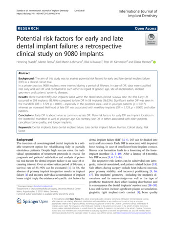
Staedt et al. International Journal of Implant Dentistry(2020) 6:81Implants of 3.75 mm were inserted more frequently (n 3360, 37%) than implants of 4.2 mm (n 2270, 25%) andlager (n 908, 10%). Early DIF occurred more often inimplants measuring 10 mm (n 134, 38.2%) in comparison to both, smaller and larger implants. Furthermore, ahigh frequency of late dental implant failure could be recorded in implants of both, 10 mm (n 51, 14.5%) and11.5 mm (n 47, 13.4%) in length (see Fig. 3). The contingency analysis showed a statistically significant correlation (χ2 13.554, p 0.004) and a medium strength ofassociation (Cramers V 0.197) between implant lengthand dental implant failure. In contrast, groups of different diameters displayed no significant correlation (χ2 2.510, p 0.474, Cramers V 0.085) with a greater incidence of early events in implants of smaller diameter(3.3 mm: n 72, 20.5%; 3.5 mm: n 88, 25.1%; 4.2 mm:n 62, 17.7%) and a maximum frequency of late lossesin implants of 3.75 mm (n 44, 12.5%) (see Fig. 4).Systemic diseasesOne hundred fifty-three patients were suffering fromcardiovascular diseases (CVD; 57.52%), 7.52% (n 20)from diabetes mellitus, and 12 patients (4.51%) showed acomorbidity of both cardiovascular diseases and diabetesmellitus.Page 5 of 10Cardiovascular diseaseIn 89 (33.46%) patients suffering from CVD, implantsfailed early, whereas late DIF happened only in 24 patients (9.02%). The contingency analysis could not showa significant correlation (χ2 1.355, p 0.244, Φ 0.071) between CVD and DIF between both groups.Diabetes mellitusFifteen patients (5.64%) suffering from diabetes mellitusshowed an early and 5 patients (1.88%) a late event without significant correlation (χ2 0.707, p 0.400, Φ 0.052) between diabetes mellitus and both, early and lateDIF.ComorbidityAnalyzing a comorbidity of CVD and diabetes mellitus, 7patients with early (2.63%) and 5 patients (1.88%) with lateDIF were recorded. The contingency analysis showed astatistically significant correlation (χ2 4.741, p 0.029)with a medium strength of association (Φ 0.134) between a comorbidity and DIF in both groups.Early and late DIF: logistic regression analysisA logistic regression analysis was performed to ascertainthe effects of the variables analyzed within this study onearly and late DIF. First, a binary logistic regressionFig. 3 Frequency of early and late dental implant failure among implants of different length. Early DIF was frequent in implants of 10 mm (n 134, 38.2%), whereas late dental implant failure showed high incidences in implants measuring 10 mm (n 51, 14.5%) and 11.5 mm (n 47,13.4%) in length
Staedt et al. International Journal of Implant Dentistry(2020) 6:81Page 6 of 10Fig. 4 Frequency of early and late dental implant failure among implants of different diameter. Groups of different diameters displayed no significantcorrelation (χ2 2.510, p 0.474, Cramers V 0.085). Implants of smaller diameter showed higher frequencies of early events (3.3 mm: n 72, 20.5%;3.5 mm: n 88, 25.1%; 4.2 mm: n 62, 17.7%) and a maximum frequency of late losses in implants of 3.75 mm (n 44, 12.5%) with no statisticallysignificant correlationanalysis was performed for the independent variables(see Table 2); thereafter, a multivariate logistic regression analysis was needed to ascertain the effects of eachfactor (see Table 3). The model for patients related factors (gender, age, jaw, CVD, diabetes mellitus) correctlyclassified 83.5% of cases with Nagelkerkes’ R2 of 0.111,which corresponds to a moderate explanatory power[31]. Analyzing the patient’s gender and age, the regression analysis did not show different probabilities forearly or late events between the groups. Maxillaryimplants showed a reduced likelihood (OR 0.260; p 0.001; CI, 0.135–0.501) for early DIF, whereas they were3.849 times as much as likely to exhibit a late event (OR 3.849; p 0.001; CI, 1.995–7.425) compared to implants placed in the mandible. Furthermore, the probabilities for early and late events did not differ betweenimplants located within the anterior or posterior area ofthe jaw. Neither for diabetes nor for CVD associationswith early and late DIF could be seen when comparingto healthy patients. The model for implant-relatedTable 2 Binary logistic regression analysisReference valueSEWaldP valueOR late DIFCI late DIFOR early DIFCI early DIFAge20-29 0.611-1.898JawMaxilla0.32816.054 gth 8 iameter3.3 VDNo Diabetes mellitusNo 1.655ComorbidityNo 77-1.1.37Abbreviations: SE Standard error, OR Odds ratio, CI Confidence interval, DIF Dental implant failure
Staedt et al. International Journal of Implant Dentistry(2020) 6:81Page 7 of 10Table 3 Multivariate logistic regression analysisReference valueSEWaldP valueOR late DIFCI late DIFOR early DIFCI early DIFAge20-29 0.611-1.898JawMaxilla0.32816.054 gth 8 iameter3.3 VDNo Diabetes mellitusNo .4490.177-1.1.37Abbreviations: SE Standard error, OR Odds ratio, CI Confidence interval, DIF Dental implant failurefactors (length, diameter) correctly classified 63.5% ofcases with Nagelkerkes’ R2 of 0.010, which correspondsto a low explanatory power. Implants of different lengthsor diameter could not be associated with an increasedlikelihood of either early or late DIF.DiscussionThe aim of the study was an investigation of potentialrisk factors for early and late dental implant failure (DIF)by including both patient and implant-related factors.Despite low failure rates [1, 32], the evaluation of potential risk factors—especially regarding their impact on themoment of implant loss—is crucial for receiving a sustainable and secure long-term provision. Limited numbers of large studies examining risk factors comparingtheir impact on early and late events are available. In thepresent study, retrospective data on 351 implants in 266routine patients inserted within a period of 10 years wereclassified into early and late DIF and compared to eachother in regard of the patient’s gender, age, site of implantation, implant geometry, and patients’ systemicdiseases.Early DIF is associated with impaired bone healing anda reduced amount of implant primary stability by insufficient bone-to-implant contact [2, 5, 8, 9]. Factors likeheat-induced necrosis and incorrect positioning maylead to impaired osseointegration [9, 16, 17] resulting inearly implant loss. Furthermore, systemic factors influencing the patient’s wound healing capability [5, 15, 20,26–29] and local inflammation could be associated withan early DIF as well [18, 25, 33]. Contrary, late DIF isdefined by a reduction of implant stability after a latencyof 6 months [5, 11, 12, 14]. This is thought to be a multifactorial process, as both implant and patient-relatedfactors influence the implants’ long-term survival. Onthe one hand, loading distribution is affected by the implant’s geometry as well as the type of prosthetic treatment in particular different occlusion concepts [18–20].On the other hand, local risk factors like plaqueaccumulation, gingivitis, bone quality and quantity, oralhygiene, periodontal disorders, and chronic occlusaltrauma determine the implants’ long-term outcome [9,21–25].The choice of a cut-off point comprises methodological problems: Event-based cut-off points like abutment connection [34]/prothesis placement [35] mightresult in an underestimation of failures due to impairedosseointegration. For example, in one-stage surgery/immediate loading, there is no time at risk. On the otherhand, a time-based cut-off point as reported by Antounet al. [36] might result in an overestimation of early DIF[13].As implant failure is more a fluent process than anevent, all cut-off points comprise of imprecisions. In alarge retrospective clinical study, Jemt differentiated between event-based and time-based cut-off points andfound a higher number of implant failures in the “timebased” group (n 81 implant failures at first year offollow-up vs. n 73 implant failures at “prothesis placement” as event-based cut-off point). Furthermore, he reported only 5% of DIF after a period of 8 months. As anevent-based cut-off point depends on the surgical protocol that might differ significantly between one patientand another, and a time-based cut-off point at first-yearresults in an overestimation of early DIF [13], this workset a cut-off point at 6 month after implant placementespecially as osseointegration takes approximately 3 to 6months (depending on implantation site and bone quality) [37, 38].As described previously [39, 40], gender did not showany correlation to DIF within this study. Within the contingency analysis, a higher age was correlated to lateevents, whereas younger patients tend to lose implantsearlier. This result corresponds with previous studies[40]. Lin et al. analyzed a total of 403 implants in anintermediate-term clinical study and detected a statistically significant OR for late implant loss for patientsolder than 40 years [14]. In case of younger patients,
Staedt et al. International Journal of Implant Dentistry(2020) 6:81Page 8 of 10early DIF may occur more often as they are more proneto an active lifestyle together with a potentially earlieroverload of the placed implants. For older patients, anetiologically correlation to the patients’ multi-morbidity,a decreased bone density, and several additional factorsassociated with age-related changes could be assumed[27, 41].In the present study, early DIF was more frequent inimplants placed in the mandible compared to the maxilla. As it impairs the process of osseointegration, thickand hard cortical bone and a reduced vascularizationmight affect the implant’s short-term prognosis. Furthermore, a significant higher rate of late DIF was seen inthe maxilla when compared to the mandible. This is inaccordance to the literature [40]. As the upper jaw consists of a wide-meshed cancellous bone, the respectivebone density is lower when compared to the mandible.This, in combination with the proximity to the sinusresulting in a reduction of bone quantity [42, 43], maycause a lower long-term stability. As studies reportcontradictory findings regarding the long-term prognosisfor implants inserted either within the anterior or posterior area of the jaw [13, 14], the present study gave evidence that posteriorly placed implants are in danger ofDIF. Even if no statistically significant correlation wasfound, early and late DIF were more frequent in the posterior jaw (early DIF, 52.63%; late DIF, 12.77%) compared to the front tooth area (early DIF, 29.32%; lateDIF, 5.26%). Impaired osseointegration resulting of thickbone and less blood supply, in combination with an occlusal overload within the early loading phase might explain these findings.Despite current research, the effect of the implants’geometry is a controversial issue. Several studies showedthat small implants failed more often and especially earlier than larger implants [24, 44, 45], whereas otherscould not show any significant correlation between implant length and early or late events [46]. The presentstudy showed implants of 10 mm in length to be positively correlated to both early and late DIF, whereas implants of bigger length were more likely to exhibit a lateevent than smaller implants. As the implants’ geometryaffects the loading distribution, a correlation to late DIFis possible [18–20]. In contrast, there was no statisticallysignificant correlation between the implant’s diameterand either early or late events within this study. Alsaadiet al. showed significantly higher late failure rates for implants of increasing diameter [24], whereas others recommended implants of wider diameter to decreasestress transferred to the surrounding bone [47]. In thestudy at hand, a heterogeneous patient collective was examined, reflecting daily praxis. Even so, this might haveled to a relevant bias as it is possible that implants of larger diameter and length were predominantly insertedinto the augmented bone of reduced quality resulting inan increased late DIF. Furthermore, it is possible thatinserted implants of predominantly used geometric features led to higher failure rates in the respective groups.Within this study, there was no statistically significantcorrelation between the occurrence of two systemic diseases and DIF even though cardiovascular diseases likehypertension are suspected to reduce bone density andhealing capacity [48–51]. Furthermore, the respectivemedication might affect bone metabolism even if in vivostudies could not show any correlation [52–56]. Diabetesmellitus is known to induce xerostomia, caries, periodontitis and deregulation of immunity [57], infectionsand a reduced healing capacity [58, 59], microvascularchanges [60], osteopenia with a 50% reduction of bonediameter and 30% less implant-bone contact [61, 62].Despite a well-documented evidence of changes in metabolism, so far, no difference could be detected in human studies [4, 39, 63–66]. In accordance to thepresented findings, Alsaadi et al. could not show an effect of cardiovascular diseases, diabetes mellitus, or thyroid dysfunction on dental implant failure [24].ConclusionThere is a lack of evidence-based information about potential risk factors for early and late DIF. However, theidentification of these risk factors is crucial for creatingindividual treatment plans. An increased likelihood ofearly DIF was present in implants located in the mandible—especially in the posterior part of the jaw. Contrary to this, a higher patient age, a localization withinthe maxilla, and a greater implant length was associatedwith late DIF. However, neither systemic diseases northe patient’s gender did show any correlation to implantfailure. As risk factors for early and late DIF differ significantly—as shown within this study—an interdisciplinary evaluation and careful analysis is crucial todevelop an individual therapeutic concept and to achievethe best possible result.AbbreviationsDIF: Dental implant failure; OR: Odds ratio; CI: Confidence interval;CVD: Cardiovascular diseasesAcknowledgementsNot applicable.Authors’ contrib
Main risk factors for early DIF are implant location in the (posterior) mandible as well as younger age. On contrary, late DIF is rather associated with older patients, cancellous bone quality, and longer implants. Keywords: Dental implants, Early dental implant failure, Late dental implant failure, Human, Cohort study, Risk factor Background