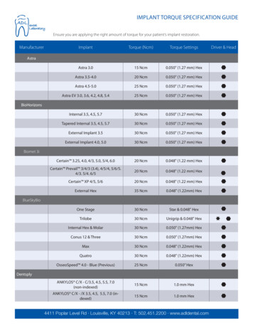
Transcription
International Journal of Research and ReviewVol.7; Issue: 10; October 2020Website: www.ijrrjournal.comE-ISSN: 2349-9788; P-ISSN: 2454-2237Original Research ArticleDental Implant Surface: Driving Force forSuccessful OsseointegrationAmit Bhandari1, Vanshika Jain2, Neha Vats3, Rashi Bhandari4, Anshu Goyal5,Vanya Srivastav61Head, 2Junior Research Fellow, 3Junior Research Fellow (Former employee),Department of Dental Research and Implantology, Institute of Nuclear Medicine and Allied Sciences, DefenceResearch and Development Organization, Timarpur, Delhi 1100544BDS, Private Practitioner, Timarpur, Delhi 1100545Scientist D, 6Scientist E,Solid State Physics Laboratory, Defence Research and Development Organization, Timarpur, Delhi 110054Corresponding Author: Vanshika JainABSTRACTBackground: Surface characteristics haveshown to influence the osseointegration propertyof dental implants. Surface alterations are donewith an aim of providing rougher surface toinduce better cell adhesion and intimateimplant-to-bone contact. Aim: To analyze thesurface characteristics of Indident DentalImplant system and to compare its surfaceproperties with three commercial dental implantsystems. Objective: To study the surfacetopography and chemical composition of fourcommercially available dental implant n of four implant systems wasstudied using optical profilometry, scanningelectron microscopy, energy dispersive . The analysis was done in tworegions; valley and top. Values and peaksobtained were studied to derive comparison andpropose reasons for success.Result: Optical profilometry showed variationin implant surface roughness at amongstdifferent location in the same implant system aswell as within different implant systems.Roughest surface was observed for Indident andAB implant systems, the findings of which wereconsistent with those of SEM images for therespective systems. SEM analysis of IndidentImplant showed amorphous pattern over thecomplete implant surface, whereas in case ofAB and Bio Horizon, top region showeddifferent morphology compared to the valley.Nobel Biocare sample showed difference inmorphology compared to rest of the implantsystems. EDX and XPS findings also correlatedwith those found via SEM analysis.Keywords: Dental Implants; Scanning ElectronMicroscopy; Energy Dispersive X-RaySpectroscopy; X-Ray PhotoelectronSpectroscopy; Optical Imaging1. INTRODUCTIONImplant based rehabilitation of losttooth has gained tremendous success overthe last decade. Newer designs are beingconstantly researched to improve clinicalsuccess compared to currently availablesystems. Dental implant surface is a criticalaspect of designing, driving towards theirsuccess in form of osseointegration.[1] Avariety of metals and metal alloys have beenused as implant material; however, titanium(Ti) and its alloys (mainly titaniumaluminium-vanadium (Ti-6Al-4V)) remainto be the material of choice.[2] on and can be achieved byusing physical, chemical or mechanicalmethods.[3] Surface modification methodscan also be classified as additive (layeredapplication of new material on implantsurface) or subtractive (removal of surfacelayer) processes, helping in achieving thedesired results.[4,5] Depending on the type oftreatment, considerable variation in theInternational Journal of Research and Review (ijrrjournal.com)Vol.7; Issue: 10; October 2020454
Amit Bhandari et.al. Dental implant surface: driving force for successful osseointegrationsurface properties have been observed,providing variable clinical results.Newer methods and chemicalcompositions have been tested to modifyimplant surface for improved properties. Toname a few, these include use of calcium(Ca),[6] magnesium (Mg),[7,8] and nanoscalediscrete crystalline deposits of calciumphosphate (CaPO4)[9] to alter surfacechemistry. Gurzawska et al foundnanocoatings of organic molecules likecarbon and graphene along with surfacemodifications with polysaccharides andglycosaminoglycans to be an effective wayto stimulate bone regeneration on boneimplant interface.[10]Many studies have individuallydiscussed the surface characteristics ofdifferent implant systems, however, fewstudies have compared the properties ofcurrently available commercial systems.The present study aimed at comparing thesurface characteristics and chemicalcomposition of four, currently marketeddental implant systems namely; IndidentTMdental implant system, AB dental implant,BioHorizons dental implant and NobelBiocareTM dental implant.2. Background of Dental Implant SystemsUsed2.1 IndidentTM Implant System (SampleA)This Implant system was developed at theInstitute of Nuclear medicine and AlliedSciences (INMAS), Defence Research andDevelopment Organization (DRDO). It is asand-blasted and acid etched implant whichhas found clinical use for single toothreplacement and implant supported denture.The implant is etched usinga patentedmethod and subjected to sandblasting withaluminium oxide powder (Al2O3) andultrasonic cleaning.[11]2.2 AB Implant System (Sample B)According to the manufacturer, this is a Ti6Al-4V implant manufactured by lasersintering. The implant undergoes specialblasting with calcium phosphate (CaPO4) toprovidemicro/nanoscaleroughness,[12]enhancing osseointegration.2.3 BioHorizon Dental Implants (SampleC)BioHorizon dental implants are LaserLoktapered internal implants, blasted withresorbable blast media. Laser-Lokmicrochannels are a series of cell-sizedcircumferential channels that are preciselycreated using proprietary laser ablationtechnology which produces extremelyconsistent micro channels, optimally sizedto attach and organize both, osteoblasts andfibroblasts.[13]2.4 Nobel BiocareTM Dental Implants(Sample D)These implants are manufactured from TiUnite, a high performance implant surfacematerial that enhances osseointegration evenunder the most challenging conditions. It ischaracterized by a moderately rough,thickened titanium oxide layer with highcrystallinity and osteoconductive propertiesleading to faster bone formation.3. MATERIALS AND METHODSThe following study was conductedin the Department of Dental Research andImplantology, INMAS, DRDO and SolidState Physics Laboratory (SSPL), DRDO.Surface characterization of four dentalimplant samples; A, B, C and D was doneusing optical profilometery, scanningelectron microscopy (SEM), energydispersive X-ray spectroscopy (EDX) andX-ray photoelectron spectroscopy (XPS).Due to lack of availability of universalimplant sizes among different commercialbrands, implant sizes of approximately thesame dimensions were selected for thepurpose of compatibility (table 1). Universalprecautions and sterility was maintained atall times to avoid surface contamination ofimplants.International Journal of Research and Review (ijrrjournal.com)Vol.7; Issue: 10; October 2020455
Amit Bhandari et.al. Dental implant surface: driving force for successful osseointegrationCOMPANYIndidentAB Implant SystemBioHorizon ImplantSystemNobelBiocareImplant SystemTable 1: Detailed description of the dental implants used in the studySURFACEDESCRIPTIONLENGTH(mm)Sand blasted and acid etchedCylindrical screw implant10mmLaser sintered surface, blasting Tapered implant with conical 10mmwith Calcium phosphateconnections groovy neckLaser-LokTapered implant using Laser 9mmAblation technologyTiUniteReplace Select Tapered10mm3.1 Optical ProfilometryEach dental implant was scanned attwo locations; top and valley region usingTaylorHobsonPrecisionOpticalProfilometer (AMETEK Inc, Germany) toassess the surface roughness. The implantswere placed on the microscope platform andresults were recorded as fringes, which wereused for analysis of roughness gradient.Surface roughness is expressed asarithmetical mean height (Sa). This devicescans the surface topography of implantsurface and quantitatively measures thesurface roughness.3.2 Scanning Electron Microscope (SEM)AnalysisCarl Zess Supra55 (Zeiss, Germany)SEM was used to observe implant surfacesat the same two positions as used in opticalprofilometry. An accelerated voltage of 20kV and vacuum maintained at 1 x 10⁻⁵ torrwas used. Implants were fixed to an Alsample holder with their long axis parallelto the holder using simple carbonconducting tape (Figure 1a). All 864.0mm15046413.5mm12102075implants were studied at a range of 70X to5000X magnification (Figure 1b).3.3 Energy-dispersive X-ray Spectroscopy(EDX)EDX was used to determine theelemental constitutes of dental implants.Implants were fixed on sample holder asdone during SEM analysis, to allow asystematic scan. 7kV accelerating voltagewas used to improve ratio for light elements.Two spectra from each implant wereacquired which were then further analysed(Figure 1c).3.4 X-ray photoelectron spectroscopy(XPS)XPS was done to study the surfacechemistry using Omicron XPS system(ScientaOmicron,Germany)usingmonochromatic A1 Kα X-ray source and abeam size of 400µm diameter. Duringspectra acquisition, electron take off anglewas fixed at 35 and vacuum pressuremaintained at below 2 x 10⁻¹⁰ torr. SurveyXPS were acquired over 1000eV andresolution of 0.6eV(Figure 1d).Figure 1: SEM setup; a: shows the placement of implants on aluminium implant holder, secured with carbon tapes, b: shows thesem analyses being done following proper sterile technique; Setup for c: EDX; d: XPSInternational Journal of Research and Review (ijrrjournal.com)Vol.7; Issue: 10; October 2020456
Amit Bhandari et.al. Dental implant surface: driving force for successful osseointegration4. RESULT4.1 Optical ProfilometrySamples B and C showed higher roughnessvalues in the valley region compared to topregions.AB (Sample B) (Sa:0.745 µm) BioHorizon(Sample C) (Sa:0.655 µm)Surface roughness values for samples A andD could not be accurately determined due tounevenness of the implant surface thusleading to failure in obtaining conclusivevalues.4.2 SEM analysisDepending on surface treatment,different peaks were obtained for eachimplant sample. Sample A showed a verycoarse, irregular and uneven surface (Figure2a). Surface of sample B too showedirregular amorphous pattern with irregularmicropores ranging from 1-2µm to even 1015µm in diameters (Figure 2b). The top areaof this implant showed microthread patternswhich were evenly distributed (Figure 3b).Sample C showed grainy surface withminute crack like patterns present along thevalley region of the implant, having a veryregular serrated pattern, with fine threadspresent all over the top region (Figure 3cand 2c). Sample D demonstrated a relativelyregular pattern with porous structures ofdifferent diameters ranging from 0.5µm to10µm with a relatively smooth top region(Figure 3d and 2d).Figure 2: SEM pictures of top areas of implants: 70 X; a:Indident Implant, b: AB Implant, c: Bio Horizon implant, d: Nobel Biocareimplant.Figure 3: EDX analysis of Implant samples at top region; a:Indident Implant, b: AB Implant, c: Bio Horizon Implant, d: NobelBiocare implantInternational Journal of Research and Review (ijrrjournal.com)Vol.7; Issue: 10; October 2020457
Amit Bhandari et.al. Dental implant surface: driving force for successful osseointegration4.3 EDX analysisThe analysis confirmed the presence of Ti in the elemental framework for allimplants. Maximum amount of Ti was found in Sample A (97.43 wt %) followed by SampleD and C with the least content found in sample B. Additional presence of Al, silicon (Si) andV along with carbon (C) and oxygen (O) was seen in sample B (Figure 2b and 4b). This alsosuggested composition of Ti-Al-V alloy sandblasted by either silicon oxide (SiO2) or siliconcarbide (SiC) microspheres. Sample C too showed presence of Al, Si, C and O in theelemental spectrum along with Ti (Figure 2c and 4c). Sample D had peaks for calcium (Ca),phosphorous (P) and O in their elemental forms all over the implant surface except the neckregion which only depicted presence of Ti (Figure 2d and 4d).Figure 4: EDX analysis of implant samples at valley region; a:Indident Implant, b: AB Implant, c: Bio Horizon Implant, d: NobelBiocare Implant4.4 XPS analysisThe survey spectra showed majorpeaks at Ti 2p, O 1s and C 1s. Presence ofTi and Al were confirmed for sample A inthe valley regions with traces of C and O.Sample B showed presence of Ti, Al, Si, Valong with C and O in the valley regionalong with presence of iron (Fe), Chromium(Cr), Nickel (Ni), Manganese (Mn)elements in the top region of the implant.Sample C depicted Ti, Al, V over the topregions and a high probability of Ti-Al-Sialloy present in the valley region. XPSanalysis of sample D was suggestive ofmaterial coating containing constituents ofCa, P, O and C. The binding energies of Caand P were found to be shifted towards thehigher side which may be an indicativetowards coating of hydroxyapatite (HA)Ca10(PO4)6(OH)2 on implant surface.5. DISCUSSIONSurface properties can be cal properties.[14] In the surfaceproperties of dental implants, topographicand physiochemical changes can beemployed to improve osseointegration andprimary implant stability.[15-17] A range ofsurface treatment techniques have beenadopted, with each resulting in implantsurface varying in morphology andchemistry. Strnad et al in their studyconcluded that SLA surfaces show strongerbone response and highest amount of boneto-implant contact.[18] Supporting to this factare the studies by Elias[19] and Ballo et al,[20]affirming that these procedure increase thesurface roughness over the implant,promoting rapid osseointegration. In thisstudy,four,clinicallysuccessful,commercially available dental implant wereInternational Journal of Research and Review (ijrrjournal.com)Vol.7; Issue: 10; October 2020458
Amit Bhandari et.al. Dental implant surface: driving force for successful osseointegrationsubjected to in-vitro analysis to comparetheir surface characteristics.The surface roughness is an essentialcomponentofsurfacetopography,enhancing osseointegration[17] and wascompared through 3D evaluation. It wasexpressed as Sa which describes 3Droughness measurement and is more reliableand advanced method than 2D measurement(Ra) of the roughness quotient. Numerousstudies have shown surface roughness oftitanium implants to affect the rate ofosseointegrationandbiomechanicalfixation. [21-23] Roughness values between 1to1.5µm have shown to provide optimalsurface for proper bone integration. In ourstudy we saw different surface roughness atdifferent locations in the same implant.Sample B and C showed increased surfaceroughness over top areas as compared to thevalley region. This can be due to thepresence of microthread pattern present inboth these implant systems. No significantdifference in surface values at the two siteswas noted for sample D.SEM study of these implant systemsrevealed the surface morphology and effectof manufacturing process on implantsurface. Sample A has been developed as acommercially pure Ti derived cylindricalimplant which is sand blasted and acidetched. Likewise, sample B has also beenstated to have undergone blasting withCaPO4. We believe, this maybe the reasonthat both these implants showed similarityin their SEM derived topographicalfindings. Irregular grainy particles of varieddimensions were seen in both the samples;however, sample B showed particles ofmore uneven shapes and sizes withprominent cracks, showing appearance ofmolten matter in between. These particleswere present in a layered pattern resulting increation of a more rough surfaced alloy withvery uneven surface. Microthread present onthe surface of sample B implant was yetanother distinguishing finding. Sample Calso showed the presence of fine, regulargroove like pattern around the top regionwith minute crack like regions in the valley.These findings were in accordance withthose of Brunette et al and Massaro et al onmachined turned or blasted implants.[24,25]Findings for sample D showed presence ofminute pores over the entire implantsurface.EDX and XPS analysis for thepurpose of surface chemistry showed Ti, Al,O and C as major elements in the elementalframework for all systems studied. Thesefindings were consistent with previousfindings by Massaro et al,[25] Olefjord etal,[26] and Kang et al.[27] AB implant sampleshowed the presence of Ti, along with Aland V, suggestive of Ti-Al-V alloy. XPSgraphs also showed the presence of Fe, Mn,Cr and Ni peaks. Similarly, BioHorizonimplant also had traces of Al and V over theimplant. Apart from these, XPS and EDXanalysis of Nobel Biocare implant suggest acoating of Ca and P. Binding energies of Caand P are seen to be shifting to higher sidesuggestive of presence of hydroxyapatitelike coating over the implant surface.6. CONCLUSIONThis study displayed the distinctsurface characteristics of four implantsystems which are actively used in clinicalpractice. Slight variations in surfaceroughness and composition were seen foreach system with samples A and B showingthe roughest surface among all. Differencesin modeling were also seen with presence ofmicrothreads in sample C. The current studyproposes for conduction of similar analysisfor other implant systems and comparison ofthe findings with clinical results to be ableto find a combination that can providesuperior holistic clinical success.ACKNOWLEDGEMENTThe project has been funded by DefenceResearch and Development Organization(DRDO), Ministry of Defence, Governmentof IndiaConflict of InterestThis research is free of any conflict ofinterest.International Journal of Research and Review (ijrrjournal.com)Vol.7; Issue: 10; October 2020459
Amit Bhandari et.al. Dental implant surface: driving force for successful osseointegrationData availabilityThe raw/processed data required to reproducethese findings cannot be shared at this time asthe data also forms part of an ongoing study.REFERENCES1. Albrektsson T, Branemark PI, Hansson HAet al. Osseointegrated titanium implants.Requirements for ensuring a long-lasting,direct bone-to-implant anchorage in man.Acta Orthop Scand. 1981;52(2):155-70.doi:10.3109/174536781089917762. Saini M, Singh Y, Arora P et al. Implantbiomaterials: A comprehensive review.World J Clin Cases. 2015 Jan 16; 3(1): 52–7. doi: 10.12998/wjcc.v3.i1.523. Kumar KA, Bhatt V, Balakrishnan M et al.Bioactivity and surface characteristics ofTitanium implants following various surfacetreatments: An in vitro study. J oi-D-13-002924. Takashima H, Shibata Y, Kim TY et al.Hydroapatite coating on a titanium metalsubstrate by a discharging method inmodified artificial body fluid. Int J OralMaxillofac Implants. 2004;19(1):66-72.5. Gaggl A, Schultes G, Muller WD et al.Scanning electron microscopical analysis oflaser-treated titanium implant surfaces-acomparative study. Biomaterials. -86. Sul YT, Byon ES, Jeong Y. Biomechanicalmeasurements of calcium-incorporatedoxidized implants in rabbit bone: effect ofcalcium surface chemistry of a novelimplant. Clin Implant Dent Relat b00032.x7. Sul YT, Johansson C, Chang BS et al. Bonetissue responses to Mg-incorporatedoxidized implants and machine-turnedimplants in the rabbit femur. J ApplBiomaterBiomech. 2005;3(1):18-28.8. Sul YT, Johansson C, Albrektsson T. Whichsurface properties enhance bone response toimplants?Comparisonofoxidizedmagnesium, TiUnite, and Osseotite implantsurfaces. Int J Prosthodont. 2006;19(4):31928.9. Mendes VC, Moineddin R, Davies JE.Discrete calcium phosphate nanocrystallinedeposition enhances osteoconduction on10.11.12.13.14.15.16.17.18.19.titanium-based implant surfaces. J BiomedMater Res A. 2009;90(2):577-85. doi:10.1002/jbm.a.32126.Gurzawska K, Svava R, Jørgensen NR et al.Nanocoating of titanium implant
of dental implants. Surface alterations are done with an aim of providing rougher surface to induce better cell adhesion and intimate implant-to-bone contact. . dental implant system, AB .