Transcription
Workshop 4 - Challenges of implant-supported rehabilitation in the aesthetic areaAngel Mangarelli1, Luis Guzzetti1, Javier Trinidad2.1Dentist. Assistant Prof., Department of Rehabilitation, Fixed Prosthodonticsand TMD. Professor at the Specialization Course in Maxillofacial Implantology. Member of the Department of Oral and Maxillofacial Implantology. Schoolof Dentistry Universidad de la República, Uruguay2 Dentist.School of Dentistry, Universidad de la República. Uruguay.INTRODUCTIONThe outcomes of the practice of replacing missing dental organs with implantshave reached such a degree of universalization and certainty(1) that it has become an everyday tool in the clinical practice. Nevertheless, the better outcomes brought about greater demands from patients and also from professionals, who strive to reach dental aesthetics and, especially, the gingival aestheticsof natural teeth.Achieving pleasing gingival aesthetics in teeth restored with implants has proven the main challenge to address in this branch of the profession. The literaturereview on this subject matter to be discussed in a workshop environment, aiming towards a consensus, was designed to make a contribution in this area.Workshop methodology. The workshop was structured in the following stages:- Literature review. The people leading the seminar accessed the Timbó,Pubmed, Medline and Lilacs portals, reviewed the literature of the last tenyears, and selected 23 studies, all of them conducted on humans, considered to be representative of the topic under discussion.
- Guiding questions. Four guiding questions representing the subject matterof the seminar and the literature selected were formulated. The questionswere:1. What are the causes of gingival recession?2. How important is the management of the provisional restoration in shaping peri-implant tissues in the aesthetic area?3. What is considered optimal management of abutments regarding design,materials and aesthetics?4. What aesthetic evaluation indices are there?- Discussion with workshop participants. The selected literature and theguiding questions for discussion were emailed to participants for them to readand evaluate, and they were invited to two prior meetings to begin the scientific exchange.- Scientific review. It was decided that a scientific reviewer should be presentat the workshop without participating in the discussion. He would evaluate thequality of the suggested literature, the representativeness of the guiding questions, the scientific level reached during the discussion on the day of the seminar, and the connection between the conclusions of the seminar and the guiding questions and bibliography.Workshop participants. The following professionals attended the workshop:Drs. Javier Trinidad, Enrique Elhordoy, Mariana Seoane, Natalia Panissa, Viviana Rocha, Sergio Montenegro, Carla Laurino, David Durán, Fernando Indart,and Susana Borrás.Workshop.
Question No. 1. What are the causes of gingival recession linked to dentalimplants? In the workshop it was determined that gingival recession is associated to the following kinds of factors: A) Intrinsic (related to the patient) andB) Extrinsic (related to technical aspects), and that they are probably closelyrelated to each other.A. Intrinsic factorsA.1. Complete or partial absence of the vestibular table at the time of implantplacement. The evidence from the literature consistently indicates that the riskof gingival recession in these cases is very high. Placing implants in sites withvestibular bone defects frequently leads to soft-tissue recession, with the potential risk of altering the harmony of the gingival margin(2,3). In this situation, it isrecommended to delay implant placement. In a literature review, Chen (2009)(4)concluded that regeneration procedures are effective in reconstructing defectsin the vestibular table, in type 1 (immediate placement) and type 2 (earlyplacement) implant placement situations. Despite this, less vestibular bone losswas observed with type 1, which has consequences in the aesthetic area. Onthe other hand, if the vestibular table is intact, and provided there is no acutedisease, the implant could be placed immediately(5,6). There is no scientific evidence that early placement guarantees a better outcome than immediateplacement when correctly indicated, whether with or without provisionalization(5,7,8). In order to reduce the treatment time, in 2008 Da Rosa(9)proposed, forcases with partial or total loss of the vestibular table, the Immediate Dentoalveolar Restoration (IDR) technique. The purpose of this technique is to repair thedefect in the socket with a corticomedullary bone graft from the maxillary tuber-
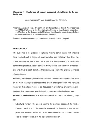
osity and, simultaneously, to place the implant and perform immediatenon-occlusal loading.A.2. Absence of soft tissue. Delaying implant placement(10) or performing softtissue grafts and the placement simultaneously is recommended. It is currentlybelieved that an adequate amount of soft tissue helps prevent marginal recession, hide the margins of the restoration and disguise the shadow in the implantplatform(11,12) (Figs. 1, 2). Moreover, from an aesthetic perspective, an adequateamount of keratinized tissue is crucial as it allows for a harmonious gingivalcontour without inflammation, light pink and spotted, synonymous with pink aes-thetics. It is therefore impossible to achieve adequate aesthetic outcomes in theaesthetic area without the right amount of keratinized tissue.Fig. 1Fig. 2A.3. Gingival biotype. A direct link between the gingival biotype and the finaloutcome has not been proven. A limited number of clinical studies which haveresearched the link between the gingival biotype and the aesthetics of implantswere found in the available literature, with some authors claiming that a thickgingival biotype does not guarantee that there will not be any gingival recession(13,14). Despite this, the literature agrees that a thick gingival biotype is a de-
sirable characteristic which will positively impact the aesthetic outcome of animplant-supported restoration since it is more resistant to mechanical and surgical insult, making it less susceptible to gingival recession. (Fig. 2). Changing thebiotype with connective tissue grafts could be considered for thin gingival biotypes. Some authors claim that the influence of the gingival biotype would manifest in the gingival margin, and not in the gingival papillae, an entity that couldbe influenced by other kinds of factors, such as the distance from the bonecrest of the adjacent tooth to the contact point of the restoration(7,15).A.4. General factors which may be contraindications for implant therapy. Asuitable assessment of the general condition of the patient is something important to be considered when planning a dental implant-based treatment. It isworth noting that the need for implants increases with the age of the patient,therefore, these treatment plans very often need to be tailored to their generalcondition. Patients who are deemed high risk due to chronic general diseases,and use of tobacco or a medication which affects bone tissue, should be treatedwith caution, since aesthetic outcomes are less predictable for them(6).B. Extrinsic factorsB.1. Correct implant selection. Aesthetic failures can occur when selecting aninappropriate implant, mainly as a consequence of using large diameter implants. The implant selected must not come into contact with the vestibular tablein order to leave a gap between them, in anticipation of the dimensional changes in the socket after extraction(8) (Figs. 3, 4). Evans and Chen (2008)(13) foundthat contact between the implant shoulder and the vestibular table was a significant factor in gingival recession. This proximity to the vestibular table can bedue either to incorrect implant placement or to the fact that the diameter was
incorrectly selected. Selecting the diameter of the implant based solely on thedimensions of the tooth to be replaced, or using implants with enlarged platforms or wide-neck implants, must be avoided. In these cases, the implantshoulder can be too close to the vestibular bone and adjacent teeth, considerably increasing the risk of recession(2) (Fig. 5).Fig. 3Fig. 4Fig. 5B.2. Three-dimensional position of the implant. There is agreement in the literature that a correct three-dimensional position of the implant is one of the main, ifnot the main, factor for good aesthetic outcome with implant-supported restorations. The relation between the implant shoulder and the planned restorationwill provide stability to hard and soft tissues. According to most authors, the correct 3D position of the implant is as follows:a. In the apical-coronal dimension, the implant platform must be located 3 or4 mm from the gingival margin expected for the future crown.b. In the buccal-palatal dimension, the emergence of the chimney should be atthe level of the cingulum of the tooth to be replaced and the labial surface ofthe implant platform should be 1 or 2 mm palatal to an imaginary line passing through the most convex portion of the two adjacent teeth at the level ofthe gingival margin.
c. In the mesial-distal dimension, at least 1.5 mm from the adjacent tooth atthe level of the bone crest and with a 3 mm distance between implants(2,13,14)(Fig.6).Fig. 6B.3. Implant design. Platform switching implants, that is, implants with a connection in which the diameter of the abutment is smaller than the diameter ofthe implant, are recommended for the aesthetic area. The gap is thereforemoved toward the center of the implant, which reduces crestal bone resorptionand allows for an increased growth of soft tissue(14,16,17) (Fig. 7).Fig. 7B.4. Surgical technique. To finish with gingival recession, it is worth noting thatalthough there is literature on it, it is not conclusive as to whether flapless techniques have better outcomes than flap techniques(18). As for immediate im-
plants, there is scientific evidence that flapless placement and immediate provisionalization result in better gingival stability.Question No. 2. How important is the management of the provisional restoration in shaping peri-implant tissues in the aesthetic area?-Gingival anatomy in natural dentition. The soft tissue that surrounds the crownsof natural teeth has a scalloped shape. This shape follows the design of thebone crests of the dental sockets –which are also scalloped– and these, in turn,mirror the scallop of the cementoenamel junction of the teeth. This junction iscloser to the apical portion in the zenith of the free surfaces, and about 2 or3 mm more toward incisal in the center of the proximal faces(19, 20).-Usefulness of implant-supported provisional restorations. Restorations on implants must meet a series of requirements, some of which are aesthetic. Significant progress has been made in ceramic restoration materials in recent times,along with the growth in the aesthetic demands of patients. But an optimal aesthetic outcome does not depend solely on correctly replicating the hard tissuesof the tooth (white aesthetics); it must be accompanied by the right anatomy ofthe surrounding soft tissue (pink aesthetics)(21). Provisional restorations are fundamental in all areas of restorative dentistry. Specifically, in implant rehabilitations, they serve different purposes in the stages prior to the final rehabilitation,making it possible to assess and decide on crown shapes and positions, curvatures and occlusal planes, incisal guide angle, among other clinical benefitswhich the dental technician will then transfer to the final restoration. They alsoplay an important part in the maturation and shaping of the soft tissues whichsurround the implant, both by attempting to maintain in the best way possible
the gingival anatomy of a tooth which was just removed, and altering the flatshape of the mucosa of an edentulous ridge, trying to replicate the naturallyscalloped shape of the gums surrounding natural teeth. Afterwards, it is possible to transfer to the laboratory the shape obtained in the soft tissues during thephase with provisional restorations, when the final impression the dental technician(22) is taken using personalized transfer copings (Figs. 6, 8).Fig. 8-The problems of dental avulsion and the peri-implant anatomy of gums. Whenteeth are missing, there is major loss of volume of the alveolar ridge in the buccal-palatal dimension(23), with minimal height bone loss(24). In the case of individual edentulous gaps, which are adjacent to natural teeth with a healthy periodontium, the gap will remain in its place thanks to the conservation of the periodontium in the adjacent tooth. In edentulous ridges that have already beenremodeled, where two or more adjacent teeth are missing, the height of thebone anatomy is approximately 1 mm more apical to where the vestibular bonecrest of the missing teeth was, and is usually flat, not scalloped, due to the lossof the proximal bone crest that was between the teeth. The oral mucosa thatlines said bone crest also has a flat shape. This is why in large gaps, after placing a provisional prosthesis on a dental implant, we find that the anatomy of softtissues is flat because the anatomy of the edentulous bone crest is also flat.
There will be a height deficit in peri-implant soft tissues in the part corresponding to the papillae. However, and depending on whether the three-dimensionallocation of the implant is correct(2), we will often have excess soft tissue in thevestibular surface. We must remember that the soft tissue can fill a maximum of5 mm vertically between two natural teeth, measuring the distance from thebone crest to the base of the contact point(25). Nevertheless, average clinicalmeasurements of soft tissue height between two implants were 3.4 mm(26). Another negative aspect to consider is the loss of height of the interdental bonecrest after the two adjacent teeth have been removed. Therefore, soft tissuebetween two adjacent implants will be approximately 1.5 mm thinner, and theinterproximal bone crest will be 3 mm more apical in relation to what happensbetween two natural teeth with a healthy periodontium.-Usefulness of provisional restorations in post-extraction immediate implants.Immediate placement techniques, performed in the hope that the functionalstimulation provided by the implant would prevent alveolar bone resorption,have been widely recommended in recent years. In the first years of this century, the research of Araujo, among others(27), made it very clear that this assumption was wrong. Similarly, it is claimed that the immediate provisionalization of the implant placed right after extraction will maintain the position of themarginal gingiva unchanged, since placing an artificial crown that supports thesoft tissues will prevent them from collapsing. It is very clear that immediateprovisionalization provides great psychological and functional advantages topatients, aside from reducing treatment times and the number of surgeries.However, clinical research does not seem to be conclusive in showing clearadvantages in final outcomes regarding the position of the vestibular marginal
gingiva and the proximal papillae (7,28,29). Also, with immediate provisional restorations after extractions, with or without occlusal contact, special attention mustbe paid to the shape and thickness of the subgingival portion of the implant.Current criteria focus on avoiding subgingival overcontours, which reduce spacefor soft tissues and are responsible for their tendency towards retraction. Otherfactors, such as the three-dimensional position of the implant, its diameter andthickness, and the position of hard and soft tissues, greatly influence the finalposition of the soft tissue that surrounds the implant and its restoration, asidefrom the placement of an immediate provisional prosthesis at the time of extraction (Figs. 7, 9, 10, 11).Fig. 9Fig. 10Fig. 11-Usefulness of provisional restorations in the modeling of peri-implant soft tissue. Starting from a flattened gingival anatomy, and having excess tissue in themiddle vestibular zone, the vestibular gingiva can be moved toward the apicalsurface very easily, overcontouring the provisional restoration subgingivally inthat zone(30). Likewise, it is also possible to move the zenith of the vestibulargingiva in the mesial or distal direction until said gingival margin is in a positionthat is correlated to the aesthetic standards established for the specific tooth
and is in harmony with neighboring teeth. The possibility of changing the position of soft tissues in the proximal area between two adjacent implants is notthat clear. In this case, the goal is not to retract the gingival margin in the apicaldirection, on the contrary, we should make it grow in the incisal direction. Techniques consisting of pressuring proximal tissues laterally in order to make themgrow have been proposed(30,31). These papers describe a compression technique wherein proximal soft tissues are compressed using the provisional restorations, after which they are reduced a few days later to allow for said tissues togrow (Figs. 6, 7, 8). In any case, we still need comparative clinical studies, withaccurate clinical measurements which scientifically prove that these techniquesachieve the desired outcomes. In contrast, papers referring to these techniquesmerely describe them, with some clinical illustrations, but nothing that wouldallow for definitive conclusions regarding whether better dimensions areachieved in interproximal tissues compared to the clinical situation in which provisionals are not used for compression. We must also consider the fact that papillae between implants have a natural tendency to grow somewhat within thefirst months after crown placement(32,33). It would be important to design clinicalstudies aimed at comparing the outcome in the final position of soft tissues between adjacent implants or between an implant and a natural tooth, havingused the compression technique with provisionals, and not having used it. Outcomes should be assessed based on objective aesthetic indices(21) and takingmeasurements to compare, whenever possible, the position of said tissues before avulsion and after the treatment has been completed. It would also be desirable to compare the position obtained in relation to the soft tissue of the contralateral teeth in order to draw valid conclusions.
-Provisional restorations and cement-retained prostheses. Provisional restorations are also essential in cement-retained restorations. Excess subgingivalcement is being cited as a risk factor for peri-implant disease(34). It has beensuggested that margins should not be located deeper than 2 mm in relation tothe free edge of the peri-implant gingiva. If the restoration is completed after asufficient tissue maturation process with screw-retained provisionals, we canleave the margins of the restoration a short distance from the free edge of thegingiva, with a small risk of them being visible in the future. Therefore, screwretained provisional restorations are recommended over cement-retained ones.When placing cement-retained crowns, a minimal perforation, measuring 1 mmin diameter, located on the occlusal or palatal surface, helps avoid the confinement effect of the cementing material, by guiding the material out through theperforation and, consequently, preventing it from projecting beyond the crownmargins. In conclusion, aside from the psychological and functional advantagesthe patient gets from having provisional fixed restorations for some time beforethe final rehabilitation, the aesthetic evaluation by both patients and professionals, for long periods of time, allows for better outcomes in the end. Moreover, byworking in this manner, at the time the final impressions are taken, soft tissueswill have a shape that is evaluated and stable enough to set the permanent positions of the final restoration, and unexpected changes in the stages afterplacement will be less likely. Aside from the limitations of interimplant soft tissues in the final outcome, provisional restorations play a vital role in the stagethat goes from the end of the osseointegration period until the placement of thefinal restoration.
Question No. 3. What is considered optimal management of abutmentsregarding design, materials and aesthetics? Different causes have beenproposed in the literature to explain the loss of the peri-implant bone crest,some of which are: accumulation of stress due to occlusal forces, thin bone tissue and peri-implant mucosa, and recovery of the biological width naturally,surgical trauma and the implant-abutment connection, among others. Thepreservation of the bone crest is a fundamental objective in the aesthetic area,as a way of achieving a harmonious gingival architecture, thus avoiding gingivalrecession. Abutment selection plays a key role in this. Based on the literaturereviewed, guidelines were established to select them correctly:-Shape. The shape of the abutment is one of the factors to consider. Usingstraight abutments or abutments with diameters smaller than the implant platform is advisable to preserve as much peri-implant soft tissue as possible. Authors such as Zuhr(35) recommend a subcontoured abutment, which favors theadaptation of gingival tissues during healing and prevents gingival recessiondue to excessive compression (Fig. 10).-Platform. Another aspect to consider to preserve the bone crest is platformswitching, in which the diameter of the abutment is smaller than the implantplatform. Thus, the implant-abutment microgap is horizontally relocated, whichcreates a healthy biological width.-Type of connection. The literature is not clear in determining whether one typeis more favorable to preserve the bone crest, and none has been shown tohave an inactive microgap since microbial contamination through the implantabutment space is found in all systems(36,37).
- Thickness of the gingival tissues. It is a significant factor in the selection of theabutment material. For gingival thicknesses smaller than 2 mm, zirconia displays better optical properties than metallic abutments(38).-Biocompatibility of the different materials. In some studies, zirconia abutmentsseemed to favor the health of gingival tissues given that there might be less biofilm adherence than with titanium abutments(39). Other authors, however, foundno differences when comparing these materials(40). Regarding epithelial adhesion, in a study conducted by Belser et al.(40) titanium and ceramic abutmentsshowed a better performance compared to gold alloy and ceramic fused to metal abutments. Other authors mention that these outcomes depend on the adhesion properties of the materials studied and their differences in terms of resistance to corrosion(39). According to the literature available today, it is difficultto reach a conclusion on the advantages of one material over others, sincethere are variables that were not considered in the different studies, includingthe degree of micro-roughness of the abutment, or the cleaning and sterilizationprocedure it requires before use. Studies that include all those variables arenecessary to reach final conclusions for selecting the ideal material for abutments. There is agreement in the aesthetic sector in indicating zirconia abutments. However, different authors have recommended caution when usingthem due to the small scientific evidence available of long-term outcomes(41).The authors of this work think that there are technical problems as a result ofusing zirconia as the material for abutments, such as the impossibility of replicating some internal connection systems, the fracture of the abutment whentorque is applied to the retention screw, or the damage to the hexagon of theimplant. To solve these problems, the industry offers zirconia abutments with
metal bases that can be cement-retained (Ti Base, Biomet), which tends to increase the volume of the abutments in the emergence at the implant level. Thiscan be a disadvantage in some clinical situations, for example, for lateral incisors (Figs. 11, 12).Fig. 12Question No. 4. What aesthetic evaluation indices are there? The literaturepresents varied attempts at objectively measuring the degree of success of thedifferent treatments on implants. Different authors have tried to develop a validmethod (using different parameters). In the opinion of the workshop participants, none covers all the variables that can be considered to measure success(42,43). Despite this, the Belser index was chosen as the most complete onein terms of evaluation criteria, since it evaluates both soft tissues: Pink EstheticScore (PES) and the restoration: White Esthetic Score (WES). The PES coversfive variables: mesial papilla, distal papilla, curvature of the facial mucosa, levelof the facial mucosa, convexity, color and texture on the facial aspect of thezone of the implant. These variables are numbered from 0 to 2, with 2 being themaximum. The WES focuses on the restoration of the implant. It is based onfive parameters: shape and volume of the clinical crown; color and texture of thesurface, and translucency. A score of 0, 1 or 2 is assigned to each parameter,therefore, for an optimal restoration, the maximum WES is 10. The five parame-
ters are evaluated by comparing them directly to the adjacent or contralateralnatural tooth. Therefore, the highest possible PES/WES score is 20, whichmeans that the peri-implant soft tissues and the clinical crown of the implant area complete match to the reference tooth(21).CONCLUSIONSThe topic of this workshop is one of the most interesting in the current literature.Achieving aesthetically pleasing outcomes is the main objective of any treatment in the anterosuperior sector, but this depends on the evaluation of multiplefactors that the professional must consider when selecting the treatment. Thesuccess of implant-supported restorations is the result of the right diagnosisand, then, of a treatment plan that considers the advantages and disadvantagesof the different options available. Despite this and even if the case has beencorrectly evaluated, small deviations occur frequently with discouraging aesthetic outcomes.Scientific review. The workshop leaders selected papers that were representative of the subject matter to be discussed, based on which they asked questionsthat covered the contents of those papers, facilitating an enriching scientific exchange among participants. The workshop followed the planning and coordination schedule. The members of the workshop showed varying degrees of leveland participation. Few participants knew the literature suggested and the valueof publications based on the type of study (systematic review, meta-analysis, aseries of clinical cases). The discussion resulted in the conclusions presented inthis paper. These conclusions are representative of the literature reviewed andthe scientific development of the workshop.
REFERENCES1. Papaspyridakos P, Chen CJ, Singh M, Weber HP and Gallucci GO. Successcriteria in implant dentistry: a systematic review. J Dent Res. 2012; 91: 2428.2. Buser D, Martin W and Belser UC. Optimizing esthetics for implant restorations in the anterior maxilla: anatomic and surgical considerations. Int J OralMaxillofac Implants. 2004; 19 Suppl: 43-61.3. Hammerle C, S.TChen and Wilson TG. Consensus statements and recommended clinical procedures regarding the placement of implants in extractionsockets. Int J Oral Maxillofac Implants. 2004; 19: 26-8.4. Chen ST and Buser D. Clinical and esthetic outcomes of implants placed inpostextraction sites. Int J Oral Maxillofac Implants. 2009; 24 Suppl: 186-217.5. Tortamano P, Camargo LO, Bello-Silva MS and Kanashiro LH. Immediateimplant placement and restoration in the esthetic zone: a prospective studywith 18 months of follow-up. Int J Oral Maxillofac Implants. 2010; 25: 345-50.6. Belser U, Buser D and Higginbottom F. Consensus statements and recommended clinical procedures regarding esthetics in implant dentistry. Int J OralMaxillofac Implants. 2004; 19 Suppl: 73-4.7. Kan JY, Rungcharassaeng K, Lozada JL and Zimmerman G. Facial gingivaltissue stability following immediate placement and provisionalization of maxillary anterior single implants: a 2- to 8-year follow-up. Int J Oral MaxillofacImplants. 2011; 26: 179-87.8. Levin BP and Wilk BL. Immediate provisionalization of immediate implants inthe esthetic zone: a prospective case series evaluating implant survival, esthetics, and bone maintenance. Compend Contin Educ Dent. 2013; 34: 35261.9. Martins Da Rosa, JC. Implantes con Carga Inmediata en Alveolos Comprometidos. Edit Santos Brazil, 2012.10.Buser D, Chen ST, Weber HP and Belser UC. Early implant placementfollowing single-tooth extraction in the esthetic zone: biologic rationale andsurgical procedures. Int J Periodontics Restorative Dent. 2008; 28: 441-51.
11.Chung DM, Oh TJ, Shotwell JL, Misch CE and Wang HL. Significance ofkeratinized mucosa in maintenance of dental implants with different surfaces.J Periodontol. 2006; 77: 1410-20.12.Schrott AR, Jimenez M, Hwang JW, Fiorellini J and Weber HP. Five-yearevaluation of the influence of keratinized mucosa on peri-implant soft-tissuehealth and stability around implants supporting full-arch mandibular fixedprostheses. Clin Oral Implants Res. 2009; 20: 1170-7.13.Evans CD and Chen ST. Esthetic outcomes of immediate implant place-ments. Clin Oral Implants Res. 2008; 19: 73-80.14.Fu JH, Lee A and Wang HL. Influence of tissue biotype on implant es-thetics. Int J Oral Maxillofac Implants. 2011; 26: 499-508.15.Chow YC, Eber RM, Tsao YP, Shotwell JL and Wang HL. Factors asso-ciated with the appearance of gingival papillae. J Clin Periodontol. 2010; 37:719-27.16.Lazzara RJ and Porter SS. Platform switching: a new concept in implantdenti
als, who strive to reach dental aesthetics and, especially, the gingival aesthetics of natural teeth. Achieving pleasing gingival aesthetics in teeth restored with implants has prov-en the main challenge to address in this branch of the profession. The literature review on this subject matter to be discussed in a workshop environment, aim-